采用IBM-SPSS 26.0统计学软件分析数据。对本研究参数值均进行描述性检验统计和采用Kolmogorov-Smirnov检验正态性;应用成对样本 t 检验或 Wilcoxon 符号秩检验来分析 SMILE术前后各项参数的变化;单因素方差分析及LSD-T检验事后多重分析不同角膜厚度组的差异性;由于观察参数变量之间存在多重共线性且样本量小,通过预测误差均方根(root mean square error of prediction,RMSEP)确认主成分数量后进行偏最小二乘回归(partial least square linear regression,PLSLR)和Spearman相关性有效性检验分析相关性。以P < 0.05为差异有统计学意义。
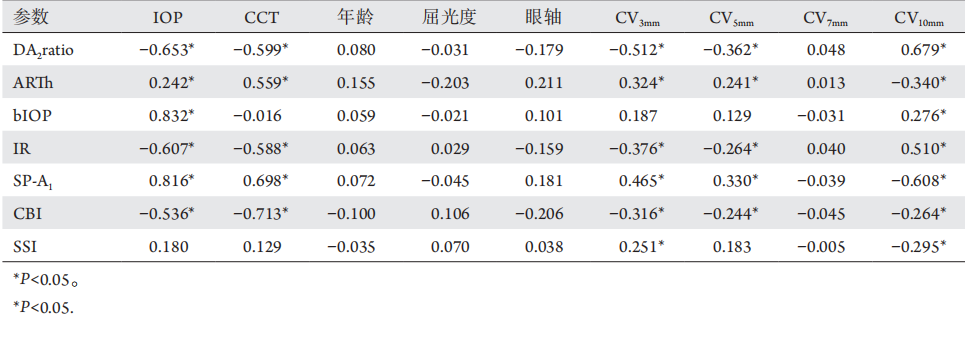
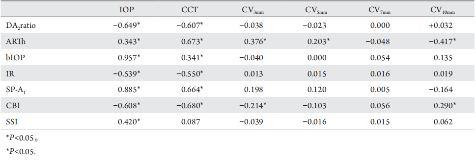
术前 bIOP 、 DA 2 ratio 、 I R 、 ARTh 、SP-A 1和 CBI 与眼压均有相关性 ( P < 0.05) ,DA2ratio、I R、ARTh、SP-A 1和CBI与CCT显著相关( P < 0.05),bIOP和SSI与CCT无相关性(P>0.05),SSI与眼压无相关性(P>0.05);DA2ratio与CV 3mm、CV 5mm呈负相关( r =?0.512、?0.362,P < 0.05) , 与 C V 10mm呈正相关 ( r = 0.679 ,P < 0.05) ; ARTh 与 CV 3mm、 CV 5mm呈正相关( r =0.324、0.241,P <0.05),与C V10mm呈负相关
2.4.2 SMILE 术后 3 个月角膜生物力学参数与 CV等形态及结构参数的相关性
SMILE术后3个月bIOP、DA2ratio、IR、ARTh、SP-A1、CBI和SSI与眼压均有相关性(P <0.05),DA2ratio、IR、ARTh、SP-A1和CBI与CCT显著相关(P<0.05),SSI与CCT无相关性(P>0.05);ARTh与CV3mm、CV5mm呈正相关(r=0.376、0.203,P<0.05),与CV10mm呈负相关(r=?0.417,P<0.05);CBI与CV3mm呈负相关( r =?0.214,P <0.05)、C V10mm呈正相关(r=0.290,P<0.05,表8)。
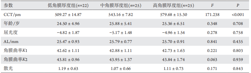
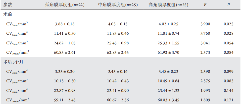
表3 不同厚度组的不同区域CV比较
Table 3 Comparison of CV in difffferent areas of difffferent thickness
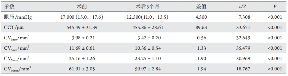
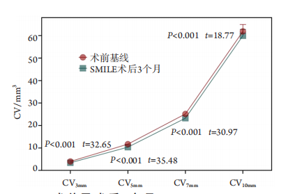
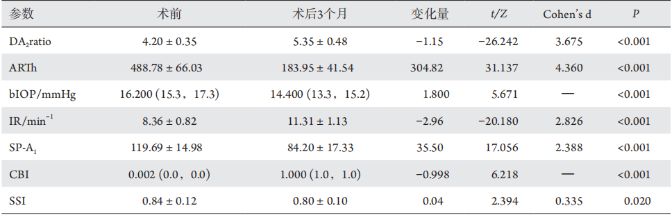
表4 SMILE术前和术后3个月角膜生物力学新型整合参数的变化
Table 4 Changes in corneal biomechanical parameters before and 3 months after SMILE
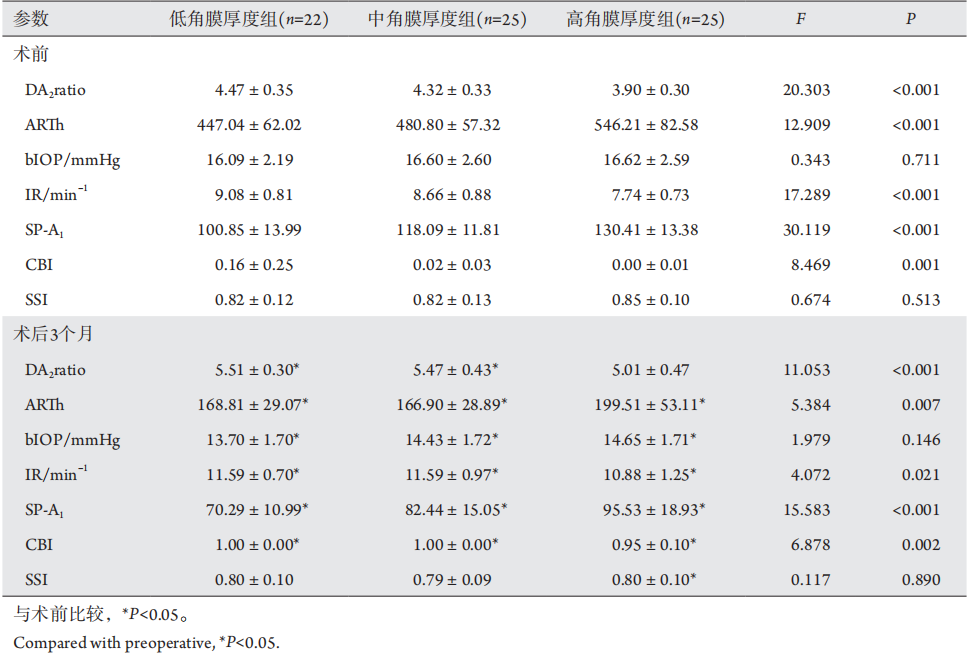
表5 不同厚度组SMILE术前和术后3个月角膜生物力学新型整合参数的比较
Table 5 Comparison of new corneal biomechanical integration parameters for SMILE of difffferent thickness groups before and 3
months after SMILE
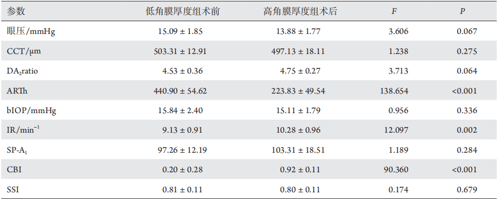
表6 16例低角膜厚度组术前角膜生物力学参数和16例高角膜厚度组术后的角膜生物力学参数对比
Table 6 Comparison of corneal biomechanical parameters between 16 patients with low corneal thickness before SMILE and 16
patients with high corneal thickness after SMILE
表7 SMILE术前角膜生物力学参数与术前角膜体积等形态及结构参数的相关性
Table 7 Correlation between corneal biomechanical parameters before SMILE and preoperative morphological and structural
parameters before SMILE
表8 SMILE术后3个月角膜生物力学参数与形态及结构参数的相关性
Table 8 Correlation of corneal biomechanical parameters with morphological and structural parameters 3 months after SMILE
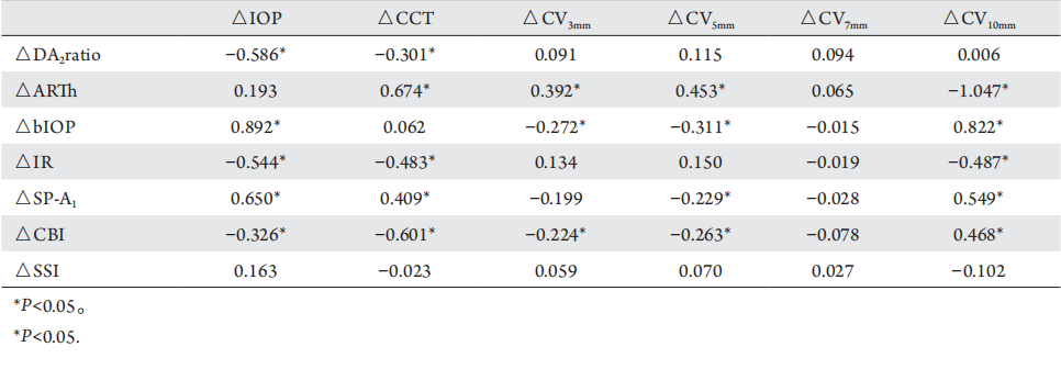
表9 SMILE术后3个月角膜生物力学参数变化量与形态及结构参数变化量的相关性
Table 9 Correlation of changes of corneal biomechanical parameters with changes of morphological and structural parameters 3
months after SMILE
2.4.3 SMILE 术后角膜生物力学参数变化量与 CV等形态及结构参数变化量的相关性
SMILE手术前后bIOP、DA2ratio、IR、SP-A1和 CBI 变化量均与眼压变化量呈显著相关 ( 均P<0.05),DA2ratio、IR、ARTh、SP-A1和CBI变化量与CCT变化量显著相关(均P <0.05);ARTh变化量与CV3mm、CV5mm变化量呈正相关(r =0.392、0.453 , P < 0.05) , 与 CV10mm变化量呈负相关(r=?1.047,P<0.05);bIOP变化量与CV3mm、CV5mm变化量呈负相关(r=?0.272、?0.311,P<0.05),与CV10mm变化量呈正相关(r=0.822,P<0.05);IR变化量与CV10mm变化量呈负相关(r=?0.487,P<0.05);SP-A1变化量与C V5mm变化量呈负相关(r=?0.229,P <0.05),与CV10mm变化量呈正相关( r =?0.549,P<0.05);CBI变化量与CV3mm、CV5mm变化量负相关(r=?0.224、?0.263,P<0.05),与CV10mm变化量呈正相关(r=0.468,P<0.05,表9)。
1. 佛山市科技创新项目医学类科技攻关项目 (2020001005825)。This work was supported by the Foshan Science and Technology Innovation Project Medical Science and Technology Project, China (2020001005825)
参考文献
1. Fernández J, Rodríguez-Vallejo M, Martínez J, et al. Corneal thickness after SMILE affects Scheimpflug-based dynamic tonometry[ J]. J Refract Surg, 2016, 32(12): 821-828.
2. Padmanabhan, P, Lopes, BT, Eliasy, A, et al. Evaluation of corneal biomechanical behavior in-vivo for healthy and keratoconic eyes using the stress-strain index[ J]. J Cataract Refract Surg, 2022. [Epub ahead of print]. doi: 10.1097/j.jcrs.0000000000000945.
3. Lu NJ, Hafezi F, Rozema JJ, et al. Repeatability of Corvis ST to measure biomechanical parameters before and after myopic refractive surgery[ J]. J Cataract Refr Surg, 2022. [Epub ahead of print]. doi: 10.1097/j.jcrs.0000000000000909.
4. Zhang Y, Wang Y, Li L, et al. Corneal stiffness and its relationship with other corneal biomechanical and nonbiomechanical parameters in myopic eyes of chinese patients[ J]. Cornea, 2018, 37(7): 881-885.
5. Eliasy A, Chen KJ, Vinciguerra R, et al. Determination of corneal biomechanical behavior in-vivo for healthy eyes using CorVis ST Tonometry: stress-strain index[ J]. Front Bioeng Biotechnol, 2019,7: 105.
6. Vinciguerra, R, Elsheikh, A, Roberts, CJ, et al. Influence of pachymetry and intraocular pressure on dynamic corneal response parameters in healthy patients[ J]. J Refract Surg, 2016, 32(8): 550-561.
7. Chen, KJ, Joda, A, Vinciguerra, R, et al. Clinical evaluation of a new correction algorithm for dynamic Scheimpflug analyzer tonometry before and after laser in situ keratomileusis and small-incision lenticule extraction[ J]. J Cataract Refr Surg, 2018, 44(5): 581-588.
8. Cao K, Liu L, Yu T, et al. Changes in corneal biomechanics during small-incision lenticule extraction (SMILE) and femtosecond-assisted laser in situ keratomileusis (FS-LASIK)[ J]. Lasers Med Sci, 2020,35(3): 599-609.
9. Fernández J, Rodríguez-Vallejo M, Martínez J, et al. New parameters for evaluating corneal biomechanics and intraocular pressure after small-incision lenticule extraction by Scheimpflug-based dynamic tonometry[ J]. J Cataract Refract Surg, 2017, 43(6): 803-811.
11. Yu M, Chen M, Dai J. Comparison of the posterior corneal elevation and biomechanics after SMILE and LASEK for myopia: a short- and long-term observation[ J]. Graefes Arch Clin Exp Ophthalmol, 2019,257(3): 601-606.
12. Sefat SM, Wiltfang R, Bechmann M, et al. Evaluation of changes in human corneas after femtosecond laser-assisted LASIK and small-incision lenticule extraction (SMILE) using Non-Contact Tonometry and Ultra-High-Speed Camera (Corvis ST)[J]. Curr Eye Res, 2016, 41(7): 917-922.
13. Han, F, Li, M, Wei, P, et al. Effect of biomechanical properties on myopia: a study of new corneal biomechanical parameters[ J]. BMC Ophthalmol, 2020, 20(1): 459.
14. 任胜卫, 杨凯丽, 徐丽妍. Corvis ST测量近视患者新型角膜生物力学参数的重复性及其影响因素[ J]. 中华实验眼科杂志, 2019,
37(12): 990-994.
REN Shengwei, YANG Kaili, XU Liyan. The reproducibility and influencing factors of corneal biomechanical parameters measured by Corvis ST in myopia[ J]. Chinese Journal of Experimental Ophthalmology, 2019, 37(12): 990-994.
15. Reinstein, DZ, Archer, TJ, Gobbe, M, et al. Lenticule thickness readout for small incision lenticule extraction compared to artemis three-dimensional very high-frequency digital ultrasound stromal measurements[ J]. J Refract Surg, 2014, 30(5): 304-309.
16. Sedaghat MR, Sharepoor M, Hassanzadeh S, et al. The corneal volume and biomechanical corneal factors: Is there any orrelation?[ J]. J Res Med Sci, 2012, 17(1): 32-39.
17. Cer v i?o A , Gonzalez -Meijome JM, Ferrer-Blasco T, et al. Determination of corneal volume from anterior topography and
topographic pachymetry: application to healthy and keratoconic eyes[ J]. Ophthalmic Physiol Opt, 2009, 29(6): 652-660.
18. Diniz CM, Hazarbassanov RM, Yamazaki E, et al. Pentacam Scheimpflug evaluation of corneal volume after LASIK[ J]. J Refract
Surg, 2010, 26(8): 600-604.
19. Wei P, Cheng GP, Zhang J, et al. Changes in corneal volume at different areas and its correlation with corneal biomechanics after SMILE and FS-LASIK surgery[ J]. J Ophthalmol, 2020, 2020: 1713979.
20. ?evik SG, K?van? SA, Akova-Budak B, et al. Relationship among corneal biomechanics, anterior segment parameters, and geometric corneal parameters[ J]. J Ophthalmol, 2016, 2016: 8418613.
21. Herber, R, Terai, N, Pillunat, KR, et al. Dynamic Scheimpflug Analyzer (Corvis ST) for measurement of corneal biomechanical parameters : A praxis-related overview[ J]. Ophthalmologe, 2018, 115(8): 635-643.
22. Esporcatte LPG, Salom?o MQ, Lopes BT, et al. Biomechanical diagnostics of the cornea[ J]. Eye Vis (Lond), 2020, 7: 9.
23. Viswanathan D, Kumar NL, Males JJ, et al. Relationship of structural characteristics to biomechanical profile in normal, Keratoconic, and crosslinked eyes[ J]. Cornea, 2015, 34(7): 791-796.
24. Schuh A, Kolb CM, Mayer WJ, et al. Comparison of changes in corneal volume and corneal thickness after myopia correction between LASIK and SMILE[ J]. PLoS One, 2021, 16(5): e0250700.
25. Abd El-Fattah EA, El Dorghamy AA, Ghoneim AM, et al. Comparison of corneal biomechanical changes after LASIK and F-SMILE with CorVis ST[ J]. Eur J Ophthalmol, 2021, 31(4): 1762-1770.
'%20fill='white'%20fill-opacity='0.01'/%3e%3cmask%20id='mask0_3477_29692'%20style='mask-type:luminance'%20maskUnits='userSpaceOnUse'%20x='0'%20y='0'%20width='16'%20height='16'%3e%3crect%20id='&%23232;&%23146;&%23153;&%23231;&%23137;&%23136;_2'%20x='16'%20width='16'%20height='16'%20transform='rotate(90%2016%200)'%20fill='white'/%3e%3c/mask%3e%3cg%20mask='url(%23mask0_3477_29692)'%3e%3cpath%20id='&%23232;&%23183;&%23175;&%23229;&%23190;&%23132;'%20d='M14%205L8%2011L2%205'%20stroke='%23333333'%20stroke-width='1.5'%20stroke-linecap='round'%20stroke-linejoin='round'/%3e%3c/g%3e%3c/g%3e%3c/svg%3e)