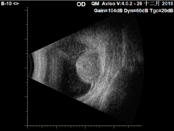
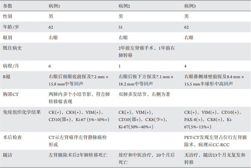
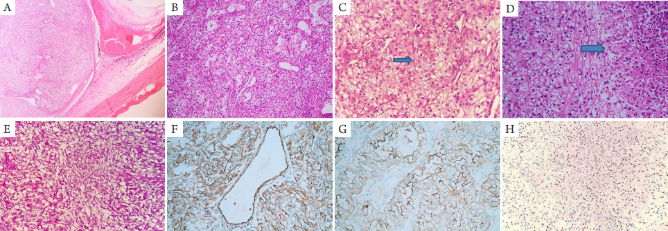
3例脉络膜转移性CC-RCC均为男性,例1和例3均为62岁,例2为51岁,平均58岁,均表现为右眼前黑影伴视力下降,病程分别为6月,1月和4月。术前检查视力均为右眼前手动,右眼底见视网膜下隆起肿块伴视网膜脱离。3例B超均显示右眼球内隆起肿物,例1和例2位于后极部,例3位于鼻侧球壁,中等回声,大小7.1~8.4 mm × 15.5~18.2 mm,均伴表面视网膜脱离,例3伴挖空征及脉络膜凹陷征,考虑脉络膜黑色素瘤(图1 )。例2两年前行左肾癌手术,1年前右肺上叶转移,术前胸部C T显示双肺多发结节,右侧为著,大者2 cm ×2 cm。例1术前胸部CT显示两肺内多个小结节影,大部分位于两肺外带,结节边界尚清晰,符合肺转移瘤表现。3例均在我院眼科全身麻醉下进行了右眼球摘除及义眼座的植入(表1)。
图1 右眼脉络膜转移性CC-RCC患者(例3):B超检查示右眼鼻侧球壁前探及8.4 mm ×15.53 mm半球形中高回声,伴挖空征及脉络膜凹陷征,考虑脉络膜黑色素瘤
Figure 1 Choroidal metastatic CC-RCC (case 3): B-scan ultrasound demonstrated an intraocular mass with medium to high internal reflectivity suspected of choroidal melanoma
表1 3例脉络膜转移性CC-RCC的临床病理资料
Table 1 Clinicopathologic data of 3 patients of choroidal metastatic clear-cell renal cell carcinoma
2.1.2 眼眶转移性CC-RCC
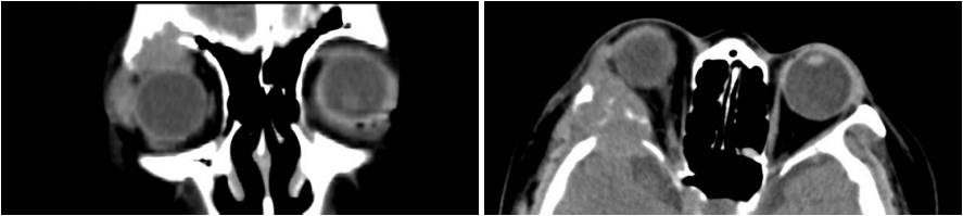
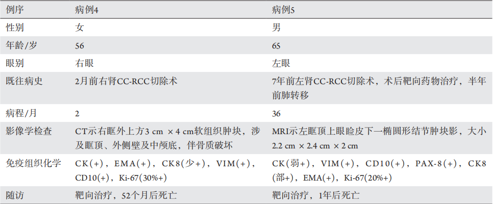
2例眼眶转移性CC-RCC中例4为女性,56岁,右眼红肿伴眼球突出2个月,2个月前外院行右肾CC-RCC切除手术,术前CT显示右眶外上方3 cm ×4 cm软组织肿块,涉及眶顶及外侧肌锥外与中颅底,伴眶顶壁、外侧壁与蝶骨大翼骨质破坏(图2 )。PET-CT提示右肾癌术区未见明显肿瘤复发,右眼眶外侧壁转移并侵及右侧额骨。术前右眼矫正视力0.6,右眼球突出,上转运动受限,眼球下移。例4在我院全身麻醉下行右眶颅联合肿物摘除术(表2)。
例5,男性,65岁,左眉弓肿物3年,渐大。7年前左肾CC-RCC切除术,术后靶向药物(多吉美)治疗,半年前“肾癌肺部转移”行胸腔镜手术。术前MRI示左眶顶上眼睑皮下一椭圆形结节肿块影,大小2.2 cm ×2.4 cm ×2 cm,紧贴眼球前上壁,眼球向下移位。术前左眼视力0.1,左眼眉弓处扪及质硬隆起肿物2 cm ×1 cm,界清,眼球下移伴上睑下垂。例5在我院全身麻醉下行左眼眶肿物摘除术(表2)。
图2 右眼眶转移性CC-RCC患者(例4):冠状位和水平位CT显示右眶外上方3 cm ×4 cm软组织肿块,涉及眶顶及外侧肌锥外与中颅底,伴眶顶壁、外侧壁与蝶骨大翼骨质破坏
Figure 2 Orbital metastatic clear-cell renal cell carcinoma (case 4): coronal and axial CT scan showed superotemporal 3 cm ×4 cm mass of the right orbit with bone destruction
表2 2例眼眶转移性CC-RCC的临床病理资料
Table 2 Clinicopathologic data of 2 patients of orbital metastatic clear-cell renal cell carcinoma
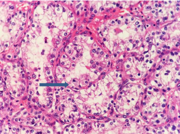
Figure 4 Orbital metastatic CC-RCC (case 5) showed some tumor cells with alveolar architecture and mitotic figure (arrow; IHC, ×400)
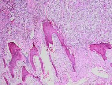
图5 光镜下观察例4患者右眼眶转移性CC-RCC,图示肿瘤侵犯骨组织(HE,×100)
Figure 5 Orbital metastatic CC-RCC (case 4) showed bone destruction (HE, ×100)
2.3 治疗和随访
2.3.1 脉络膜转移性CC-RCC
例1术后腹部CT示左肾下部见一椭圆形软组织影,大小6.5 cm ×5.8 cm ×8.9 cm,左肾门下部肾静脉内见充盈缺损影,诊断左肾癌伴左肾静脉癌栓形成,后在当地医院行左肾摘除术,病理检查证实为左肾CC-RCC。术后2年患者因肺转移死亡。例2眼部手术后因同时肺部转移行放疗和中医治疗,20个月后死亡。例3术后PET-CT检查发现左肾占位,在当地医院行左肾摘除术,术后病理示左肾CC-RCC,肿瘤大小约4.5 cm ×4 cm ×3 cm。术后未行特殊治疗,随访至今2 3个月无复发和转移(表1)。CC-RCC
1. Flanigan RC, Campbell SC, Clark JI, et al. Metastatic renal cell
carcinoma[ J]. Curr Treat Options Oncol, 2003, 4(5): 385-390.
2. Rai R , Jakobiec FA, Fay A . Ocular metastatic renal carcinoma
presenting with proptosis[ J]. Ophthalmic Plast Reconstr Surg, 2015,
31(4): e100-8.
3. Ferry AP, Font RL. Carcinoma metastatic to the eye and orbit. I. A
clinicopathologic study of 227 cases[ J]. Arch Ophthalmol, 1974,
92(4): 276-286.
4. Shields CL, Welch RJ, Malik K, et al. Uveal metastasis: clinical features
and survival outcome of 2214 tumors in 1111 patients based on primary
tumor origin[ J]. Middle East Afr J Ophthalmol, 2018, 25(2): 81-90.
5. Shields CL, Shields JA, Gross NE, et al. Survey of 520 eyes with uveal
metastases[ J]. Ophthalmology, 1997, 104(8): 1265-1276.
6. Shields JA, Shields CL, Brotman HK, et al. Cancer metastatic to
the orbit: the 2000 Robert M. Curts Lecture[ J]. Ophthalmic Plast
Reconstr Surg, 2001, 17(5): 346-354.
7. 廖松林. 肿瘤病理诊断与鉴别诊断学[M]. 福州: 福建科学技术
出版社, 2006: 405-407.
LIAO SL. Histopathology for diagnosis and differential diagnosis
of tumor[M]. Fuzhou: Fujian Science and Technology Publishing
House, 2006: 405-407.
8. Shome D, Honavar SG, Gupta P, et al. Metastasis to the eye and orbit
from renal cell carcinoma—a report of three cases and review of
literature[ J]. Surv Ophthalmol, 2007, 52(2): 213-223.
9. Alasil T, Khazai B, Loredo L, et al. Renal cell carcinoma metastasis
to the ciliary body responds to proton beam radiotherapy: a case
report[ J]. J Med Case Rep, 2011, 5: 345.
10. Arepalli S, Kaliki S, Shields CL. Choroidal metastases: Origin, features,
and therapy[ J]. Indian J Ophthalmol, 2015, 63(2): 122-127.
11. Komanski CB, Rubino SM, Meyer JC, et al. Choroidal melanoma
mimicker: a case of metastatic clear-cell renal cell carcinoma[ J]. Ocul
Oncol Pathol, 2017, 3(4): 279-282.
12. Goldberg RA, Rootman J, Cline RA. Tumors metastatic to the orbit: a
changing picture[ J]. Surv Ophthalmol, 1990, 35(1): 1-24.
'%20fill='white'%20fill-opacity='0.01'/%3e%3cmask%20id='mask0_3477_29692'%20style='mask-type:luminance'%20maskUnits='userSpaceOnUse'%20x='0'%20y='0'%20width='16'%20height='16'%3e%3crect%20id='&%23232;&%23146;&%23153;&%23231;&%23137;&%23136;_2'%20x='16'%20width='16'%20height='16'%20transform='rotate(90%2016%200)'%20fill='white'/%3e%3c/mask%3e%3cg%20mask='url(%23mask0_3477_29692)'%3e%3cpath%20id='&%23232;&%23183;&%23175;&%23229;&%23190;&%23132;'%20d='M14%205L8%2011L2%205'%20stroke='%23333333'%20stroke-width='1.5'%20stroke-linecap='round'%20stroke-linejoin='round'/%3e%3c/g%3e%3c/g%3e%3c/svg%3e)