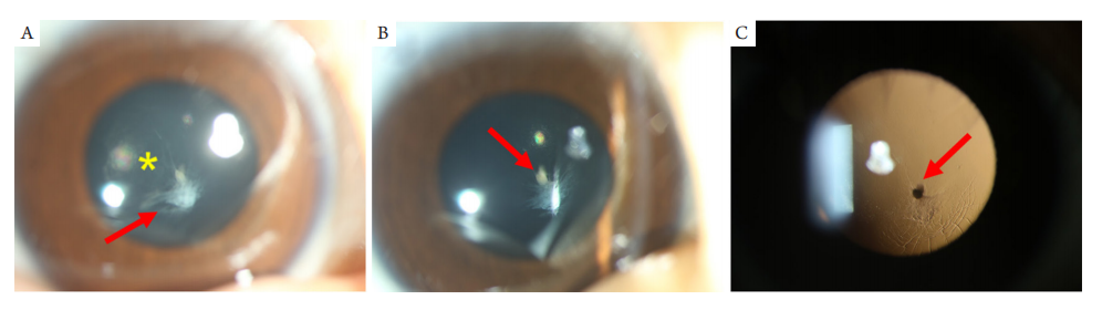
(A,B) Posterior capsular opacities (yellow asterisk) of right eye; in the inferonasal of posterior capsule, a white circular opacity tissue extends to the vitreous cavity with obvious terminal (red arrow); (C) Mitterndorf dot is observed under retro-illumination view (red arrow).
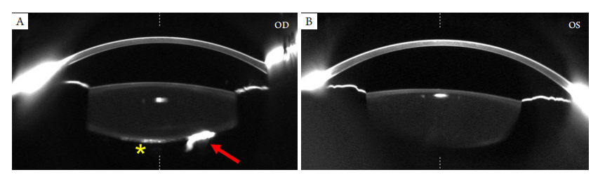
(A) Posterior subcapsular opacification of the central region of the lens of the right eye, and on the nasal side, a compact short strip of cord pointing to the vitreous cavity with a stop (red arrow); (B) There is no significant abnormality in the left eye.
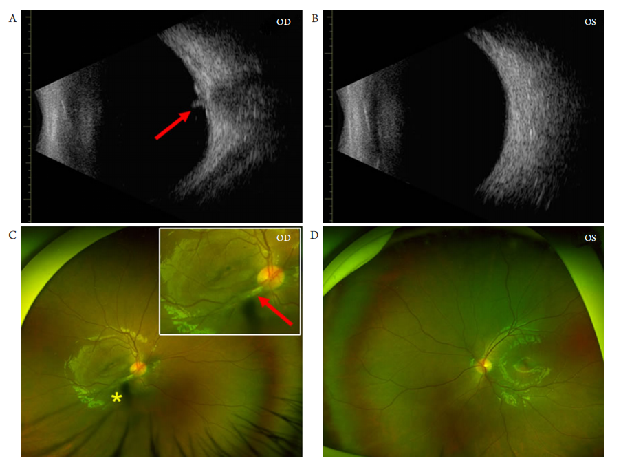
(A) B-mode ultrasonography of right eye reveals a short tubular membrane adheres to the optic disk (red arrow) and temporal eyeball wall is thickening; (B) There is no obvious abnormality in the B-mode ultrasonography of left eye; (C) In SLO images of right eye: the posterior capsular opacities present a dark shadow (yellow asterisk), and the short tubular membrane adheres to the temporal optic disk, accompanied with macular structure disorders; (D) There is no obvious abnormality in the SLO image of left eye.
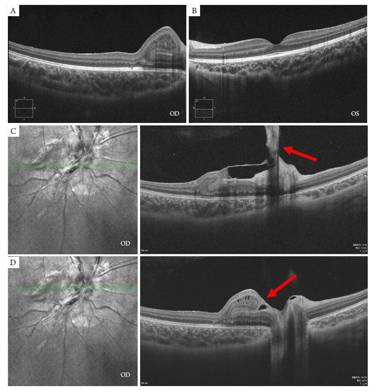
OCT scanning indicates (A) the disorders of macular structure in right eye and (B) the normal structure of posterior segments in left eye. Macular intensive scanning with OCT reveals (C) the short tubular membrane adheres to optic disk in the right eye (red arrow), leading to (D) the abnormal macular structure.
1. 国家自然科学基金 (82171035)。This work was supported by the National Natural Science Foundation of China (82171035)
参考文献
1. Zou Y, Liu Z, Liu Y. Pigmented posterior lenticonus in unilateral development cataract[ J]. Am J Ophthalmol, 2022, 240: e3-e4.
2. De la Huerta I, Mesi O, Murphy B, et al. Spectral domain optical coherence tomography imaging of the macula and vitreomacular interface in persistent fetal vasculature syndrome with posterior involvement[ J]. Retina, 2019, 39(3): 581-586.
3. Sun MH, Kao LY, Kuo YH. Persistent hyperplastic primary vitreous:magnetic resonance imaging and clinical findings[ J]. Chang Gung Med J, 2003, 26(4): 269-276.
4. Goldberg MF, Mafee M. Computed tomography for diagnosis of persistent hyperplastic primary vitreous (PHPV)[ J]. Ophthalmology,1983, 90(5): 442-451.
5. Ranchod TM, Ho LY, Drenser KA, et al. Clinical presentation of familial exudative vitreoretinopathy[ J]. Ophthalmology, 2011,
118(10): 2070-2075.
6. Neudorfer M, Waisbourd M, Buzi S, et al. Color Doppler imaging of eyes with persistent fetal vasculature[ J]. Pediatr Radiol, 2012, 42(10):1229-1234.
7. Perrimon N, Bernfield M. Specificities of heparan sulphate proteoglycans in developmental processes[ J]. Nature, 2000, 404(6779): 725-728.
8. Hu A, Yuan M, Liu F, et al. Ultrasonographic feature of persistent hyperplastic primary vitreous[ J]. Eye Sci, 2014, 29(2): 100-103.
9. Galal AH, Kotoury AI, Azzab AA. Bilateral persistent hyperplastic primary vitreous: an Egyptian family supporting a rare autosomal dominant inheritance[ J]. Genet Couns, 2006, 17(4): 441-447.
10. Mackeen LD, Nischal KK , Lam WC, et al. High-frequenc y ultrasonography findings in persistent hyperplastic primary vitreous[ J]. J AAPOS, 2000, 4(4): 217-224.
11. Azcarate PM, Grace SF, Shi W, et al. B-scan echography in cases of confirmed persistent fetal vasculature[ J]. J Pediatr Ophthalmol Strabismus, 2016, 53(4): 252-253.
12. Hu A, Pei X, Ding X, et al. Combined persistent fetal vasculature: a classification based on high-resolution B-mode ultrasound and color Doppler imaging[ J]. Ophthalmology, 2016, 123(1): 19-25.
13. Goldberg MF. Persistent fetal vasculature (PFV): an integrated interpretation of signs and symptoms associated with persistent hyperplastic primary vitreous (PHPV). LIV Edward Jackson Memorial Lecture[ J]. Am J Ophthalmol, 1997, 124(5): 587-626.
14. Tartarella MB, Takahagi RU, Braga AP, et al. Persistent fetal vasculature:
ocular features, management of cataract and outcomes[ J]. Arq Bras
Oftalmol, 2013, 76(3): 185-188.
15. Sisk RA , Berrocal AM, Feuer WJ, et al. Visual and anatomic
outcomes with or without surgery in persistent fetal vasculature[ J].
Ophthalmology, 2010, 117(11): 2178-83.e1-2.
16. Khandwala N, Besirli C, Bohnsack BL. Outcomes and surgical
management of persistent fetal vasculature[ J]. BMJ Open Ophthalmol,
2021, 6(1): e000656.
17. Sanghvi DA , Sanghvi CA , Purandare NC. Bilateral persistent
hyperplastic primary vitreous[ J]. Australas Radiol, 2005, 49(1): 72-74.
18. Ding X. Surgery of congenital cataracts associated with persistent fetal
vasculature[M]//Liu Y. Pediatric lens diseases. Singapore: Springer
Nature, 2016: 255-269.
'%20fill='white'%20fill-opacity='0.01'/%3e%3cmask%20id='mask0_3477_29692'%20style='mask-type:luminance'%20maskUnits='userSpaceOnUse'%20x='0'%20y='0'%20width='16'%20height='16'%3e%3crect%20id='&%23232;&%23146;&%23153;&%23231;&%23137;&%23136;_2'%20x='16'%20width='16'%20height='16'%20transform='rotate(90%2016%200)'%20fill='white'/%3e%3c/mask%3e%3cg%20mask='url(%23mask0_3477_29692)'%3e%3cpath%20id='&%23232;&%23183;&%23175;&%23229;&%23190;&%23132;'%20d='M14%205L8%2011L2%205'%20stroke='%23333333'%20stroke-width='1.5'%20stroke-linecap='round'%20stroke-linejoin='round'/%3e%3c/g%3e%3c/g%3e%3c/svg%3e)