





INTRODUCTION
The whole lacrimal passage intubation is widely used in lacrimal surgery, including puncta, lacrimal canaliculi, nasolacrimal duct stenosis, inflammatory diseases and nasolacrimal duct obstruction.[1-3] Two ends of the bicanalicular silicone tube reach the inferior nasal meatus through puncta, canaliculi, lacrimal sac and nasolacrimal duct, and the free ends of the silicone tube will be fixed by a knot.[4] The role of the whole lacrimal passage intubation is to maintain the patency of newly formed channels, and prevent anastomotic scar formation and stenosis.[5-6] However, the prolapse of the silicone tube from the medial canthus is one of the most common complications in the whole lacrimal passage intubation.[2-3,7]
When the bicanalicular silicone tube is completely prolapsed from medial can thus, the distal part of the silicone tube and the enter suck into the nasolacrimal duct or is embedded in the lacrimal sac, and the silicone tube cannot be found in the inferior meatus and the opening of nasolacrimal duct.[5] In this case, it would be a challenge to reposition or remove. A new method with modified suture-probe and silk thread traction is developed to reposition the bicanalicular silicone tube that has completely prolapsed from the medial canthus.
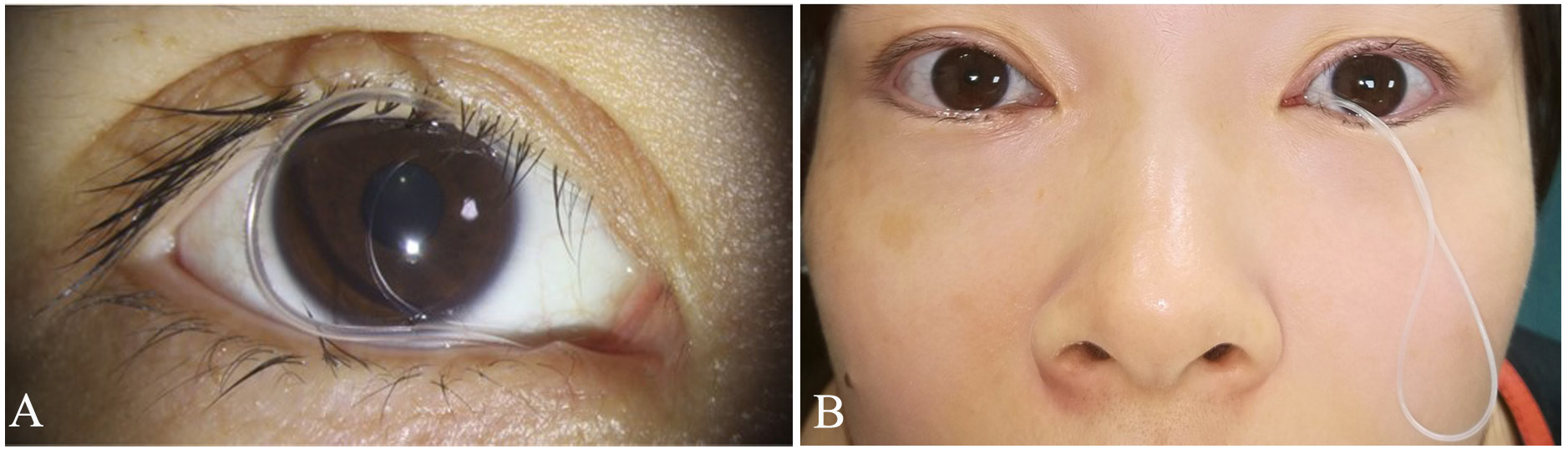
Figure 1 The prolapsed silicone tube
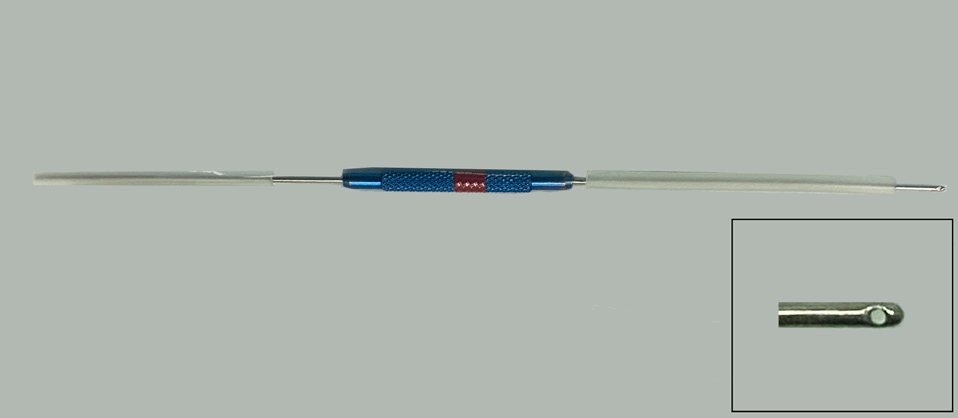
Figure 2 7-8 gauge stainless steel lacrimal suture-probe with side opening at one end
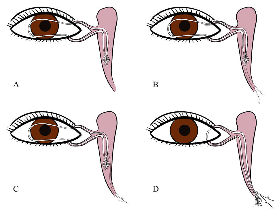
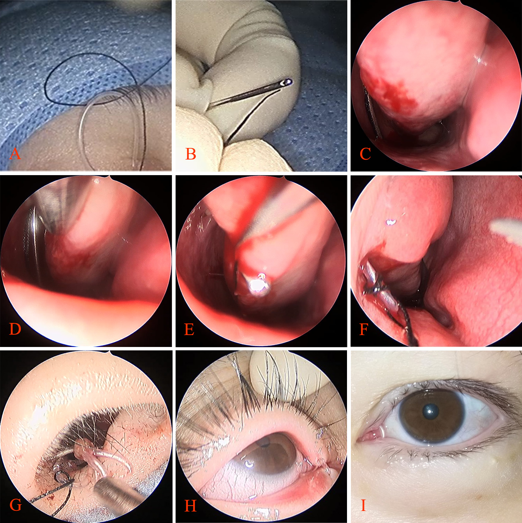
Figure 4 The surgical procedures
The complete prolapse of the bicanalicular silicone tube from the whole lacrimal passage, originating from the medial canthus, was successfully repositioned with no complications, using the modified suture-probe and silk thread traction method in this study. The results unequivocally showed that the effectiveness and safety of the modified approach in treating complete dislocations of the bicanalicular silicone tune from the medial canthus. From 2018 to 2023, our hospital encountered only two such cases, indicating the rarity of this condition. This finding contrasts with previous research.[4] To prevent such complete prolapses during whole lacrimal passage intubation surgery, a unique double tube knot technique for the silicone tube was introduced in our hospital. This knot is deliberately designed to be too large to fit through the nasolacrimal duct opening, thus preventing accidental dislocation. Additionally, after the operation, our nursing team will educate patients carefully, emphasizing silicone tube care measures, such as keeping eyes closed during face washing avoiding vigorous eye rubbing, and refraining from forceful nose blowing.[8] These measures, combined with our surgical techniques, contribute to the low incidence of complete prolapses in our hospital.
The dislocation of the bicanalicular silicone tube after whole lacrimal duct intubation can vary from partial to complete, depending on the extent of the displacement.[4] In cases of partial dislocation, where only a small portion of the silicone tube from the inner canthus with, the distal end of the tube and knot remaining visible in the nasal cavity, the tube can be easily repositioned by pulling the distal end down with forceps under the nose or gently pushing it back in place using forceps around the inner canthus. The management of complete dislocation of the silicone tube, where the tube knot becomes lodged in the nasolacrimal duct or dacryocyst with no visible portion in the nasal cavity, poses a significant changing in terms of repositioning.[4] Fortunately, the incidence of such complete prolapse of silicone tube is relatively are. Byun Z et al.[9] reported 11 cases of complete prolapse of bicanalicular silicone tube from medial canthus in 2 years, and Chu Z et al.[4] reported 12 cases in 5 years. The protrusion of the silicone tube from the medal canthus can readily lead to corneal trauma and conjunctivitis,[4] therefore, it is imperative to promptly reposition or remove the prolapsed silicone tube to prevent any further complications.
There are several ways to take out the silicone tube. Yeh H et al.[10] described an approach involving removal a skin dacryocystorhinostomy,[10] but this method can enlarge the wound, leave behind a visible skin, and increase the cost. Additionally, they also discussed the technique of removing the tube through the trans nasal endoscopic dacryocystorhinostomy, but this method can also result in additional trauma and expenses. Patel BC et al.[11] reported clipping the silicone tubes and gently pulling the knot out of the lacrimal puncta for tube removal. However, we found that the forcibly pulling the prolapsed silicone tube from the lacrimal puncta can easily lead to tears in the lacrimal passage and residual lacrimal duct.
There are various approaches to reposition a completely prolapsed silicone tube. Byun Z et al.[9] reported a method that they obliquely cut the prolapsed tube with scissors, creating a small semicircular hole. A probe is then inserted into this hole, entered into the inferior nasal meatus, and the silicone tube is clamped with pliers. Subsequently, the probe is withdraw from the puncta. However, this method is only applicable for hollow silicone tubes. He J et al.[5] reported a different method where the probe with memory wire is passing through the lacrimal puncta and into the inferior nasal meatus. The suture is threaded through the head of the memory wire. After removing the probe from the puncta, the suture is cut and looped around the silicone tube. The two ends of the suture were then knotted. Finally, by pulling the suture from the nasal cavity, the prolapsed tube can be repositioned. Chu Z et al.[4] introduced another method utilizing a suture-probe. They cut the suture, wrapped one end around the silicone tube, and tied it to the other end of the thread to create a suture knot. Pulling the suture from the nasal cavity resets the prolapsed tube. Additionally, they also suggested attaching 30 mm sutures to the distal end of the silicone tube as a preventive measure against re-prolapse. However, the suture knot formed by the two ends of the sutures poses a risk of loosening as it is withdrawn from the nasal cavity. Additionally, the sutures reserved in the inferior meatus may cause irritation and promote the deposition of secretions.
In this study, patients who experienced complete dislocation of the bicanalicular silicone tube from medial canthus following whole lacrimal duct intubation were treated with modified suture-probe and silk thread traction method. This innovative method addresses the limitation associated with previous surgical methods. It is not only simple and safe, but also cost-effective, effectively preventing surgical failures or the need for silicone tube replacement due to premature removal of the prolapsed tube. The modified suture-probe and silk thread traction method introduced in this study offers a novel and effective solution for managing the complete prolapse of the bicanalicular silicone tube from the medial canthus after intubation of the entire lacrimal passage. Furthermore, the choice of suture is discretionary. While 5-0 black silk thread is thick and lacks smoothness, resulting in greater friction against the lacrimal duct mucosa, it exhibits a lesser cutting force. Conversely, the 6-0 prolene suture is thinner and smoother, minimizing friction with the lacrimal duct mucosa but possessing a higher cutting force. The safety of employing various suture types for repositioning the completely prolapsed silicone tube remains to be thoroughly investigated within the context of suture-probe and silk thread traction method. Given the infrequent occurrence of silicone tube prolapse and the limited sample size in this study, additional research is warranted to establish the safety of this method and identify potential intraoperative complications.
Correction notice
NoneAcknowledgement
NoneAuthor Contributions
(I) Conception and design: Xuanwei Liang and Rongxin Chen(II)Administrative support: None
(III) Provision of study materials or patients: Xuanwei Liang
(IV) Collection and assembly of data: Jing Li
(V) Data analysis and interpretation: Jing Li and Xuanwei Liang
(VI) Manuscript writing: Jing Li
(VII) Final approval of manuscript: All authors