AD患者存在多种异常眼球运动类型。和正常对照相比,AD患者扫视潜伏期延长,扫视速率下降、幅度降低[46-48],但上述各指标在不同AD患者间变异度较大[43,49]。其中扫视潜伏期和扫视速率的异常程度和简易精神状态评价(Mini-Mental State Examination,MMSE)量表评分呈正相关,提示扫视测试或许可以和MMSE评分一同作为AD诊断和严重程度分级的依据之一[50]。神经影像学研究结果[51]显示:扫视潜伏期延长可能与AD患者双侧顶叶和枕叶及右侧颞叶容积减少相关。其中顶叶容积的减少可导致视觉注意力下降,进而影响正常扫视功能[51-52]。在反向扫视测试中,AD患者扫视潜伏期延长,扫视方向错误率升高,且校正率低[47,53-54]。虽然类似的发现也存在于非AD类型的痴呆患者中[55],但AD患者的反向扫视的错误率与包括MMSE、阿尔茨海默病评定量表、彩色表格排序测试等神经心理学评分量表的结果相关性更强[12,54-57]。在微扫视和扫视性侵扰检查中,AD患者的眼球运动方向并非与注视点相平行,而是存在显著倾斜[30],且扫视性侵扰的发生频率明显升高,也与MMSE评分相关[58]。因此,微扫视和扫视侵扰也是区分被检查者是否存在认知功能障碍的眼球运动指标之一。AD患者平滑追随眼球运动异常表现和扫视异常类似。在观察目标移动时,AD患者进行平滑眼球运动追随的潜伏期延长,移动速率下降,移动速率的加速度下降,正确捕捉移动物体的时间占比也下降[51,59-61]。由于眼球运动速度常落后于目标物的移动速度,AD患者可频繁出现补偿性扫视以重新捕捉目标物。有研究[62]认为补偿性扫视出现的频率与MMSE评分呈负相关,但也有研究[63]结果显示AD组受试者的平滑追随功能依然位于正常范围。平滑追随眼球运动的异常程度也可能与AD患者病情进展严重程度相关,但目前尚缺乏被学界广泛认可的研究结果,相关病理生理机制也有待进一步研究。扫视性侵扰出现的频率也与MMSE评分呈负相关。对AD患者瞳孔对光反射功能研究相对较少,已有研究[64-65]提示:与正常对照相比,AD患者瞳孔对光反射变化的幅度和速率均有降低。
近十年来,随着机器学习算法的突破,人工智能技术可以使计算机在极短时间习得人类的既得知识和经验并进行相应推理判断。在海量存储空间和强大计算能力的硬件基础上,深度学习算法在图像识别领域发展尤为迅猛。由于人类专家的受训水平有波动性,且存在应对任务的疲劳曲线,在某些定性定量的图像识别场景下,优秀调制的深度学习算法模型可以达到人类专家水平,甚至可以准确判断某些人眼无法识别的特征[67-68],例如Poplin等[68]采用深度学习技术开发的心血管风险预测模型,仅通过受试者的眼底彩照进行5年内心血管不良事件发病风险预测,其接收者操作特征曲线下面积(area under curve,AUC)可达到0.70(95%CI:0.65~0.74),该表现不亚于业内公认的欧洲心血管风险预测计算公式的表现(AUC 0.72,95%CI:0.67~0.76,且该模型通过眼底彩照判断受试者性别的AUC可高达0.98(95%CI:0.97~0.99),超出了人眼可以判断的特征范围。
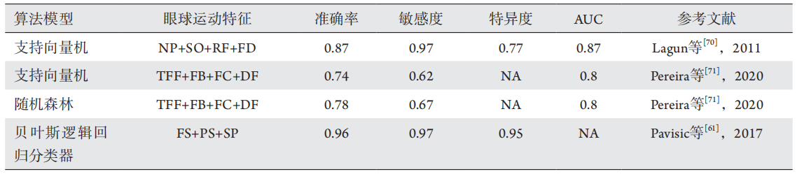
表1 既往研究中基于眼球运动的机器学习相关认知功能分类模型表现总结 Table 1 Summary of the performance of the classification model of cognitive function related to machine learning based on eye movement in previous studies
NP: novelty preference, the fraction of the total looking time spent gazing at the novel image region; SO: saccade orientation, the
corresponding endpoints of the fixations; RF: re-fixations, the times when the gaze position re-visits (re-fixates) on previously seen parts of the stimuli; FD: fixation duration, the duration of fixations during the test phase; TFF: time to first fixation; FB: fixations before, number of fixations before the first fixation on any regions of interest for the first time; FC: fixation count, number of fixations made in a specific region of interest; DF: duration of fixations, total duration of fixations within an ROI; FS: fixation stability; PS: pro-saccade; SP: smooth pursuit; NA: not available in the original literature.
1. 广州市科技计划项目基础研究计划市重点实验室建设项目(202002010006)。This work was supported by Guangzhou Key Laboratory Project, China (202002010006).
参考文献
1. Willvonseder R, Goldstein NP, McCall JT, et al. A hereditary disorder
with dementia, spastic dysarthria, vertical eye movement paresis, gait
disturbance, splenomegaly, and abnormal copper metabolism[ J].
Neurology, 1973, 23(10): 1039-1049.
2. Dix MR. Clinical observations upon the vestibular responses in certain
disorders of the central nervous system[ J]. Adv Otorhinolaryngol,
1970, 17: 118-128.
4. Bayne T, Brainard D, Byrne RW, et al. What is cognition[ J]. Curr Biol,
2019, 29(13): R608-R615.
5. Petersen RC. Mild cognitive impairment as a diagnostic entity[ J]. J
Intern Med, 2004, 256(3): 183-194.
6. Hill NT, Mowszowski L, Naismith SL, et al. Computerized cognitive
training in older adults with mild cognitive impairment or dementia: a
systematic review and meta-analysis[ J]. Am J Psychiatry, 2017, 174(4):
329-340.
7. Martin M, Clare L, Altgassen AM, et al. Cognition‐based interventions
for healthy older people and people with mild cognitive impairment[ J].
Cochrane Database Syst Rev, 2011(1): CD006220.
8. Arvanitakis Z, Shah RC, Bennett DA. Diagnosis and management of
dementia: review[ J]. JAMA, 2019, 322(16): 1589-1599.
9. Lane CA, Hardy J, Schott JM. Alzheimer’s disease[ J]. Eur J Neurol
2018, 25(1): 59-70.
10. Prince MJ. World Alzheimer Report 2015: the global impact of
dementia: an analysis of prevalence, incidence, cost and trends:
Alzheimer’s Disease International[R]. London: Alzheimer’s Disease
International, 2015.
11. Xu J, Wang J, Wimo A, et al. The economic burden of dementia in
China, 1990–2030: implications for health policy[ J]. Bull World
Health Organ, 2017, 95(1): 18.
12. Livingston G, Sommerlad A, Orgeta V, et al. Dementia prevention,
intervention, and care[ J]. Lancet, 2017, 390(10113): 2673-2734.
13. Petersen RC, Morris JC. Mild cognitive impairment as a clinical entity
and treatment target[ J]. Arch Neurol, 2005, 62(7): 1160-1163.
14. Winblad B, Palmer K, Kivipelto M, et al. Mild cognitive impairment--
beyond controversies, towards a consensus: report of the International
Working Group on Mild Cognitive Impairment[ J]. J Intern Med, 2004,
256(3): 240-246.
15. Harper L, Barkhof F, Scheltens P, et al. An algorithmic approach to
structural imaging in dementia[ J]. J Neurol Neurosurg Psychiatry,
2014, 85(6): 692-698.
16. Duara R, Loewenstein DA, Potter E, et al. Medial temporal lobe atrophy
on MRI scans and the diagnosis of Alzheimer disease[ J]. Neurology,
2008, 71(24): 1986-1992.
17. Burton EJ, Barber R, Mukaetova-Ladinska EB, et al. Medial temporal
lobe atrophy on MRI differentiates Alzheimer’s disease from dementia
with Lewy bodies and vascular cognitive impairment: a prospective
study with pathological verification of diagnosis[ J]. Brain, 2009,
132(Pt 1): 195-203.
18. Knopman DS, DeKosky ST, Cummings JL, et al. Practice parameter:
diagnosis of dementia (an evidence-based review). Report of the
Quality Standards Subcommittee of the American Academy of
Neurology[ J]. Neurology, 2001, 56(9): 1143-1153.
19. Anderson TJ, MacAskill MR . Eye movements in patients with
neurodegenerative disorders[ J]. Nat Rev Neurol, 2013, 9(2): 74-85.
20. Bridgeman B. Conscious vs unconscious processes: the case of
vision[ J]. Theory Psychol, 1992, 2(1): 73-88.
21. Deubel H, Schneider WX. Delayed saccades, but not delayed manual
aiming movements, require visual attention shifts[ J]. Ann N Y Acad
Sci, 2003, 1004(1): 289-296.
22. Girard B, Berthoz A. From brainstem to cortex: computational
models of saccade generation circuitry[ J]. Prog Neurobiol, 2005,
77(4): 215-251.
23. Pierrot-Deseilligny C, Rivaud S, Gaymard B, et al. Cortical control of
saccades[ J]. Ann Neurol, 1995, 37(5): 557-567.
24. Coiner B, Pan H, Bennett ML, et al. Functional neuroanatomy of the
human eye movement network: a review and atlas[ J]. Brain Struct
Funct, 2019, 224(8): 2603-2617.
25. Stuphorn V, Taylor TL, Schall JD. Performance monitoring by the
supplementary eye field[ J]. Nature, 2000, 408(6814): 857-860.
26. Parton A , Nachev P, Hodgson TL, et al. Role of the human
supplementary eye field in the control of saccadic eye movements[ J].
Neuropsychologia, 2007, 45(5): 997-1008.
27. Quaia C, Lefèvre P, Optican LM. Model of the control of saccades by
superior colliculus and cerebellum[ J]. J Neurophysiol, 1999, 82(2):
999-1018.
28. Pierrot-Deseilligny C, Müri RM, Nyffeler T, et al. The role of the human
dorsolateral prefrontal cortex in ocular motor behavior[ J]. Ann N Y
Acad Sci, 2005, 1039: 239-251.
29. Tulunay-Keesey U. Fading of stabilized retinal images[ J]. J Opt Soc
Am, 1982, 72(4): 440-447.
30. Kapoula Z, Yang Q, Otero-Millan J, et al. Distinctive features
of microsaccades in Alzheimer’s disease and in mild cognitive
impairment[ J]. Age, 2014, 36(2): 535-543.
31. Abadi RV, Gowen E. Characteristics of saccadic intrusions[ J]. Vision
Res 2004, 44(23): 2675-2690.
32. Martinez-Conde S. Fixational eye movements in normal and
pathological vision[ J]. Prog Brain Res, 2006, 154: 151-176.
33. Otero-Millan J, Macknik SL, Serra A, et al. Triggering mechanisms in
microsaccade and saccade generation: a novel proposal[ J]. Ann N Y
Acad Sci, 2011, 1233: 107-116.
34. Martinez-Conde S, Otero-Millan J, Macknik SL. The impact of
microsaccades on vision: towards a unified theory of saccadic
function[ J]. Nat Rev Neurosci, 2013, 14(2): 83-96.
35. Thier P, Ilg UJ. The neural basis of smooth-pursuit eye movements[ J].
Curr Opin Neurobiol, 2005, 15(6): 645-652.
36. Fukushima K. Frontal cortical control of smooth-pursuit[ J]. Curr Opin
Neurobiol, 2003, 13(6): 647-654.
37. Petit L, Haxby JV. Functional anatomy of pursuit eye movements in
humans as revealed by fMRI[ J]. J Neurophysiol, 1999, 82(1): 463-471.
38. Yan YJ, Cui DM, Lynch JC. Overlap of saccadic and pursuit eye
movement systems in the brain stem reticular formation[ J]. J
Neurophysiol, 2001, 86(6): 3056-3060.
41. Tschanz JT, Welsh-Bohmer KA, Lyketsos CG, et al. Conversion to
dementia from mild cognitive disorder: the Cache County Study[ J].
Neurology, 2006, 67(2): 229-234.
42. Petersen RC. Mild cognitive impairment: current research and clinical
implications[ J]. Semin Neurol, 2007, 27(1): 22-31.
43. Yang Q, Wang T, Su N, et al. Specific saccade deficits in patients with
Alzheimer’s disease at mild to moderate stage and in patients with
amnestic mild cognitive impairment[ J]. Age, 2013, 35(4): 1287-1298.
44. Chehrehnegar N, Nejati V, Shati M, et al. Behavioral and cognitive
markers of mild cognitive impairment: diagnostic value of saccadic eye
movements and Simon task[ J]. Aging Clin Exp Res, 2019, 31(11):
1591-1600.
45. Alichniewicz KK, Brunner F, Klünemann HH, et al. Neural correlates
of saccadic inhibition in healthy elderly and patients with amnestic mild
cognitive impairment[ J]. Front Psychol, 2013, 4: 467.
46. Fletcher WA, Sharpe JA. Saccadic eye movement dysfunction in
Alzheimer’s disease[ J]. Ann Neurol, 1986, 20(4): 464-471.
47. Shafiq-Antonacci R, Maruff P, Masters C, et al. Spectrum of saccade
system function in Alzheimer disease[ J]. Arch Neurol, 2003, 60(9):
1272-1278.
48. Kahana Levy N, Lavidor M, Vakil E. Prosaccade and antisaccade
paradigms in persons with Alzheimer’s disease: a meta-analytic
review[ J]. Neuropsychol Rev, 2018, 28(1): 16-31.
49. Yang Q, Wang T, Su N, et al. Long latency and high variability in
accuracy-speed of prosaccades in Alzheimer’s disease at mild to
moderate stage[ J]. Dement Geriatr Cogn Dis Extra, 2011, 1(1):
318-329.
50. Peltsch A, Hemraj A, Garcia A, et al. Saccade deficits in amnestic mild
cognitive impairment resemble mild Alzheimer’s disease[ J]. Eur J
Neurosci, 2014, 39(11): 2000-2013.
51. Garbutt S, Matlin A, Hellmuth J, et al. Oculomotor function in
frontotemporal lobar degeneration, related disorders and Alzheimer’s
disease[ J]. Brain, 2008, 131(Pt 5): 1268-1281.
52. Posner MI, Petersen SE. The attention system of the human brain[ J].
Annu Rev Neurosci, 1990, 13: 25-42.
53. Kaufman LD, Pratt J, Levine B, et al. Executive deficits detected in mild
Alzheimer’s disease using the antisaccade task[ J]. Brain Behav, 2012,
2(1): 15-21.
54. Crawford TJ, Higham S, Mayes J, et al. The role of working memory
and attentional disengagement on inhibitory control: effects of aging
and Alzheimer’s disease[ J]. Age, 2013, 35(5): 1637-1650.
55. Currie J, Ramsden B, McArthur C, et al. Validation of a clinical
antisaccadic eye movement test in the assessment of dementia[ J]. Arch
Neurol, 1991, 48(6): 644-648.
56. Abel LA, Unverzagt F, Yee RD. Effects of stimulus predictability and
interstimulus gap on saccades in Alzheimer’s disease[ J]. Dement
Geriatr Cogn Disord, 2002, 13(4): 235-243.
57. Heuer HW, Mirsky JB, Kong EL, et al. Antisaccade task reflects cortical
involvement in mild cognitive impairment[ J]. Neurology, 2013,
81(14): 1235-1243.
58. Bylsma FW, Rasmusson DX, Rebok GW, et al. Changes in visual
fixation and saccadic eye movements in Alzheimer’s disease[ J]. Int J
Psychophysiol, 1995, 19(1): 33-40.
59. Boxer AL, Garbutt S, Rankin KP, et al. Medial versus lateral frontal lobe
contributions to voluntary saccade control as revealed by the study of
patients with frontal lobe degeneration[ J]. J Neurosci, 2006, 26(23):
6354-6363.
61. Pavisic IM, Firth NC, Parsons S, et al. Eyetracking metrics in young
onset Alzheimer’s disease: a window into cognitive visual functions[ J].
Front Neurol, 2017, 8: 377.
63. Moser A, K?mpf D, Olschinka J. Eye movement dysfunction in
dementia of the Alzheimer type[ J]. Dementia, 1995, 6(5): 264-268.
64. Fotiou DF, Stergiou V, Tsiptsios D, et al. Cholinergic deficiency in
Alzheimer’s and Parkinson’s disease: evaluation with pupillometry[ J].
Int J Psychophysiol, 2009, 73(2): 143-149.
65. Fotiou F, Fountoulakis KN, Tsolaki M, et al. Changes in pupil reaction
to light in Alzheimer’s disease patients: a preliminary report[ J]. Int J
Psychophysiol, 2000, 37(1): 111-120.
66. Crawford TJ, Higham S, Renvoize T, et al. Inhibitory control of saccadic
eye movements and cognitive impairment in Alzheimer’s disease[ J].
Biol Psychiatry, 2005, 57(9): 1052-1060.
67. LeCun Y, Bengio Y, Hinton G. Deep learning[ J]. Nature, 2015,
521(7553): 436-444.
68. Poplin R, Varadarajan AV, Blumer K, et al. Prediction of cardiovascular
risk factors from retinal fundus photographs via deep learning[ J]. Nat
Biomed Eng, 2018, 2(3): 158-164.
69. Long E, Liu Z, Xiang Y, et al. Discrimination of the behavioural
dynamics of visually impaired infants via deep learning[ J]. Nat Biomed
Eng, 2019, 3(11): 860-869.
70. Lagun D, Manzanares C, Zola SM, et al. Detecting cognitive
impairment by eye movement analysis using automatic classification
algorithms[ J]. J Neurosci Methods, 2011, 201(1): 196-203.
71. Pereira ML, Camargo M, Bellan AFR, et al. Visual search efficiency in
mild cognitive impairment and Alzheimer’s disease: an eye movement
study[ J]. J Alzheimers Dis, 2020, 75(1): 261-275.
'%20fill='white'%20fill-opacity='0.01'/%3e%3cmask%20id='mask0_3477_29692'%20style='mask-type:luminance'%20maskUnits='userSpaceOnUse'%20x='0'%20y='0'%20width='16'%20height='16'%3e%3crect%20id='&%23232;&%23146;&%23153;&%23231;&%23137;&%23136;_2'%20x='16'%20width='16'%20height='16'%20transform='rotate(90%2016%200)'%20fill='white'/%3e%3c/mask%3e%3cg%20mask='url(%23mask0_3477_29692)'%3e%3cpath%20id='&%23232;&%23183;&%23175;&%23229;&%23190;&%23132;'%20d='M14%205L8%2011L2%205'%20stroke='%23333333'%20stroke-width='1.5'%20stroke-linecap='round'%20stroke-linejoin='round'/%3e%3c/g%3e%3c/g%3e%3c/svg%3e)