
Clinical Analysis of the Incidence and the Treatment of Pediatric Cataract Patients with Optic-nerve Maldevelopment
阅读量:852
DOI:10.3969/j.issn.1000-4432.2014.01.003
发布日期:2025-07-17
作者:
Cancan Lv ,Wei Xiao
展开更多 '%20fill='white'%20fill-opacity='0.01'/%3e%3cmask%20id='mask0_3477_29692'%20style='mask-type:luminance'%20maskUnits='userSpaceOnUse'%20x='0'%20y='0'%20width='16'%20height='16'%3e%3crect%20id='&%23232;&%23146;&%23153;&%23231;&%23137;&%23136;_2'%20x='16'%20width='16'%20height='16'%20transform='rotate(90%2016%200)'%20fill='white'/%3e%3c/mask%3e%3cg%20mask='url(%23mask0_3477_29692)'%3e%3cpath%20id='&%23232;&%23183;&%23175;&%23229;&%23190;&%23132;'%20d='M14%205L8%2011L2%205'%20stroke='%23333333'%20stroke-width='1.5'%20stroke-linecap='round'%20stroke-linejoin='round'/%3e%3c/g%3e%3c/g%3e%3c/svg%3e)
关键词
pediatric cataract
optic nerve maldevelopment
pattern visual evoked potential(P-VEP)
murine nerve growth factor (mNGF)
摘要
Purpose: To interpret the incidence of optic-nerve maldevelopment in postoperative pediatric cataract patients, and evaluate the clinical efficacy of administration of murine nerve growth factor (mNGF) in such patients.
Methods: Pattern visual evoked potential (P-VEP) was used to measure the visual pathway function in 28 cases (56 eyes) with bilateral congenital cataract and 13 cases (13 eyes) with unilateral congenital cataract who underwent cataract extraction and intraocular lens implantation surgeries. The results were compared with 25 age-sex-matched healthy children (50 eyes). mNGF was administered in 13 cases (23 eyes) who had visual pathway disorder. The efficacy of mNGF injection was observed. P100 latencies, which were used as a main parameter in P-VEP measurement , were analyzed statistically.
Results: When compared with normal children, the P100 latency was significantly prolonged in the congenital cataract group (P<0.05). A significant improvement was noted in the visual pathway of subjects treated with mNGF (P<0.05).
Conclusion: Compared with normal children, the congenital cataract patients are more vulnerable to optic-nerve maldevelopment. Murine NGF likely plays a protective and nutritive role in the development of optic nerve in cases of optic-nerve maldevelopment foloowed by congenital cataract surgery.
全文
Introduction
Congenital cataract is a hereditary or developmental congenital disorder that occurs before or after birth. It is the most common cause of childhood blindness and accounts for 5 to 20% of blindness in children worldwide1. The prevalence of congenital cataract was reported to be 0.01-0.04% in developing countries2. Lensectomy, intraocular lens implantation, and anterior vitrectomy are the main ways of dealing with congenital cataract3-5.However, even when surgery is performed in time and a series of visual function rehabilitation therapy has been foloowed, children with postoperative congenital cataract still have a relatively poor prognosis6-8. The visual acuity is still worse than that in normal children of scho01 age6. Some studies indicate that the amblyopia associated with congenital cataract is a neurodevelopmental disorder9. Our study, which uses P-VEP measurement, focuses on the evaluation of optic-nerve development in cases of pediatric cataract after cataract surgery. Meanwhile, the efficacy of mNGF was also observed and assessed in these patients.
Materials and methods
Participants
Forty-one postoperative pediatric cataract patients were enroloed in our study. There were also 25 age-sex-matched healthy volunteers who were selected as controls in the study.
The subjects were divided into 3 groups: Group 1 consisted of 28 cases (56 eyes) of bilateral cataract which included 16 males and 12 females ranging in age from 3 to 10 years. Of these 28 cases, 10 cases (20 eyes) were treated with mNGF, 11 cases (22 eyes) were not, and 7 cases (14 eyes) were lost during follow-up. Group 2 consisted of 13 cases (13 eyes) of unilateral cataracts which included 6 males and 7 females, 3 to 10 years of age. Group 3 (control group) consisted of 25 normal children (50eyes); 12 males and 13 females; aged from 3 to 10 years old.
All the postoperative pediatric cataract patients (groups 1 and 2) in the present study were carefully selected and had no other ocular anomalies, such as microcornea, microphthalmia, chorioretinopathy, and PHPV, or postoperative complications such as poste rior capsule opacification, glaucoma, and nystagmus.
All data were coloected with informed consent from the participants. Written informed consent was obtained from the parents of each child. All experiments carried out with human subjects were in compliance with the Helsinki Declaration.
Surgical procedures
All the patients had undergone phacoemulsification and anterior vitrectomy surgery within 3 months after birth in our hospital. The aphakic eyes were corrected with spectacles 1 month foloowing cataract surgery. When the axial lengths of the eyeballs reached 21 mm(around 2 years of age), the intraocular lenses (IOLs) were implanted.
P-VEP examination
An NBI-200P+ automatic visual electrophysiological examination system (Shanghai Haishen Science and Technology Company) was used in the present study for P-VEP examination. The patient was asked to remain quietly seated and gaze horizontally at the midpoint of the screen with 1.0 m. The reference electrodes were placed in the middle of the forehead and at approximately 1-2 cm above occipital protuberance after ethanol disinfection. An electrode at the mastoid was connected to the earth. The light was turned off and the unexamined eye was covered. The subject eye with corrected visual acuity was asked to fix at the central red dot marking of the screen and figure out the pattern reversal. The stimulation parameters were assigned as black and white squares at the contrast of 80% intensity. The stimulation frequency was 2 Hz. The data of P100 peak latency were recorded. We repeated the measurement for each eye to ensure the stability and reliability of the statistical analysis.
Administration of mNGF
Thirteen cases (23 eyes) from groups 1 and 2 who had abnormal P100 latency received a 30 μg mNGF intramuscular injection+2 ml sterile water as a singledose injection, once a day. A 10-day course was regarded as a single therapeutic course. The entire therapy consisted of 2 therapeutic courses.
Statistical analysis
P100 latencies of PVEP in all patients of groups 1 and 2 were measured as postoperative routine examinations. Twenty-five age/sex-matched healthy children (50 eyes) in group 3 were given PVEP examination as controls. P100 latencies of PVEP were measured in subjects in groups 1 and 2 after mNGF intramuscular injection. The P100 latencies of PVEP of subjects without mNGF injection in group 1 were also measured with an interval of at least 1 month. All data were coloected and recorded using Microsoft Office Excel.
Data were statistically analyzed using SPSS16.0 software. The measurement data were expressed as mean±standard deviation. The parametric tests(t test) were applied when normality (and homogeneity of variance) assumptions were satisfied; Otherwise, the equivalent non-parametric test was used. Differences were considered statistically significant when P<0.05. The mNGF intervention cases were analyzed using a paired-t test.

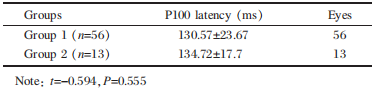
P100 latency of group 1 and group 2 was analyzed by homogeneity test of variances (Levene test). The t-test was used under an equal condition (F=2.207, P=0.142 >0.05).The statistical comparison between groups 1 and 2 showed no significant difference (t=-0.594, df=67, P=0.555>0.05)(Table 2).
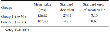
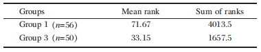
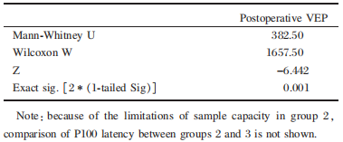
statistical analysis was performed using Mann- whitney U tests and estimation of the median with 95% CI was calculated (U =382.5,w =l657.5). The comparison of p100 latency between group l and group 3 showed statistically significant differences. The p100 latency was much longer in group l than in group 3(P=0.000l), which implies that the con- genital cataract patients are more vulnerable to optic- nerve malformation. (Table 4, Table 5)


The observation data of group 1 before and after medication were analyzed by a paired sample t test. Table 9 shows that these variables have a dependency relation (correlation=0.628, P (sig)=0.003). After the medication, the P100 latency is much shorter than before (Table 9) and the difference is statistically significant (t=2.314, df=19, P=0.032<0.05), which implies that mNGF improves the visual pathway function.

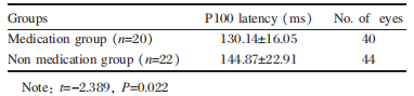
After mNGF intervention, the observation data of both groups were analyzed by an independent-sample t test, which was used under an equal condition (F=0.97, P=0.331>0.05). The P100 latency was much shorter for the medication group than for the non-medication group(Table 11). The difference was statistically significant ( t=-2.389, df =40, P=0.022<0.05).
Results
The incidence of visual pathway disorder
The total incidence of visual pathway disorder in groups 1 and 2 was 86.96% (60 eyes). 83.93% (47 eyes) of the cases in group1 had visual pathway disorder with varying degrees, 100% (13 eyes) of the cases in group 2 had visual pathway disorder with varying degrees (Table 1).The severity distribution of visual pathway disorders in groups 1 and 2
The results of P-VEP measurement were divided into four levels according to the severity of P100 latency; i.e., normal, mild, moderate, and severe. P100 latency ≤110 ms is regarded as normal, 110 ms<P100 latency≤120 ms as mild, 120 ms<P100 latency≤l50 ms as moderate,and P100 latency >150 ms as severe (Table 1).Table 1 severity distribution of visual pathway disorders between groups l and 2 (4l cases, 69 eyes)
Table 2 Mean values of P100 latency between groups 1 and 2(ms,x±5)
Comparison of visual pathway function between groups 1 and 3
The Levene test indicated that the two sets of data had unequal variances (F=64.421, P<0.05). Therefore, the data in Table 3 were analyzed by a nonparametric test for a systematic analysis of the visual pathway function of the groups 1 and 3 pediatric cataract patients (Table 3).Table 3 Comparison of P100 latency between groups 1 and 3(ms,x±5)
statistical analysis was performed using Mann- whitney U tests and estimation of the median with 95% CI was calculated (U =382.5,w =l657.5). The comparison of p100 latency between group l and group 3 showed statistically significant differences. The p100 latency was much longer in group l than in group 3(P=0.000l), which implies that the con- genital cataract patients are more vulnerable to optic- nerve malformation. (Table 4, Table 5)
Table 4 Mann-whitney U tests of P100 latency between groups 1 and 3
Table 5 Test statistics
Comparison of visual pathway function between congenital cataract eyes and contralateral eyes in group 2
The observation data of group 2 were analyzed by a paired sample t test. According to Table 6, these variables do not have the dependency relation (correlation=0.265,P (sig)=0.381). The P100 latency is much longer on the affected side than on the contralateral side. The difference was statistically significant (t=5.952, df=12, P=0.001<0.05), which implies that the congenital cataract eye are more vulnerable to optic-nerve malformation.Table 6 Comparison of P100 latency of congenital cataract eyes and contralateral eyes in group 2(n=13, ms)
Comparison of visual pathway function before and after medication
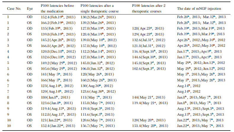
Thirteen cases(23 eyes) in the experimental group (10 cases in group 1 and 3 cases in group 2) which were diagnosed with visual pathway disorder (P100 latency>110 ms) received mNGF. (Table7, Table 8).Table 7 P100 latencies (ms) of group 1 before and after medication and the date of mNGF injection
Table 8 P100 latencies (ms) of group 2 before and after medication and the date of mNGF injection
The observation data of group 1 before and after medication were analyzed by a paired sample t test. Table 9 shows that these variables have a dependency relation (correlation=0.628, P (sig)=0.003). After the medication, the P100 latency is much shorter than before (Table 9) and the difference is statistically significant (t=2.314, df=19, P=0.032<0.05), which implies that mNGF improves the visual pathway function.
Table 9 Comparison of P100 latency of group 1 before and after medication(n=20, ms)
Comparison of visual pathway function with and without mNGF intervention in group 1
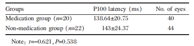
Ten cases (20 eyes) who were diagnosed with visual pathway disorder received mNGF intervention (medication group), while 11 cases (22 eyes) who also had visual pathway disorder were not medicated (non-medication group). Before medication, the P100 latency of both groups was analyzed by homogeneity test of variances (Levene test), and a t test was used under an equal condition (F=0.058,P=0.81>0.05). The statistical comparison between the medication and non-medication group showed no significant difference (t=-0.621, df=40, P=0.538>0.05)as shown in Table 10.
Table 10 Mean values of P100 latency of intervention andcontrol group before medication (ms, x±5)
Table 11 Mean values of P100 latency of intervention andcontrol group after medication (ms,x±5)
Side-effects related to mNGF treatment
Two cases of local skin swelling and ipsilateral limb pain were observed during the treatment. No other severe allergic reactions, such as gastrointestinal reactions and convulsions, appeared at any time during the treatment.Discussion
The postoperative visual function of congenital cataract patients depends on the age of onset, whether the cataract is unilateral or bilateral, the type of cataract, preexisting ocular abnormalities or diseases, complications following surgery, and outcome of the amblyopic treatment6,10-12. Optic nerve maldevelopment is one of the most important factors related to postoperative amblyopia, which has a considerable effect on the quality of life3,13. Therefore, we suggest that the optic nerve function be evaluated in postoperative congenital cataract children. Meanwhile, amblyopic treatment should be initiated as early as possible.
The present study indicates that a majority of postoperative pediatric cataract patients have complications of visual pathway dysfunction. A total of 83.93% (47 eyes) of bilateral pediatric cataract patients had visual pathway disorder to various degrees (Table 1); 100% of unilateral pediatric cataract patients (13 eyes) had visual pathway disorder (Table 1). We believe that the visual pathway disorder in postoperative pediatric cataract patients plays an important role in the impairment of their visual function.
Visual evoked potential (VEP) is the most com mon means of evaluating visual pathway function14. VEP is a group of electrical signals which predominantly reflects the activation of the macular pathway and the foveal representation at the occipital pole15,16. VEP can be considered to estimate the development of the optic-nerve when other retina diseases are eliminated17. The decline in retina sensitivity and the increment in the retina stimulation areas caused by the light scattering of the flash visual evoked potential (FVEP) can be eliminated by applying pattern visual evoked potentials (P-VEP) to children with visual fixation18.
Our study shows that the P100 latencies of P-VEP are significantly longer in children with postoperative congenital cataract than in normal children (P=0.0001, Table 3). We found that the majority of cases in groups 1 and 2 had visual pathway disorders of various degrees, which were expressed mainly in mild or moderate degrees (Table 1). A significant statistical difference was also noted between congenital cataract eyes and contralateral eyes in unilateral congenital cataract children (P=0.001<0.05, Table 6). These clinical findings support our hypothesis that children with congenital cataract are more vulnerable to optic-nerve malformation.
The critical period for visual acuity improvement and optic nerve development is widely believed to be within the first six months after birth19. During this sensitive period of visual development, form deprivation inhibits the afferent impulse, which leads to abnormal development of the lateral geniculate nucleus and the striate cortex20. Thus, the visual acuity was impacted. However, all the subjects in our study with optic nerve maldevelopment underwent cataract surgery within 3 months after birth. This should have relieved form deprivation and the patients should have resumed normal visual function. However, optic-nerve conduction anomalies were still present in 85.71% of our cases. Therefore, we deduce that other causes, like optic nerve malformation, might exist in addition to form deprivation in amblyopia of congenital cataract.
The present study indicates that a majority of postoperative pediatric cataract patients have complications of visual pathway dysfunction. A total of 83.93% (47 eyes) of bilateral pediatric cataract patients had visual pathway disorder to various degrees (Table 1); 100% of unilateral pediatric cataract patients (13 eyes) had visual pathway disorder (Table 1). We believe that the visual pathway disorder in postoperative pediatric cataract patients plays an important role in the impairment of their visual function.
Visual evoked potential (VEP) is the most com mon means of evaluating visual pathway function14. VEP is a group of electrical signals which predominantly reflects the activation of the macular pathway and the foveal representation at the occipital pole15,16. VEP can be considered to estimate the development of the optic-nerve when other retina diseases are eliminated17. The decline in retina sensitivity and the increment in the retina stimulation areas caused by the light scattering of the flash visual evoked potential (FVEP) can be eliminated by applying pattern visual evoked potentials (P-VEP) to children with visual fixation18.
Our study shows that the P100 latencies of P-VEP are significantly longer in children with postoperative congenital cataract than in normal children (P=0.0001, Table 3). We found that the majority of cases in groups 1 and 2 had visual pathway disorders of various degrees, which were expressed mainly in mild or moderate degrees (Table 1). A significant statistical difference was also noted between congenital cataract eyes and contralateral eyes in unilateral congenital cataract children (P=0.001<0.05, Table 6). These clinical findings support our hypothesis that children with congenital cataract are more vulnerable to optic-nerve malformation.
The critical period for visual acuity improvement and optic nerve development is widely believed to be within the first six months after birth19. During this sensitive period of visual development, form deprivation inhibits the afferent impulse, which leads to abnormal development of the lateral geniculate nucleus and the striate cortex20. Thus, the visual acuity was impacted. However, all the subjects in our study with optic nerve maldevelopment underwent cataract surgery within 3 months after birth. This should have relieved form deprivation and the patients should have resumed normal visual function. However, optic-nerve conduction anomalies were still present in 85.71% of our cases. Therefore, we deduce that other causes, like optic nerve malformation, might exist in addition to form deprivation in amblyopia of congenital cataract.
Genetic research indicates that PAX6 gene mutations are not only responsible for the congenital cataracts but also lead to optic nerve maldevelopment21,22. The PAX6 gene is involved in ocular morphogenesis and is expressed in the developing central nervous system and numerous ocular tissues during its development 23-25. PAX6 mutations have been de tected in various ocular anomalies, including congenital cataracts, aniridia, Peters’ anomaly, and foveal hypoplasia21. In light of the theories mentioned above, we consider that congenital cataract
patients with optic-nerve malformation might harbor PAX6 muta tions. Further study is still necessary.
patients with optic-nerve malformation might harbor PAX6 muta tions. Further study is still necessary.
Murine NGF is a 26.5 kD molecular weighted protein with two amino acid peptide chains, and is isolated and purified from mouse submandibular glands26. It is a kind of nerve cell growth regulatory factor which not only has neuron nutritional function but also promotes nerve growth. It has been shown to modulate the development, differentiation, regeneration, and function of the central and peripheral neurons27.
Murine NGF modulates the development and dif ferentiation of retina and optic nerve, and promotes the survival and recovery of retinal ganglion cells (RGCs) by binding with mNGF receptor TrkA, which is widely expressed in the optic nerve tract28. some animal experiments have revealed that intraocular injection of mNGF promotes recovery of dam aged RGCs after ischemic injury, optic-nerve transaction, and ocular hypertension29. The exogenous mNGF activated the TrkA receptors through the axon and up-regulated the Bcl-2 protein, which protects cells from apoptosis by preventing caspase activation30.
Application of mNGF has been reported to treat central nervous system diseases in infants and young children31-33. Few side effects have been observed so far. Murine NGF is believed to be safe for use in children.
In the present study, we demonstrated that intrmuscular injection of mNGF could improve visual pathway function and optic nerve development in postoperative pediatric cataract patients, particularly in cases with moderate and severe visual pathway disorder. Our clinical observations revealed that after a single therapeutic course, 10% of the cases (2 eyes) showed visual pathway improvement (10 ms≤P100 latency shortening<20 ms) and 15%(3 eyes) had obvious visual pathway improvement (20 ms≤P100 latency shortening). After2 therapeutic courses, 33.33% of the cases (4 eyes) showed visual pathway improvement (Table 7).
However, after 2 therapeutic courses of mNGF injection, P100 latency in two cases (2 eyes) in group 1 became longer than in their single therapeutic course (case 4 OS and case 8 OD, as shown in Table 7). We recognized that the interval between therapeutic courses in case 4 was more than 6 months, which may have reduced the additive effects of mNGF, thereby leading to a variation in the P100 latency. In case 8, although the P100 latency after 2 therapeutic courses was relatively longer than after the single therapeutic course, it was still much im proved over the unmedicated (Table 7). Our preference is 2 therapeutic courses rather than a single course in our treatment of optic nerve maldevelopment in postoperative pediatric cataract patients.
We also determined the P100 latency for 11 cases in group 1 who had visual pathway disorder and were not medicated with mNGF. The P100 latency of PVEP showed no significant differences between the mNGF medication and non-medication groups before mNGF intervention. However, the P100 latency of PVEP was improved significantly in the medication group compared with non-medication group after mNGF intervention, which implies that mNGF medication could improve the optic neural development in postoperative pediatric cataract children.
It should be noted that mNGF has been confirmed effective only in a small number of our cases (less than 20%) in the present study. The efficacy of mNGF still needs to be foloowed and verified clinically in large sample studies.
Compared with the normal children, postoperative pediatric cataract patients are more susceptible to optic-nerve maldevelopment. Murine NGF is likely to prove beneficial in promoting the development of the optic nerve as well as improving visual function.
Murine NGF modulates the development and dif ferentiation of retina and optic nerve, and promotes the survival and recovery of retinal ganglion cells (RGCs) by binding with mNGF receptor TrkA, which is widely expressed in the optic nerve tract28. some animal experiments have revealed that intraocular injection of mNGF promotes recovery of dam aged RGCs after ischemic injury, optic-nerve transaction, and ocular hypertension29. The exogenous mNGF activated the TrkA receptors through the axon and up-regulated the Bcl-2 protein, which protects cells from apoptosis by preventing caspase activation30.
Application of mNGF has been reported to treat central nervous system diseases in infants and young children31-33. Few side effects have been observed so far. Murine NGF is believed to be safe for use in children.
In the present study, we demonstrated that intrmuscular injection of mNGF could improve visual pathway function and optic nerve development in postoperative pediatric cataract patients, particularly in cases with moderate and severe visual pathway disorder. Our clinical observations revealed that after a single therapeutic course, 10% of the cases (2 eyes) showed visual pathway improvement (10 ms≤P100 latency shortening<20 ms) and 15%(3 eyes) had obvious visual pathway improvement (20 ms≤P100 latency shortening). After2 therapeutic courses, 33.33% of the cases (4 eyes) showed visual pathway improvement (Table 7).
However, after 2 therapeutic courses of mNGF injection, P100 latency in two cases (2 eyes) in group 1 became longer than in their single therapeutic course (case 4 OS and case 8 OD, as shown in Table 7). We recognized that the interval between therapeutic courses in case 4 was more than 6 months, which may have reduced the additive effects of mNGF, thereby leading to a variation in the P100 latency. In case 8, although the P100 latency after 2 therapeutic courses was relatively longer than after the single therapeutic course, it was still much im proved over the unmedicated (Table 7). Our preference is 2 therapeutic courses rather than a single course in our treatment of optic nerve maldevelopment in postoperative pediatric cataract patients.
We also determined the P100 latency for 11 cases in group 1 who had visual pathway disorder and were not medicated with mNGF. The P100 latency of PVEP showed no significant differences between the mNGF medication and non-medication groups before mNGF intervention. However, the P100 latency of PVEP was improved significantly in the medication group compared with non-medication group after mNGF intervention, which implies that mNGF medication could improve the optic neural development in postoperative pediatric cataract children.
It should be noted that mNGF has been confirmed effective only in a small number of our cases (less than 20%) in the present study. The efficacy of mNGF still needs to be foloowed and verified clinically in large sample studies.
Conclusion
基金
暂无基金信息
参考文献
1. Li LH ,Li N ,zhao JY ,et al.Findings of perinatal ocular examination performed on 3573 ,healthy full-term new- borns.British Journal of ophthalmology ,2013 ,97 (5) : 588-591.
2. Foster A ,Gilbert C ,Rahi J.Epidemiology of cataract in childhood :a global perspective.Journal of cataract and Refractive surgery ,1997 ,23(supplyment 1):601-604.
3. Zhu XN ,Yu F ,Xing XY. Comparison of effects of secondary in-the-bag and sulcus intraocular lens implantation in pediatric aphakia aftercongenital cataract operation. Zhonghua Yan Ke Za Zhi ,2013 ,49(8):700-705.
4. Amon M.Surgical management challenges and clinical results of bimanual micro-incision phacoemulsification cataractsurgery in children with congenital cataract.Nepal Journal of ophthalmology ,2011,3(1):3-8.
5. Alexandrakis G ,Peterseim MM ,Wilson ME.clinical outcomes of pars plana capsulotomy with anterior vitrectomy in pediatric cataract surgery.American Association for Pediatric ophthalmology and strabismus ,2002 ,6 (3) : 163-167.
6. Ventura Mc ,Sampaio VV ,Ventura BV.Congenital cataract surgery with intraocular lens implantation in microphthalmic eyes:visual outcomes and complications. Arquivos Brasileiros de oftalmologia ,2013 ,76 (4):240- 243.
7. Bao YZ ,Chen Y.Long-term outcome after bilateral cataract surgery in infants with congenital cataract. Zhonghua Yan Ke Za Zhi ,2013 May ,49(5):395-398.
8. Shah MA ,Shah SM ,Shah AH.Visual outcome of cataract in pediatric age group: does etiology have a role.visual outcome of cataract in pediatric age group: does etiology have a role.European Journal of Ophthalmology ,2013 May ,24(1):76-83.
9. Mansouri B ,stacy Rc ,kruger J ,et al.Deprivation amblyopia and congenital hereditary cataract.Semin Ophthal-mol ,2013 Sep-Nov ,28(5-6):321-326.
10. Chan WH ,Biswas S ,Ashworth JL.Congenital and infantile cataract:aetiology and management.European Journal of Pediatrics ,2012 Apr ,171(4):625-630.
11. Pechereau A ,Paire V ,Raffin L.Amblyopia treatment of unilateral and bilateral cataract with visual acuity result. Journal of French Ophtalmology , 2010 Mar ,34(3):2008- 212.
12. Kim DH ,Kim JH.Long-term results of bilateral congenital cataract treated with early cataract surgery ,aphakic glasses and secondary IOL implantation.Acta ophthalmologica scandinavica ,2012 May ,90(3):231-236.
13. Zhou LH ,Wang S ,Xing YQ.Evaluation of visual function after congenital cataract extraction.Zhonghua Yan Ke Za Zhi ,2008 Feb ,44(2):135-137.
14. Fan YW ,Li XQ ,Yan XM.The transient PVEP acuity assessment for 2 to 5 years old normal children.Zhonghua Yan Ke Za Zhi ,2010 May ,46(5):423-426.
15. Liu RJ ,zhu GY ,Fan LH.Visual electrophysiology and objective visual function.Fa Yi Xue Za Zhi ,2002 May ,18 (2)115-117.
16. Brecelj J.From immature to mature pattern ERG and VEP. Documenta ophthalmologica ,2003 Nov,107(3):215-224.
17. Holder GE.The pattern electroretinogram in anterior visual pathway dysfunction and its relationship to the pattern vi- sual evoked potential: a personal clinical review of 743
eyes.Eye (Lond) 1997 ,11(6):924-934.
18. Luo ZW ,Sun ZH.The observation of visual evoked potential (VEP) in patients with single congenital cataract. China journal of practical ophthalmology ,2004 May ,22 (5):135-138.
19. Brδmond-Gignac D ,Copin H ,Lapillonne A ,et al.Visual development in infants:physiological and pathological mechanisms.Current Opinion in Ophthalmology ,2010 Apr ,22(supplyment):1-8.
20. Wright KW. Visual development ,amblyopia ,and sensory adaptations.Pediatric Ophthalmology and Strabismus , 1995 :119-138.
21. Noriyuki Azuma ,Yuki Yamaguchi ,Hiroshi Handa.Mutations of the PAX6 Gene Detected in Patients with a variety of optic-Nerve Malformations.American Journal of Human Genetics ,2003 Jun ,72(6):1565-1570.
22. Shaham O ,Gueta K ,Mor E.PAX6 regulates gene expression in the vertebrate lens through miR-204.PLoS Genetics ,2013 ,9(3):e1003357-e1003357.
23. Walcher T ,Xie Q ,Sun J.Functional dissection of the paired domain of Pax6 reveals molecular mechanisms of coordinating neurogenesis and proliferation.Development , 2013 Mar ,140(5):1123-1136.
24. Lleras-Forero L ,Tambalo M ,christophorou N.Neuropeptides:developmental signals in placode progenitor formation.Developmental cell ,2013 Jul ,26(2):195-203.
25. Xie Q ,Yang Y ,Huang J.Pax6 interactions with chromatin and identification of its novel direct target genes in lens and forebrain.PLoS One ,2013 ,8(1):e54507-e54507.
26. Aloe L ,Rocco ML ,Bianchi P ,et al.Nerve growth factor:from the early discoveries to the potential clinical use. Journal of Translational Medicine ,2012 Nov ,29 (10) : 239-243.
27. Sofroniew MV ,Howe CL ,Mobley WC.Nerve growth factor signaling ,neuroprotection ,and neural repair. Annual Review of Neuroscience ,2001 ,24:1217-1281.
28. Maffei L ,Berardi N ,Domenici L ,et al.Nerve growth factor (NGF) prevents the shift in ocular dominance distribution of visual cortical neurons in monocularly deprived rats.Journal of Neuroscience ,1992 ,12(12):4651-4662.
29. Lambiase A ,Mantelli F ,Bonini S.Nerve growth factor eye drops to treat glaucoma.Drug News perspect ,2010 ,23(6):311-367.
30. Colafrancesco V ,Parisi V ,Sposato V ,et al.Ocular Application of Nerve Growth Factor protects degenerating retinal ganglion cells in a rat model of glaucoma.Journal of glaucoma ,2011 ,20(2):100-108.
31. Xing LH ,Zhang LX ,Zhang LL.Umbilical cord blood stem cell transplantation combined with mouse neural growth factor application and physical rehabilitation therapy for infantile cerebral palsy. Chinese Journal of Tissue Engineering Research, 2012 Oct ,7(16):7777-7781.
32. Mao QQ ,Gao YX.NGF and IVIG treatment and prognosis in Guillain-Barre
syndrome.Chinese Journal of Practical ,2012 Dec ,27(12):944—949.
33. David M Holtzman ,R Ann Sheldon.Nerve growth factor protects the neontal brain against hypoxic-ischemic injury. The American Neurological Association ,1996 ,39 (1) : 114—122.
相关文章
刘思源;张怡萌;梁飞雁;柳夏林;郑丹莹;范舒欣;黄圣松,Non-fundus visual prognosis indicators in cataract patients with congenital iridochoroidal colobomaYan Zhang;Xiaolei Liang;Shihui Wei;Hongyang Li,Differential Diagnosis for Multiple Sclerosis-related Optic NeuritisMingyue Wang;Wei Xiao,Congenital Cataract: Progress in Surgical Treatment and Postoperative Recovery of Visual Function