





In the realm of life sciences, the convergence of biological complexity and technological innovation has reached a pivotal juncture as biomedical research has long relied on a reductionist approach—dissecting tissues into cells and cells into molecules. While this strategy has advanced our understanding of diseases such as cataract, which is the leading global cause of blindness, it has also created a fragmented view of biology. We now possess vast catalogs of genes and proteins, yet we often struggle to predict how these components interact dynamically across spatial and temporal scales to produce complex phenotypes or respond to therapeutic interventions.
Traditional biological models, such as in-vitro cell models and animal models, often suffer from low translational success rates because they cannot fully recapitulate the complex, patient-specific spatiotemporal environment of human pathology. Digital twin (DT) technology, a computational paradigm that creates dynamic, multiscale virtual replicas of physical entities using longitudinal or real-time data, has emerged as a powerful approach to address these limitations. Operationally, a lens DT is defined by two core features: iterative updating of the virtual model as new patient or experimental data are acquired, and feedback from the model to the physical system through predictions, risk estimates, or experimental hypotheses. For example, a DT for posterior capsule opacification (PCO) risk could be initialized with perioperative clinical and imaging data, updated at each slit-lamp visit with observed capsular changes, and used to provide revised individualized risk estimates or optimize follow-up intervals, rather than relying on the static estimates generated by conventional predictive models. By enabling bidirectional mapping between biological systems and their digital counterparts, DTs bridge the gap between macroscopic and microscopic scales, offering new capabilities to simulate, analyze, and optimize cataract pathophysiology and treatment within a virtual environment (Figure 1). This perspective outlines how DT technology could advance cataract therapeutics through multiscale integration, from molecular interactions to clinical outcomes, while establishing a framework for broader biomedical applications.
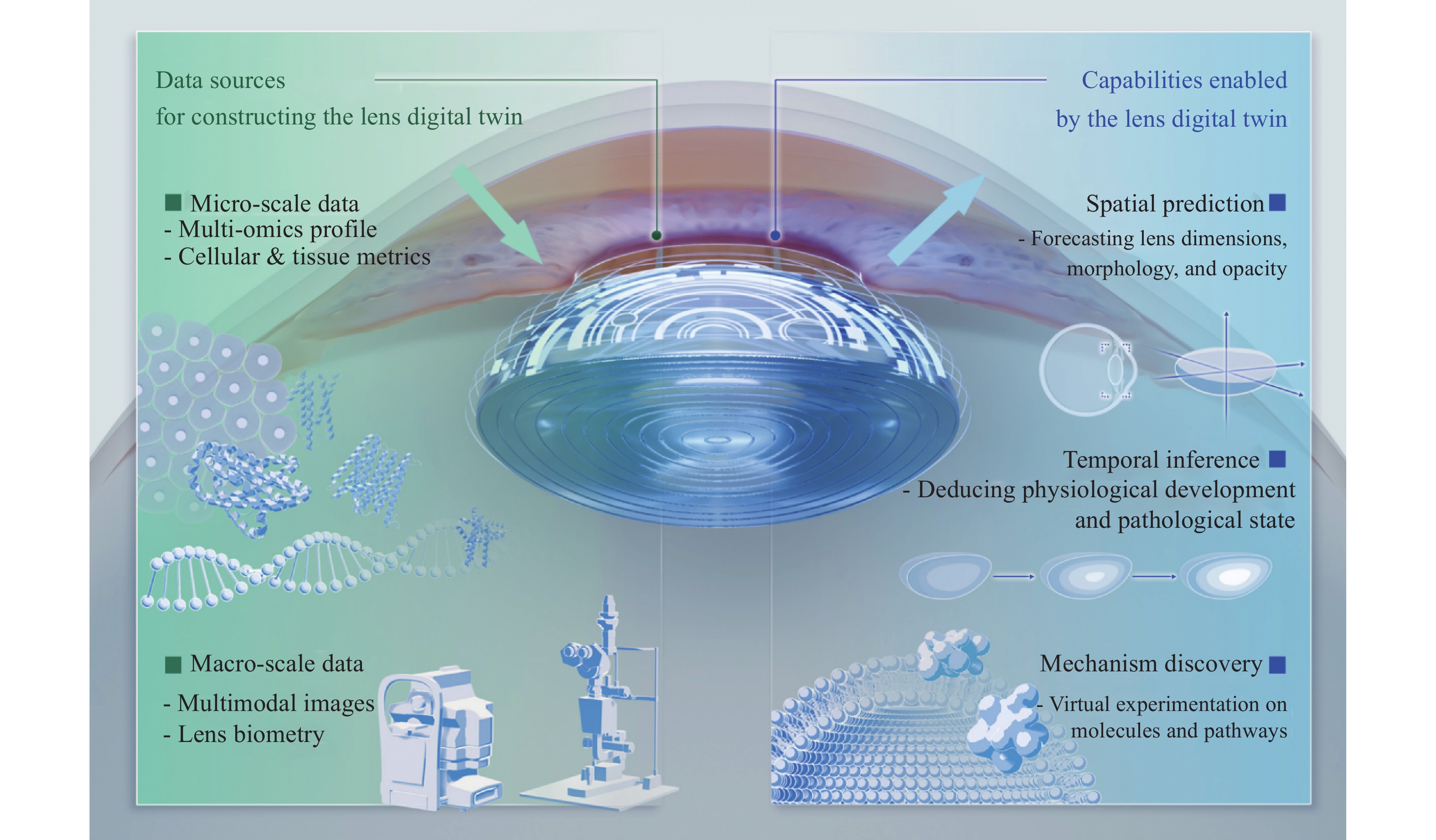
This schematic depicts the integrative framework for constructing and applying a lens digital twin. The central virtual lens model and directional arrows represent the continuous bidirectional mapping between physical and virtual systems. Longitudinal observations update the model, while its predictions guide subsequent clinical or experimental assessment. Left panel: Data sources are categorized into micro-scale inputs (multi-omics profiles, cellular and tissue- metrics) and macro-scale inputs (multimodal imaging, lens biometry) to construct a hierarchical biological representation. Right panel: Core computational capabilities enabled by the digital twin include spatial prediction, temporal inference, and mechanism discovery. By integrating discrete biological scales through spatiotemporal modeling, the framework supports precision diagnostics and therapeutic optimization in cataract management.
Lens disorders: pending challenges demand innovative solutions
The rationale for adopting DT technology is best understood by first recognizing the limitaton of current therapeutic paradigms. The ocular lens is a transparent, biconvex tissue that relies on precise cellular organization to maintain optical clarity. Age-related cataract affects 65–74% of individuals aged 85–89, and congenital cataract remains a substantial contributor to infant blindness worldwide.[1-3] Although surgical extraction and intraocular lens (IOL) implantation represent the clinical gold standard, long-term outcomes are frequently compromised by postoperative complications, most notably posterior capsule opacification (PCO, or “secondary cataract”). PCO is driven by epithelial-mesenchymal transition (EMT) and subsequent fibrosis of residual lens epithelial cells (LECs). Nd:YAG laser capsulotomy remains the standard treatment for established PCO; however, it carries clinically relevant risks of macular edema and retinal detachment. Consequently, a dual clinical gap persists: preoperative risk stratification for PCO remains unfeasible, and targeted early interventions to prevent or halt its progression are lacking. These limitations fundamentally stem from an incomplete understanding of the dynamic pathophysiology of the lens and the microscale spatiotemporal mechanisms governing PCO formation. [4]
Regenerative approaches harnessing lens epithelial cells (LECs) show promise, as demonstrated by Lin et al.’s work on in situ lens regeneration.[5] However, clinical outcomes remain inconsistent due to individual variability in regenerated lens morphology, stem cell dynamics, and spatiotemporal signaling.[6] These challenges, including unpredictable fibrosis and disorganized regeneration, underscore the need for a systems-level approach to decode lens physiology and pathology. DT technology is uniquely positioned to address these complexities.
DT technology: a parade for biomedical transformation. Given the multifactorial determinants underlying the mechanistic and phenotypic heterogeneity of the lens, relying solely on traditional biological models is highly resource-intensive and time-consuming. A practical goal is to use digital technologies to mathematically represent the microscopic and macroscopic processes of lens regeneration, ultimately simulating these processes in silico. This strategy forms the basis of a “digital twin” (DT). The DT concept originated at National Aeronautics and Space Administration (NASA) during the Apollo mission in the United States. Since then, DTs, defined as dynamic virtual replicas of physical entities updated with real-time data, have been widely adopted in manufacturing and healthcare to simulate predictive and counterfactual scenarios.
The application of DTs in medicine differs fundamentally from traditional bioinformatics. While bioinformatics primarily analyzes static datasets to identify statistical correlations, a DT enables virtual experiments on biological processes, helping to overcome two fundamental limitations in disease research. First is the spatiotemporal disconnect: standard histology provides only a snapshot in time, failing to capture dynamic processes such as cell differentiation or migration rates. A DT integrates time-series data to reconstruct developmental trajectories, allowing researchers to "rewind" or "fast-forward" disease progression in silico. Second is the micro-macro disconnect, which stems from the difficulty of linking complex multi-omics data directly to macroscale tissue function. DTs transcend conventional modeling by integrating real-time data streams across biological hierarchies (molecular, cellular, tissue, and organ) into predictive simulations. For the ocular lens, this enables the creation of a model where a change in a specific gene expression pattern (micro) propagates through the simulation to predict alterations in the refractive index or transparency of the lens (macro). Ultimately, DT modeling can provide unprecedented insights into underlying disease mechanisms.[7]
Building the lens DT model: exploration and practice
Developing a comprehensive DT model of the lens is an ambitious goal that builds upon decades of computational biology. Prior studies have laid the groundwork by computationally modeling isolated lens features. For instance, digital modeling techniques have been used to reconstruct three-dimensional (3D) spatial architecture of the lens, characterizing phenotypic heterogeneity across the anterior, central, and posterior cortical regions.[8] Finite element modeling (FEM) has also been employed to simulate the 3D biomechanics of the lens during accommodation[9] and to map its internal microcirculation system.[10] Similarly, systems biology and computational approaches have facilitated the construction of time-series models that capture temporal phenotypic changes across various developmental and aging stages.[11-12] Crucially, the mapping between genetic variations and phenotypes outcomes is not static: rather, it evolves dynamically throughout lens development. Beyond the computational characterization of static tissue microarchitecture, comprehensive modeling of dynamic behaviors, such as the morphological changes the lens undergoes during accommodation, is essential. This integration is required to establish a unified digital framework that spans from the macroscale, effectively bridging static structure with dynamic function.[13-14] Ultimately, a fully realized Lens DT must synthesize these disparate models into a cohesive, interactive system.
Building the lens DT model: a three-step strategy
To transition from fragmented models to a fully evolving and iterative lens DT, we propose a three-step implementation strategy. This framework progresses from data acquisition to dynamic modeling, and finally to translational application. It should be viewed as a staged research roadmap rather than a ready-to-use clinical product.
Step 1: multiscale data acquisition
We propose integrating sub-tissue cellular assays (e.g., evaluating LEC proliferation, differentiation, migration, and apoptosis) with single-cell/spatial multi-omics to resolve molecular-microanatomical relationships across disease and regeneration models. Macroscopic parameters (e.g., lens structure, refractive index, transparency) are captured and correlated with these microscopic features. For instance, by employing single-cell sequencing in tandem with spatial transcriptomics, specific markers governing LEC behaviors or EMT can be mapped to precise anatomical coordinates within the lens. Subsequently, these molecular maps can be coregistered with clinical imaging data, linking gene expression patterns to macroscopic physical attributes. This multimodal integration generates a structured map that connects optical, anatomical, and molecular information in a clinically interpretable manner.
Step 2: spatiotemporal modeling
Simulating these spatiotemporal dynamics requires advanced computational algorithms. Leveraging the intrinsic relationships between temporal and spatial attributes, adaptive dynamic programming or spatiotemporal interpolation algorithms can be applied to infer the missing data points. Furthermore, physics-informed neural networks (PINNs) can embed physical laws (e.g., fluid dynamics, tissue mechanics, diffusion equations) into the machine learning process, ensuring that the simulations respect the biophysical constraints of the lens. Subsequently, deep reinforcement learning can be employed to predict future states. By treating lens development or PCO progression as a sequential state-transition process, the model can learn the optimal pathways for cell migration and differentiation. A critical objective is to align serial observations to predict clinically meaningful changes, such as opacity progression, aberrant cell migration, EMT-related fibrosis, or regenerated lens morphology. Model outputs should be rigorously validated against held-out experimental data and, when feasible, prospective clinical follow-up.
Step 3: cross-species transfer learning
Human clinical data are often sparse and limited in dimensionality compared to experimental models. Obtaining comprehensive, time-series molecular data from living human lenses is ethically and practically unfeasible. To bridge the species gap, we propose utilizing transfer learning. First, a high-data-density DT model is established using experimental animals (e.g., rabbits or mice), where invasive longitudinal sampling is permissible. The mature DT model learns the fundamental biological rules of lens and is subsequently fine-tuned using sparse, lower-dimensional human data (e.g., slit-lamp images and aqueous humor proteomics). While animal models provide the dense longitudinal data required for initial model architecture, human images and clinical phenotypes serve for calibration. Transfer success should be assessed on held-out human data using prespecified calibration and prediction-error metrics. The calibrated model must be benchmarked against human-only models and direct, uncalibrated transfer from animal models. Crucially, this step should be regarded as cross-species calibration rather than direct species substitution, and all inferred human predictions require independent validation.
Beyond the lens: implications for biomedical discovery
The lens DT paradigm extends far beyond cataract treatment. By establishing a template for multiscale digital twins, this approach can accelerate the development of analogous systems for retinal, neural, and cardiac tissues. Three key implications emerge for broader biomedical research. First, regarding personalized intervention, DTs enable "virtual clinical trials" that tailor therapies to individual molecular profiles. Second, for mechanistic discovery, hypotheses regarding molecular pathways can be iteratively tested in silico prior to wet-lab validation. Third, this approach fosters a new research paradigm in biomedicine—one where digital and physical experimentation coexist synergistically.
Clinical translation considerations
Clinical translation must proceed cautiously. Early applications will likely be confined to research-support tools, such as estimating PCO risk or prioritizing laboratory experiments, before being integrated into direct clinical decision-making. Patient data must be de-identified, securely stored, and governed by informed consent and institutional review board (IRB) oversight. If deployed as clinical decision-support software, a lens DT would require rigorous documentation, continuous performance monitoring, and regulatory review under applicable Software as a Medical Device (SaMD) and Good Machine Learning Practice (GMLP) frameworks.[15] Furthermore, integration into routine cataract care should prioritize simple, interpretable outputs, such as risk categories or recommended follow-up intervals, over complex, opaque black-box predictions.
Limitations and outlook
Several limitations should be acknowledged. First, biological DTs face challenges in ground-truth validation. As many microscopic states cannot be measured longitudinally in living human lenses. Second, aligning imaging, clinical, cellular, and multi-omics data across temporal scales is technically challenging and resource-intensive. Third, highly complex models often lack interpretability, which may hinder clinical adoption and trust. Finally, genetic and epigenetic heterogeneity dictates that a generalized lens DT will require population-specific calibration and continuous updating. Given these constraints lens DTs should initially be developed as hypothesis-generating and risk-stratification tools, necessitating rigorous prospective validation before routine clinical deployment.
Conclusion
DT technology is more than a technical advance; it fundamentally changes how we decode biological complexity. For cataracts, DTs bridge the gap between molecular mechanisms and clinical phenotypes, enabling predictive, preventive, and personalized ocular therapeutics. Rigorously validated, DTs can powerfully complement experimental and clinical research. As the technology matures, computational modeling will evolve from a supplementary tool into an integral partner in bench research. Ultimately, the Lens DT will drive the shift from reactive medicine to proactive, simulation-based precision health—paving the way from digital twins of the eye to whole-body models.
Correction notice
None
Acknowledgements
The authors gratefully acknowledge the Global Ophthalmic AI and Technology Society (GOATS) and the Guangdong Basic Research Center of Excellence for Major Blinding Eye Diseases Prevention and Treatment for their support of this work.
Author contributions
(I) Conception and design: Xi Chen, Xiaohang Wu, Zhenzhen Liu, and Haotian Lin
(II) Administrative support: Yu-Wai-Man Patrick, Carol Cheung, Zhenzhen Liu, and Haotian Lin
(III) Provision of study materials or patients: None
(IV) Collection and assembly of data: Wenben Chen, Yuanjun Shang, Lixue Liu
(V) Data analysis and interpretation: Xi Chen, Xiaohang Wu
(VI) Manuscript writing: All authors
(VII) Final approval of manuscript: All authors
Conflict of interests
The authors declare that they have no conflicts of interest. All authors have completed the ICMJE uniform disclosure form.
Patient consent for publication
None
Ethics approval and consent to participate
None
Data availability statement
None
Open access
This is an Open Access article distributed in accordance with the Creative Commons Attribution Non Commercial-No Derivs 4.0 International License(CC BY-NC-ND 4.0), which permits the non-commercial replication and distribution of the article with the strict proviso that no changes or edits are made and the original work is properly cited(including links to both the formal publication through the relevant DOI and the license).
Declaration of generative AI use
No generative artificial intelligence (GenAI) tools were used in the preparation of this manuscript.