





HIGHLIGHTS
1. Critical Discoveries and Outcomes
• As myopia becomes increasingly prevalent, the number of children with early-onset high myopia (eoHM) is also rising. The majority of existing studies on eoHM focus on genetics and hereditary factors, with very few studies examining the refractive and visual development in children with eoHM.
2. Methodological Innovations
• Based on a large hospital-based dataset, we found that two-thirds of children aged 2 to 8 years with eoHM could achieve stability or even regression in their myopia severity. Children with older age, boys, and less myopic SE at baseline tended to have a higher risk of faster myopia progression. Half of the children with eoHM could not reach best corrected visual acuity (BCVA) >0.5 (20/40) by age 6 years. Children with the same BCVA exhibited different myopia severity, while those with the same myopia severity could have substantial variations in their BCVA.
3. Prospective Applications and Future Directions
• Future larger-scale and long-term studies are needed to investigate the subtype-specific impacts of strabismus on refractive changes in children with eoHM.
The escalating prevalence of myopia, especially high myopia, has become a major global concern. By 2050, an estimated 938 million people worldwide are projected to be diagnosed with high myopia.[1] This condition can lead to many irreversible ocular complications, including myopic macular degeneration (MMD), retinal detachment (RD), cataract, open angle glaucoma (OAG), and diffuse chorioretinal atrophy, all of which are associated with vision loss and diminished quality of life.[2-4] For every 1 diopter (D) increase in myopia, the risk of MMD, RD, posterior subcapsular cataract, and OAG increases by 58%, 30%, 21%, and 20% respectively.[5] Moreover, the prevalence of early-onset high myopia (eoHM), defined as high myopia manifesting before the age of 7, has been reported to be rising across various countries and regions.[1,6-8] Children with eoHM face a prolonged progression period, increasing the likelihood of more severe myopia and a higher risk of vision-threatening complications in adulthood.[9-11]
Monitoring myopia progression in children with eoHM is crucial; however, existing studies have primarily focused on genetic factors,[12-14] leaving the longitudinal patterns of myopia progression and visual acuity (VA) development in this population largely unexplored. Children with eoHM are at significantly elevated risk for amblyopia and irreversible vision loss due to myopic retinopathy.[8] Therefore, monitoring VA changes in this population is equally important. Although previous research has shown that myopia progression in preschool children is influenced by factors such as age, baseline myopia severity, family history of myopia, and astigmatism, [15-17] little is known about the specific risk factors associated with myopia progression in children with eoHM. To date, only two studies have addressed this issue, both limited by small sample sizes.[18-19] To address this gap, our study utilizes a larger hospital-based dataset with an extended follow-up period to investigate longitudinal changes in refraction and VA among Chinese children with eoHM, while also exploring potential factors influencing these changes.
Materials and methods
Study population
We retrospectively reviewed clinical records to identify children with eoHM, defined as a cycloplegic SE ≤−6.00 D in both eyes before 7 years of age, based on the electronic medical records (EMR) at Zhongshan Ophthalmic Center (ZOC) between 2009 and 2023. Patients with available cycloplegic refraction data and BCVA from baseline visit and at least two subsequent annual follow-up visits were included in this study. Children meeting any of the following exclusion criteria were omitted: (1) a history of ocular surgery, such as Laser-assisted in situ keratomileusis, strabismus correction or other internal intraocular surgery, (2) a history of vision-threatening ocular diseases, such as congenital cataract and retinal diseases, (3) a history of ocular trauma. The study was approved by the Ethics Committee of Zhongshan Ophthalmic Center, Sun Yat-sen University, Guangzhou and conducted in accordance with the Declaration of Helsinki.
For each participant at every hospital visit, we retrospectively extracted data on age, sex, cycloplegic refraction, BCVA (expressed in LogMAR), and presence of strabismus from EMR. At ZOC, cycloplegic refraction was measured using an autorefractor (Topcon, Tokyo, Japan) after complete cycloplegia, defined as the absence of a light reflex and a dilated pupil at least 6 mm. In our hospital, 0.5% tropicamide eye drops were routinely used for cycloplegia, with one drop administered every 5 minutes for a total of three applications. For children under 6 years of age at their visit, as well as those with moderate-to-high hyperopia, esotropia, or significant variability in tropicamide refraction results, 1% atropine eye ointment was typically administered. The ointment was applied at bedtime for three consecutive nights, with autorefraction performed on the fourth day. This practice was followed by the Chinese Expert Consensus on Cycloplegic Refraction and Safe Medication Use in Children (2019).[20] BCVA was measured following subjective refraction by optometrist or ophthalmic nurse using an early treatment diabetic retinopathy study (ETDRS) tumbling E chart at a standard testing distance of 5 meters and recorded in Snellen decimal notation. The presence of strabismus was assessed and diagnosed by ophthalmologists.
The first visit of each child with eoHM at ZOC was designated as the baseline visit. The mean myopia progression rate was calculated as the change in SE between the final and baseline visits divided by the intervening follow-up duration. We categorized the study population into three groups based on the rate of refractive change. The myopia progression group was defined as those with an average myopic shift of ≥ 0.5D per year (i.e.,an SE change rate of ≤ −0.5D/year).[21] The myopia regression group was defined as those with an SE change rate of ≥ +0.5D/year, while those falling between these thresholds were classified as the myopia stable group.[19] Strabismus was considered present if a diagnosis was recorded in the EMR during any hospital visit.
Statistical analysis
SE was calculated as the sum of the spherical power and half the cylindrical power. Given the high correlations between the right and left eyes within the same individual, only data from the right eyes were included in the analysis. The Kolmogorov–Smirnov test and histogram were used to assess the normality of continuous variables. The baseline SE distribution was expressed as median (IQR) across different age groups, while the distributions of sex, SE categories, and strabismus were expressed as absolute and relative frequencies.
Children were categorized by age (<3 years, 3 to 4 years, and 5 to 6 years), baseline SE (>−8 D to ≤−6 D, >−10 D to ≤−8 D, and ≤−10 D), and the presence of strabismus (yes or no). Independent t-tests were used to examine the differences in the mean myopia progression rate by sex and strabismus status at baseline. Trend analyses were performed to detect any differences across different baseline SE and age subgroups, as well as differences in BCVA among age subgroups. Linear mixed-effects models were used to analyze longitudinal changes in SE. The model incorporated time since the initial visit as a continuous variable. Fixed effects included sex, baseline age, baseline SE, and the presence of strabismus, along with their interaction terms with time, to assess their impact on the rate of SE progression. To account for the within-subject correlation of repeated measures and inter-subject variability in both baseline refractive error and progression rates, the model included random intercepts for each participant and random slopes for time. Individual SE change trajectories and linear mixed-effects regression lines for SE associated with age were plotted to visualize longitudinal changes. The distribution of BCVA across children with different SE level was represented as stacked bar graphs, incorporating BCVA from all follow-up visits for each participant (excluding visits with under-correction).
Statistical analyses were performed using SPSS software version 26.0 (SPSS, Inc., Chicago, IL, USA) and STATA version 18.0 (Stata Corporation, College Station, TX, USA). A two-sided P value < 0.05 was considered statistically significant.
Results
From 2009 to 2023, a total of 4,876 children with eoHM were identified from the EMR. Of these, 3,416 (70.07%) were excluded due to lack of at least one annual follow-up visit, and 530 (10.87%) were excluded due to a history of ocular surgery, vision-threatening ocular diseases, or ocular trauma. Ultimately, 930 children (19.07%) were eligible for the current analysis. The median age of the included children at baseline was 4.33 years (IQR, 3.42 to 5.25 years). Among them, 603 (64.84%) were boys, 170 (18.28%) were diagnosed with strabismus. Participants were followed for a median of 2.85 years (IQR, 2.03 to 3.57 years). The median baseline SE was −8.25D (IQR, −10.00D to −7.00D). Detailed baseline characteristics of the study participants stratified by age are provided in Table 1.
|
|
Baseline Age (years) |
|||||
|
Total |
1-2 |
3 |
4 |
5 |
6 |
|
|
Number |
930 |
70 |
214 |
272 |
222 |
152 |
|
Sex, boy n (%) |
603 (64.84) |
42 (60.00) |
145 (67.76) |
180 (66.18) |
135 (60.81) |
101 (66.45) |
|
Baseline SE |
|
|||||
|
Median (IQR), diopters |
|
−9.375(−10.781, −8.000) |
−8.375(−9.906, −7.250) |
−8.250(−10.000, −7.000) |
−8.063(−9.875, −6.875) |
−8.313(−9.969, −6.656) |
|
>−8 D and ≤−6 D, n (%) |
389 (41.83%) |
16 (22.86) |
89 (41.59) |
112 (41.18) |
103 (46.40) |
69 (45.39) |
|
>−10 D and ≤−8 D, n (%) |
297 (31.94%) |
25 (35.71) |
72 (33.64) |
90 (33.08) |
65 (29.28) |
45 (29.61) |
|
≤−10 D, n (%) |
244 (26.24%) |
29 (41.43) |
53 (24.77) |
70 (25.74) |
54 (24.32) |
38 (25.00) |
|
Strabismus, n (%) |
170 (18.28%) |
14 (20) |
42 (19.63) |
53 (19.49) |
37 (27.41) |
26 (17.11) |
|
Abbreviations: eoHM: early-onset high myopia; IQR: interquartile range; SE: spherical equivalent; D: diopter. |
||||||
Across all 930 participants, the mean myopia progression rate was −0.33 D/year (SD, 0.37). Over the follow-up period, 10 participants (1.08%) demonstrated myopia regression, 622 (66.88%) showed stable refraction, and 298 (32.04%) exhibited myopia progression. Table 2 presents the mean myopia progression rates across different subgroups. Older children exhibited a faster mean myopia progression rate (P=0.029), and boys progressed faster than girls (−0.35 vs. −0.28 D/year, P=0.008). Those with a less myopic baseline SE showed a faster rate of myopia progression (P=0.002). Individual SE change trajectories and linear regression lines for SE associated with age are shown in Figure 1. The presence of strabismus was not associated with the rate of myopia progression.
|
Characteristics |
Number |
Mean rate of myopia progression (D/year), Mean (SD) |
|
Sex |
|
|
|
Boys |
603 |
−0.35 (0.36) |
|
Girls |
327 |
−0.28 (0.39) |
|
P |
|
0.008 |
|
Baseline age (years) |
|
|
|
≥1 and <3 |
70 |
−0.25 (0.38) |
|
≥3 and <5 |
486 |
−0.32 (0.37) |
|
≥5 and <7 |
374 |
−0.36 (0.38) |
|
P for trend |
|
0.029 |
|
Baseline SE (D) |
|
|
|
>−8 and ≤−6 |
389 |
−0.38 (0.35) |
|
>−10 and ≤−8 |
297 |
−0.29 (0.38) |
|
≤−10 |
244 |
−0.29 (0.40) |
|
P for trend |
|
0.002 |
|
Strabismus at Baseline |
|
|
|
Yes |
170 |
−0.35 (0.40) |
|
No |
760 |
−0.32 (0.37) |
|
P |
|
0.310 |
|
Abbreviations: SD: standard deviation; SE: spherical equivalent; eoHM: early-onset high myopia; D: diopter. |
||
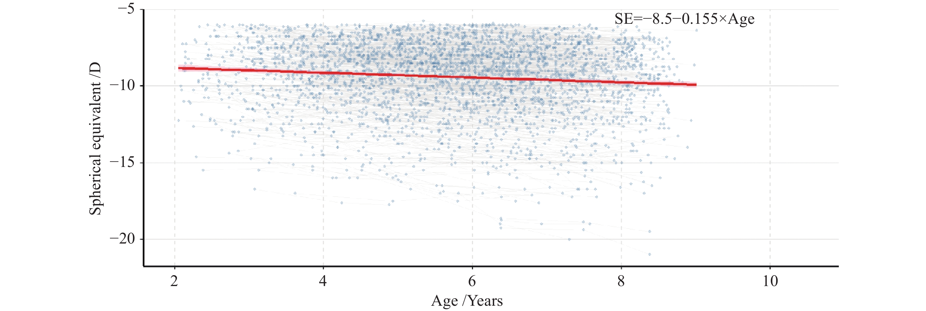
The blue-colored trajectories illustrate individual participant’s SE change curve with age, whereas the red line represents the linear, mixed-effects regression lines for SE associated with age, with the corresponding regression equation provided in the upper-right inset.
As shown in Table 3, older baseline age (β = −0.041, P<0.001), boys (β= −0.067, P=0.012), and a less myopic SE at baseline (β = −0.013, P=0.021) were associated with faster myopia progression. In addition, the baseline BCVA for the stable, progression, and regression groups was 0.37±0.20, 0.33±0.19, and 0.33±0.15, respectively (P<0.05).
|
Characteristics |
Mean rate of myopia progression (D/year) |
|||
|
Model 1* |
Model 2† |
|||
|
ß (95%CI) |
P |
ß (95%CI) |
P |
|
|
Baseline age (years) |
−0.041 (−0.062, −0.019) |
<0.001 |
−0.041 (−0.063, −0.019) |
<0.001 |
|
Female, vs male |
−0.065 (−0.118, −0.013) |
0.015 |
−0.067(−0.120, −0.015) |
0.012 |
|
Baseline SE (D) |
−0.013 (−0.024, −0.002) |
0.024 |
−0.013 (−0.024, −0.002) |
0.021 |
|
Strabismus, yes vs no |
|
|
0.051 (−0.014, 0.116) |
0.122 |
|
Model 1* adjusted for baseline age, sex, and initial SE. Abbreviations: SE: spherical equivalent; D: diopter Model 2† adjusted for baseline age, sex, initial SE and strabismus at baseline. |
||||
Table 4 presents the mean BCVA values and the proportion of children with a LogMAR BCVA < 0.3 across different ages. Overall, older children exhibited better BCVA (P<0.001). Figure 2 illustrates the distribution of SE among children with eoHM, stratified by BCVA. Each BCVA category encompassed a broad range of SE values; likewise, a given SE level was associated with varying BCVA outcomes.
|
Baseline Age |
Number |
Percentage of children with LogMAR BCVA < 0.3 (%) |
LogMAR BCVA, Mean (SD) |
|
4 |
426 |
11.27 |
0.50 (0.25) |
|
5 |
661 |
30.56 |
0.39 (0.23) |
|
6 |
748 |
50.27 |
0.31(0.23) |
|
7 |
629 |
63.28 |
0.25 (0.23) |
|
8 |
405 |
73.58 |
0.19 (0.17) |
|
P for trend |
|
|
<0.001 |
|
Abbreviation: BCVA: best corrected visual acuity; SD: standard deviation. BCVA was recorded and analyzed in Snellen decimal. BCVA from each follow-up of each participant were placed into the corresponding age subgroups for calculation (except for underfitting). |
|||
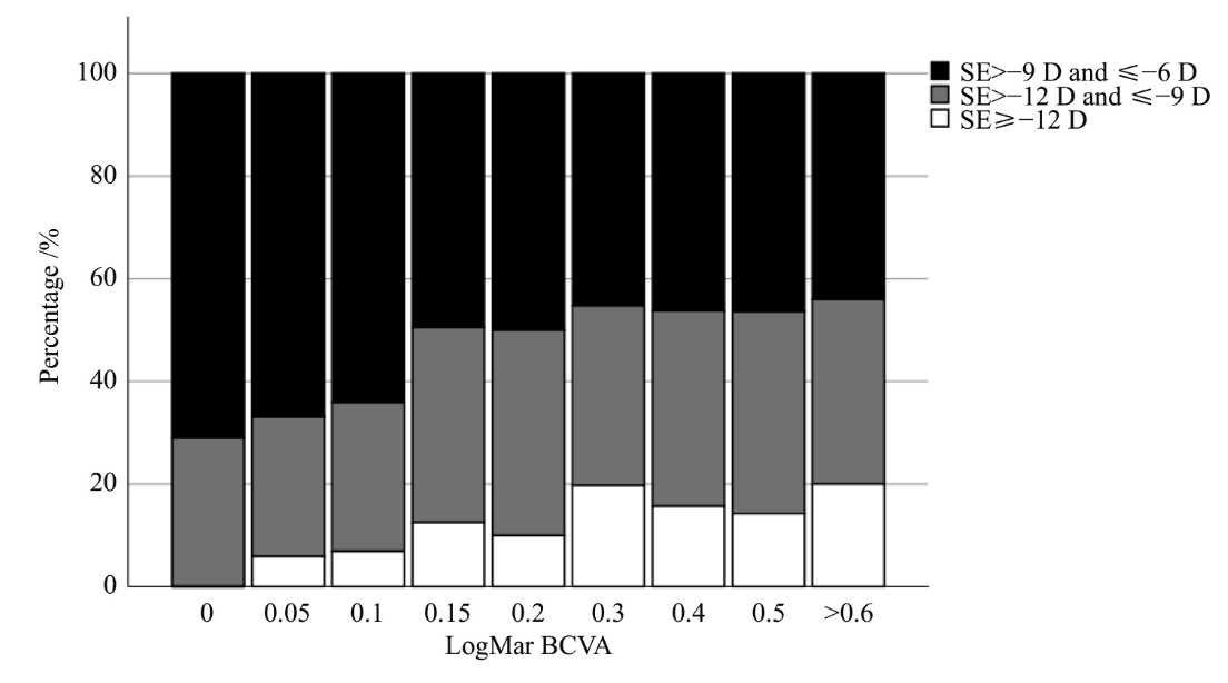
BCVA and SE from each follow-up of each participant were included (except for underfitting). Abbreviation: BCVA: best corrected visual acuity; SE: spherical equivalent.
Discussion
In the current study, we reported longitudinal changes in SE and the distributions of BCVA in Chinese children with eoHM based on a large hospital dataset. The mean myopia progression rate was −0.33 D/year (SD, 0.37), with two-thirds of children with eoHM exhibiting myopic stability or regression. Children who were older age, male, and had a less myopic SE at baseline appeared to experience faster myopia progression. Overall, BCVA improved with age, but only half of these children achieved LogMAR BCVA < 0.3 by the age of 6 years.
In a 3-year longitudinal study of 60 children aged ≤ 6 years with eoHM in Korea, the myopia progression rate was −0.37D/year (SD, 0.39), with 40% of the children exhibiting myopic progression.[19] Another study included 57 children under 5 years of age with full-correction spectacles from Taiwan, China with an SE ≤−5.0D; the myopia progression rate for those with SE in the ≥−7.75 D to ≤−5.0 D, ≥−10.75 D to ≤−8.0 D, ≤−11.0 D group were −0.47 (SD 1.33) D/year, −0.21 (SD,1.56) D/year, and −0.07 (SD 1.38) D/year, respectively.[18] Roughly 45% of the children showed binocular or monocular myopia progression, 23% showed binocular stability, the remaining 32% showed binocular or monocular regression.[18] The myopia progression rate in our study was similar to these two studies, which collectively suggest that children with older age and less myopic SE at baseline tend to progress faster. Another study also reported associations between older age, less myopic SE and faster myopia progression among preschool children with myopia.[15] This may be because children in this age group experience more schooling as they grow older, and those with less severe myopia may have more room for the eyeball growth triggered by environmental risk factors. Furthermore, in this preschool cohort, the faster progression rate in children with less myopia is attributed to the superimposed process of emmetropization. A similar phenomenon has been observed in populations without eoHM, where individuals with a more hyperopic baseline demonstrated a faster myopic shift in SE. In contrast, since emmetropization is largely completed in school-aged children, higher baseline myopia typically predicts faster subsequent progression in that demographic. These findings contrast with those in school-aged myopia, where older age or less myopic initial SE is associated with slower myopia progression.[22-25]
Moreover, 55%-65% of children with eoHM remained relatively stable or even showed regression in the aforementioned studies, which could be partly attributed to the effectiveness of timely interventions. The study from Taiwan, China, prescribed full-correction, whereas the South Korea studies did not specify this detail. Although our data does not directly capture this, we speculate that the majority of children with eoHM presenting to ZOC adhere to medical recommendations regarding spectacle wear and other treatments. This underscores the critical importance of early detection and intervention for children with eoHM. Additionally, our findings that two-thirds of children maintained refractive stability is encouraging, indicating that such stability is achievable under current clinical management. Communicating this stability can help reassure patients and their parents regarding treatment outcomes. The critical next step is to identify which children are likely to remain stable and those at risk of rapid progression. For stable cases, follow-up intervals could be adjusted to reduce healthcare burdens and associated costs. In contrast, for children at risk of progression, it is essential to determine the underlying drivers and implement targeted strategies to prevent further deterioration.
In our study, the mean myopia progression rate was higher in boys than in girls; however, but the South Korean study reported no association between sex and myopic progression rate. Previous research has indicated that boys exhibit faster AL elongation than girls in the preschoolers,[26] but further studies are needed to clarify whether sex differences in myopia progress truly exist.
Previous longitudinal studies of preschool children with low to moderate myopia reported a myopia progression rate of −0.79 to −0.59 D/year,[15,27] which is faster than the rates observed in children with eoHM in our study, however, the exact mechanisms underlying this discrepancy remain unclear. It may be due to that children with eoHM are more likely to be diagnosed and treated at an early years, or because the already elongated AL in these children has its biological growth constraints. Analyzing the corresponding changes in biometric factors during the onset and progression of eoHM may help elucidate these mechanisms, yet such changes remain unclear.[28-29] Further studies investigating longitudinal changes in ocular biometric parameters and refractive error in children with eoHM are warranted to better understand these underlying mechanisms.
In our study, the BCVA of children with eoHM was lower than that of age-matched children without high myopia,[30-32] indicating a higher risk of amblyopia in the children with eoHM.[33] Children with a less myopic baseline SE exhibited better VA than those with a higher myopic baseline SE. The findings align with a previous study from Taiwan, China,[18] and are consistent with the physiological pattern of visual development. This suggests that SE is not the sole determinant of BCVA (Figure 2). To date, our understanding of the visual prognosis and the factors influencing visual outcomes in children remains limited, highlighting the need for further research.
The strengths of this study include its large sample size of children with eoHM , and the availability of cycloplegic SE and VA data at each study visit. However, several limitations should be noted. First, a retrospective study conducted at a single institution, we are unable to fully explain why the myopia progression rate was lower than that reported in school-aged children and adolescents; further dedicated prospective studies are needed. Second, due to the retrospective study design, data on ocular biometrics, parental myopia, genetics, and time spent outdoors were unavailable. Additionally, there was an insufficient follow-up duration for the oldest participants in our cohort. Consequently, longitudinal data beyond the age of 9 were unavailable for analysis, which may limit the assessment of long-term trends in older children. Regarding parental myopia, a previous retrospective study demonstrated that it was not associated with an increased risk of developing eoHM, suggesting its absence in our dataset may not severely bias the primary outcomes.[34] Third, different cycloplegic agents may have been used across different visits. However, previous studies suggest that the differences in refractive outcomes between atropine and cyclopentolate in highly myopic eyes are minimal,[35] and full pupil dilation was ensured before refraction regardless of the cycloplegia agent used. Therefore, we believe the impact of using different agents was minimal. Fourth, information on whether the children had received myopia correction was unavailable in this dataset. We assume that most, if not all, with eoHM presenting to ZOC received optical correction. However, data regarding amblyopia diagnosis and specific therapeutic interventions were lacking; thus, the observed BCVA changes may not solely reflect natural visual maturation. Nevertheless, the effects of various myopia interventions on the SE and VA changes in children with eoHM warrant further investigation. Lastly, the mean follow-up period of the study was relatively short; prospective studies with longer follow-up durations are encouraged. Additionally, the analysis of strabismus was limited to a binary variable due to the relatively small sample size of the strabismus cohort in our dataset. Future studies with larger sample sizes are warranted to investigate the subtype-specific impacts of strabismus on refractive changes in children with eoHM.
Conclusions
In conclusion, our findings suggest that children with eoHM generally exhibited slower myopia progression and poorer BCVA compared to those with low to moderate myopia. The progression of refractive error in children with eoHM differs significantly from that typically observed in school-age children. Two-thirds of children with eoHM maintained refractive stability or even regression. Male sex, older age, and a less myopic baseline SE were associated with an increased risk of myopia progression. Further studies are needed to elucidate the longitudinal changes in SE, ocular biometrics, and VA in children with eoHM to optimize myopia management and improve visual outcomes.
Correction notice
None
Acknowledgements
None
Author contributions
(I) Conception and design: Wenlu Yu, Shiran Zhang, Xuhua Tan
(II) Administrative support: Wenlu Yu, Shiran Zhang, Yangfa Zeng
(III) Provision of study materials or patients: Wenlu Yu, Jiaqing Zhang
(IV) Collection and assembly of data: Wenlu Yu, Shiran Zhang, Xiaohang Xie
(V) Data analysis and interpretation: Wenlu Yu
(VI) Manuscript writing: All authors
(VII) Final approval of manuscript: All authors
Fundings
This study was supported by the National Natural Science Funds of China (82571189, 82571188); Natural Science Foundation of Guangdong Province(2025A1515012910); Guangzhou Major Difficult and Rare Diseases Project (2024MDRD05).
Conflict of interests
The authors declare that they have no conflicts of interest. All authors have completed the ICMJE uniform disclosure form.
Patient consent for publication
None
Ethics approval and consent to participate
The study adhered to the tenets of the Declaration of Helsinki and was approved by the Ethics Committee of Zhongshan Ophthalmic Center, Sun Yat-sen University (2023KYPJ143).
Data availability statement
None
Open access
This is an Open Access article distributed in accordance with the Creative Commons Attribution NonCommercial-NoDerivs 4.0 International License(CC BY-NC-ND 4.0), which permits the non-commercial replication and distribution of the article with the strict proviso that no changes or edits are made and the original work is properly cited(including links to both the formal publication through the relevant DOI and the license).
Declaration of generative AI use
No generative artificial intelligence (GenAI) tools were used in the preparation of this manuscript.