Focal choroidal excavation (FCE) can be defined as an excavation into the choroid along the retinal pigment epithelium (RPE) based on optical coherence tomography (OCT) without evidence of corresponding staphyloma or other scleral changes. The pathogenesis is usually congenital choroidal dysplasia, inflammation or infection. Based on optical coherence tomography images, FCE can be classified in three ways: whether the photoreceptor cell layer is separated from the retinal pigment epithelium, morphological characteristics or central choroidal thickness. FCE is often associated with choroidal neovascularization and other diseases. This article will review the clinical research status of FCE.
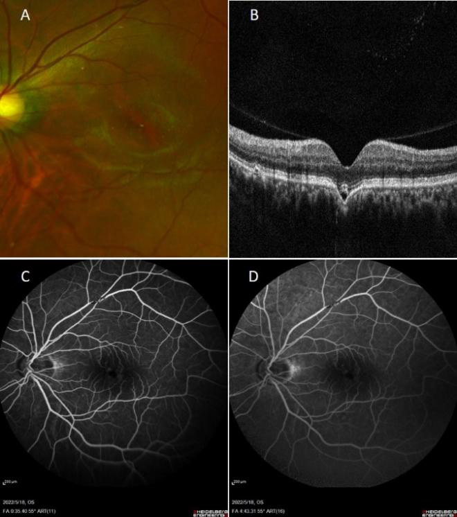
(A) Corlor fundus of the left eye showed pigment irregularity in the macular area; (B) The contour of cone-shaped of unconfirming type FCE; (C&D) Early- and late-phase FFA images showed patches of window defects corresponding to FCE.
Klien BA. The pathogenesis of some atypical colobomas of the choroid[ J]. Am J Ophthalmol, 1959, 48(5): 597-607.
Pur DR , Pereira AJ, Choudhry N. Diagnostic and therapeutic challenges[ J]. Retina, 2006, 26(9): 1072-1076.
Margolis R. The expanded spectrum of focal choroidal excavation[ J]. Arch Ophthalmol, 2011, 129(10): 1320.
Kumano Y, Nagai H, Enaida H, et al. Symptomatic and morphological differences between choroidal excavations[ J]. Optom Vis Sci, 2013, 90(4): e110-e118.
Hashimoto Y, Saito W, Noda K , et al. Acquired focal choroidal excavation associated with multiple evanescent white dot syndrome: obser vations at onset and a pathogenic hypothesis[ J]. BMC Ophthalmol, 2014, 14: 135.
Galvin JC, Fung AT. Focal choroidal excavation and choroidal neovascularization: the chicken and the egg[ J]. Clin Exp Ophthalmol, 2020, 48(6): 839-840.
Sato T, Yasukawa T, Hayashi K. Focal choroidal excavation disappearing after successful treatment of type 2 choroidal neovascularization with intravitreal aflibercept[ J]. Am J Ophthalmol Case Rep, 2021, 22: 101078.
Ellabban AA, Tsujikawa A, Ooto S, et al. Focal choroidal excavation in eyes with central serous chorioretinopathy[ J]. Am J Ophthalmol, 2013, 156(4): 673-683.e1.
Matsubara H, Uchiyama E, Suzuki K, et al. A case of focal choroidal excavation development associated with multiple evanescent white dot syndrome[ J]. Case Rep Ophthalmol, 2018, 9(2): 388-394.
Shinojima A, Kawamura A, Mori R, et al. Morphologic features of focal choroidal excavation on spectral domain optical coherence tomography with simultaneous angiography[ J]. Retina, 2014, 34(7): 1407-1414.
王振, 王应利, 周玉梅, 等. 局限性脉络膜凹陷39例的临床和OCT特征[ J]. 国际眼科杂志, 2017, 17(5): 912-916.Wang Z, Wang
YL, Zhou YM, et al. Clinical characteristics and OCT findings of focal choroidal excavation in 39 cases[ J]. Int Eye Sci, 2017, 17(5): 912-916.
Capellan P, Gonzalez LA, Abdallah Mahrous M, et al. Primary and
secondary focal choroidal excavation morphologic phenotypes,
associated ocular disorders and prognostic implications[ J]. Br J
Ophthalmol, 2023, 107(3): 373-379.
Chen YC, Chou YB, Lin CK, et al. Characterization and functional
correlation of multiple imaging modalities with focal choroidal
excavation[ J]. J Chin Med Assoc, 2018, 81(5): 487-495.
Park KA, Oh SY. The absence of focal choroidal excavation in children
and adolescents without retinal or choroidal disorders or ocular
trauma[ J]. Eye (Lond), 2015, 29(6): 841-842.
Olguin-Manríquez F, Enríquez AB, Crim N, et al. Multimodal imaging
in choroidal osteoma[ J]. Int J Retina Vitreous, 2018, 4: 30.
Gan Y, Ji Y, Zuo C, et al. Correlation between focal choroidal excavation
and underlying retinochoroidal disease: a pathological hypothesis from
clinical observation[ J]. Retina, 2022, 42(2): 348-356.
罗丹, 金明. 脉络膜新生血管抑制剂最新研究进展[ J]. 眼科新进展, 2017, 37(3): 285-288.
Luo D, Jin M. Research advances in inhibitors for choroid neovascularization[ J]. Recent Adv Ophthalmol, 2017, 37(3): 285-288.
Tang WY, Zhang T, Shu QM, et al. Focal choroidal excavation
complicated with choroidal neovascularization in young and middle
aged patients[ J]. Int J Ophthalmol, 2019, 12(6): 980-984.
Zhu RL, Gu XP, Zhang YD, et al. Focal choroidal excavation
complicated with choroidal neovascularization: a case report and
review of the literature[ J]. Int J Ophthalmol, 2022, 15(10): 1717-1719.
Rajabian F, Arrigo A, Jampol LM, et al. Optical coherence tomography
angiography features of focal choroidal excavation and the choroidal
stroma variations with occurrence of excavation[ J]. Retina, 2020,
40(12): 2319-2324.
Mahendrakar P, Bhende M, Menon R. Development of focal choroidal
excavation in a case of idiopathic choroidal neovascular membrane[ J].
RETINAL Cases Brief Rep, 2022.
Suzuki M, Gomi F, Hara C, et al. Characteristics of central serous
chorioretinopathy complicated by focal choroidal excavation[ J].
Retina, 2014, 34(6): 1216-1222.
Wang S, Zhao P. Another form of focal choroidal excavation based on
multimodality imaging[ J]. Optom Vis Sci, 2016, 93(10): 1296-1303.
Luk FOJ, Fok ACT, Lee A, et al. Focal choroidal excavation in patients with central serous chorioretinopathy[ J]. Eye (Lond), 2015, 29(4):
453-459.
叶祖科, 尹小芳, 罗书科, 等. 局限性脉络膜凹陷患眼多模式影像特征观察及发生并发症的危险因素分析[ J]. 中华眼底病杂志, 2019, 35(4): 342-347.
Ye ZK, Yin XF, Luo SK, et al. Multimodal imaging characteristics of focal choroidal excavation and risk factors analysis of its complications[ J]. Chin J Ocular Fundus Dis, 2019, 35(4): 342-347.
Docherty G, Sidiqi A, Martens R, et al. Conversion of focal choroidal
excavation with the onset of central serous chorioretinopathy: report
of 2 cases and review of the literature[ J]. Retin Cases Brief Rep, 2021,
15(4): 376-385.
Parodi MB, Zucchiatti I, Fasce F, et al. Bilateral choroidal excavation in
best vitelliform macular dystrophy[ J]. Ophthalmic Surg Lasers Imaging
Retina, 2014, 45 Online: e8-e10.
Liu J, Zhang Y, Xuan Y, et al. Novel BEST1 mutations and special
clinical features of best vitelliform macular dystrophy[ J]. Ophthalmic
Res, 2016, 56(4): 178-185.
Kumar V, Chatra K. Fibrotic pillar leads to focal choroidal excavation
in Best vitelliform dystrophy[ J]. Graefes Arch Clin Exp Ophthalmol,
2018, 256(11): 2083-2087.
Bessho H, Imai H, Azumi A. The histopathological finding of the
surgically extracted atypical dome-shaped choroidal osteoma[ J]. Case
Rep Ophthalmol Med, 2017, 2017: 2874823.
Bernabei F, Pellegrini M, Schiavi C, et al. Choroidal osteoma associated
with focal choroidal excavation[ J]. J Fr Ophtalmol, 2021, 44(7): 1105-
1107.
Rajabian F, Arrigo A, Grazioli A, et al. Focal choroidal excavation and
pitchfork sign in choroidal neovascularisation associated with choroidal
osteoma[ J]. Eur J Ophthalmol, 2021, 31(2): NP67-NP70.