





There has been a significant advancement in the field of biomedical artificial intelligence (AI) since 2012, particularly in the medical applications of deep learning (DL) algorithms. Medical AI plays an increasingly important role in the development of medicine and improvement of the health and longevity of the population. This period witnessed the transition from the maturity of DL algorithms to their incorporation into various medical domains, marking a complete phase for biomedical AI development. Ophthalmology has experienced significant growth and development in the field of DL, with both early-stage and well-established DL applications being utilized in real-world scenarios.[1] With the technical breakthrough of DL algorithms in the last decade, the field have gradually achieved intelligent diagnosis of different ocular diseases such as diabetic retinopathy (DR),[2-3] cataract[4] and glaucoma,[5] and have expanded the coverage of different image modalities from retinal photograph to optical coherence tomography (OCT) and other modalities. Therefore, further clarification and refinement are needed to fully comprehend the developmental patterns and trends in the field of DL in ophthalmology.
Recent publications on DL in ophthalmology has shown explosive growth. These publications provide wealth of valuable information about the changing trends in DL-assisted diagnostic systems for ocular diseases, along with a comparative analysis of the development status in different countries worldwide. This field acts as an excellent demonstration of the evolution of medical AI, showcasing changes in various aspects and providing insights into future trends. Nonetheless, published literature reviews or expert consensuses have largely focused on specific research areas or highly cited articles,[6–9] limiting their scope and providing only a narrow perspective on the overall changes in the field of DL in ophthalmology. Consequently, comprehensive insights into the broader dimensions of ophthalmic AI have been difficult to obtain, such as the trend of research in ocular diseases, changes in data modalities, quantities and quality of research data, along with the factors affecting the DL model performance.[10] Additionally, manually reading and analyzing through such a large volume of literature is a time-consuming and challenging task, and might lead to a biased representation of the overall perspective.[11] It is urgently needed to conduct a comprehensive and practical overview of this fast-evolving field.
Recently, large language models (LLM) have shown significant success in following instructions and producing human-like responses.[12-13] Using LLM for natural language processing and applying the state-of-the-art bibliometric analysis in joint with human experts, this study presented a comprehensive overview of the evolution in this rapid changing research field. Base on this, we have identified trends and challenges among common ophthalmic DL research and further provided prospects for future applications. Additionally, this study offers a practical approach to comprehensively investigate current status and future trends in the field, making it a valuable reference for other researchers.
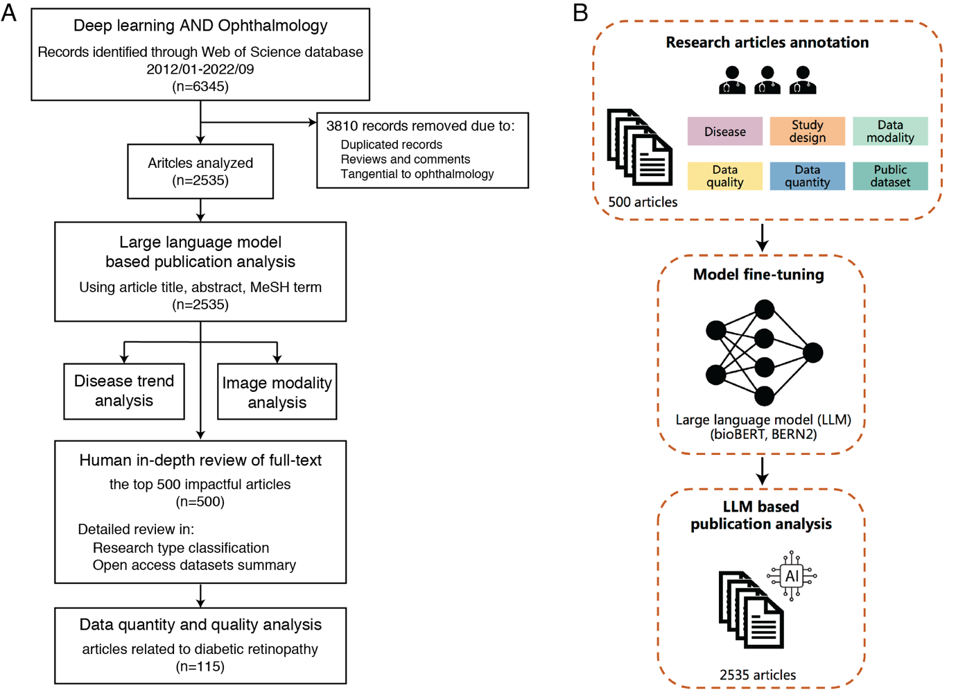
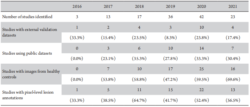
Using the LLM-based text analysis method, we successfully extracted the studied eye diseases from 2,300 of 2,535 (90.7%) articles in the field of DL ophthalmology and categorized numerous disease types based on PubMed's disease MeSH terms, and the data modalities used were extracted and summarized from 1,611 of 2,535 (63.5%) articles. Assessing the quality and quantity of study data was relatively challenging, and required manual analysis given the limitations of LLMs in mathematical processing. To ensure accuracy, the data quality and quantity analysis included results from manual review of the 500 highest impact articles. In addition, 115 DR-related DL studies were further selected to analyze the dataset used including whether the studies had external validation datasets, used public datasets, a dataset with images of healthy controls and provided pixel-level annotations (identifying the boundaries of disease lesions).
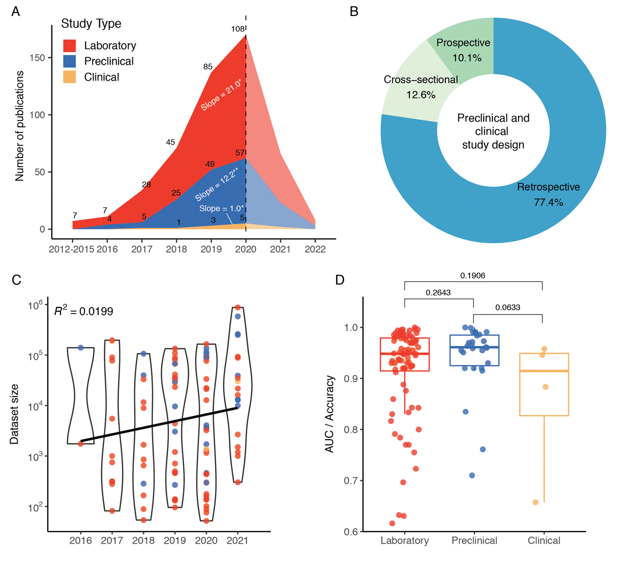
The articles were classified into three categories: laboratory, preclinical and clinical research. Laboratory studies involve constructing and validating algorithms and models using public data. Clinical studies utilize revalidated AI algorithms and models in real clinical scenarios to assist in disease diagnosis and intervention. Preclinical studies, a type between laboratory and clinical studies, focus on the algorithm or model validation through public or limited private datasets to assess their future suitability for large-scale clinical use. Preclinical and clinical research studies were classified into three categories according to their design: retrospective, cross-sectional, and prospective. Study types were initially classified by two junior researchers (>3 years research experience). In cases of disagreement, adjudication was performed by a senior researcher (>8 years research experience) to finalize the study type.
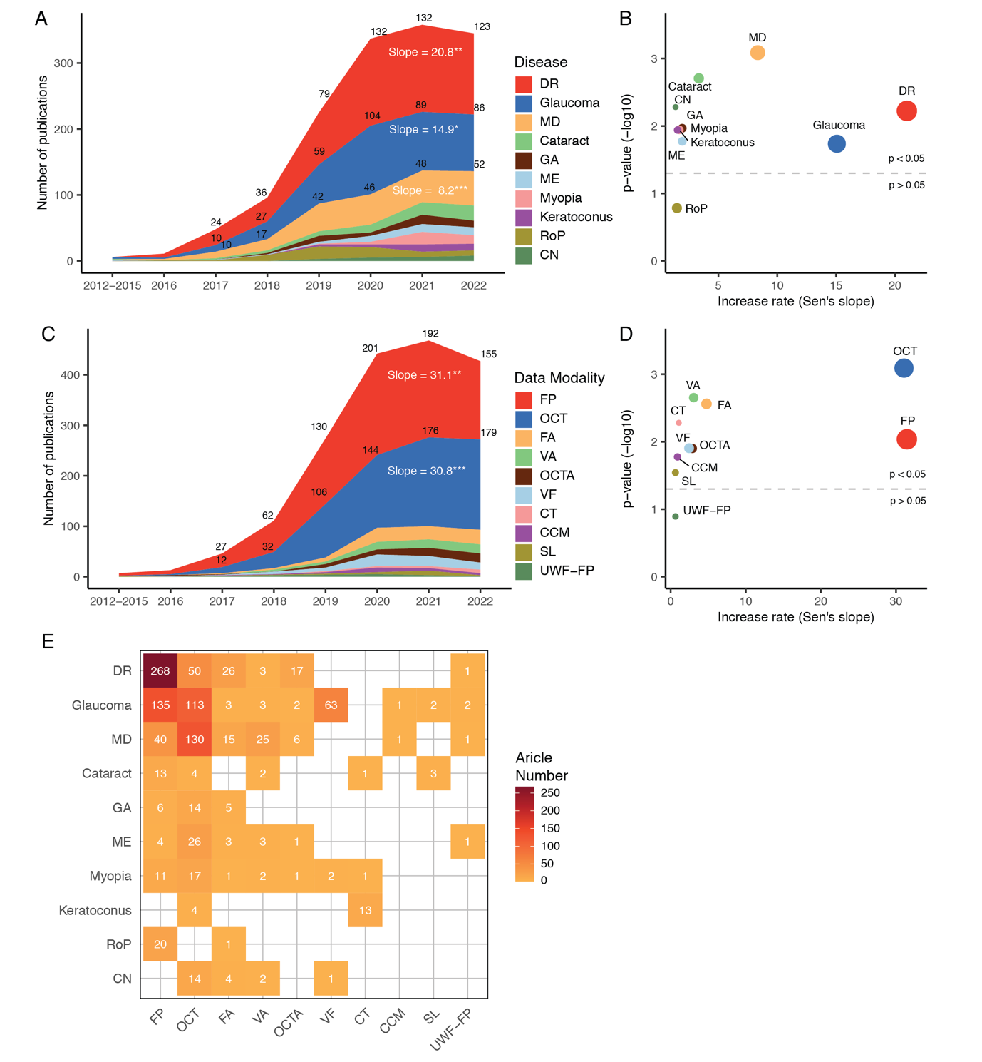
Diseases and Data Modalities in Ophthalmic DL Research
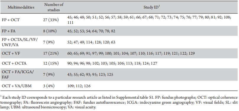
The 10 most commonly studied diseases were shown in Figure 3A, including DR, glaucoma, macular degeneration, cataract and fundus diseases. As stated here, researchers related to various ocular diseases began to gradually increase in 2016 and showed a significant increase between 2017 and 2020. The most studied on ocular diseases are DR, glaucoma and macular degeneration, which have also shown an increasing trend in the last 10 years, with Sen’s slopes of 20.8 (P = 0.000 3), 14.9 (P = 0.001 1) and 8.2 (P = 0.000 1), respectively (Figure 3B).With similar methods, the data modalities used were extracted and summarized. The top 10 data modalities in terms of growth rate were ranked on Sen’s slope (Figure 3C). As stated, DL studies using fundus photographs (FP) and OCT began to rapidly increase in number and gain prominence in 2018, with Sen’s slopes of 31.1 (P = 0.001 5) and 30.8 (P = 0.000 1), respectively (Figure 3D). Many other data modalities including fluorescein angiography (FA), optical coherence tomography angiography (OCTA), visual acuity (VA), visual fields (VF) and corneal topography (CT), have been increasing applied since 2020. The data modalities used in studies of different ocular diseases varied considerably (Figure 3E). Specifically, studies on DR mainly used OCT and FP; studies on glaucoma relied mainly on OCT, FP, and VF; and macular degeneration studies relied on OCT and FP. Additionally, several DL studies used multimodal ophthalmic data (Table 1). 27 (33%) multimodality studies involved both FP and OCT images, 17 (21%) used OCT and VF, and 7(9%) studies utilized FP and one of the modalities such as OCTA, slit lamp (SL), and VA.
The dataset size and fnal model performance used in the 115 DR studies were compiled. This is to determine whether a large data size is necessary to train a model with adequate performance (Figure 4C). The interquartile range (IQR) of the data size for DR classification models is 462–74,198 images, and the average data size has grown from 70,817 images in 2016 to 122,810 in 2021, with an average annual increase of 10,398 (14.7%) images. By dividing the presented performances in these studies according to the study design, it was revealed that the average accuracy of the models declined consistently (Figure 4D).
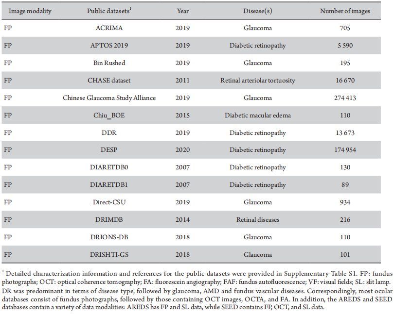
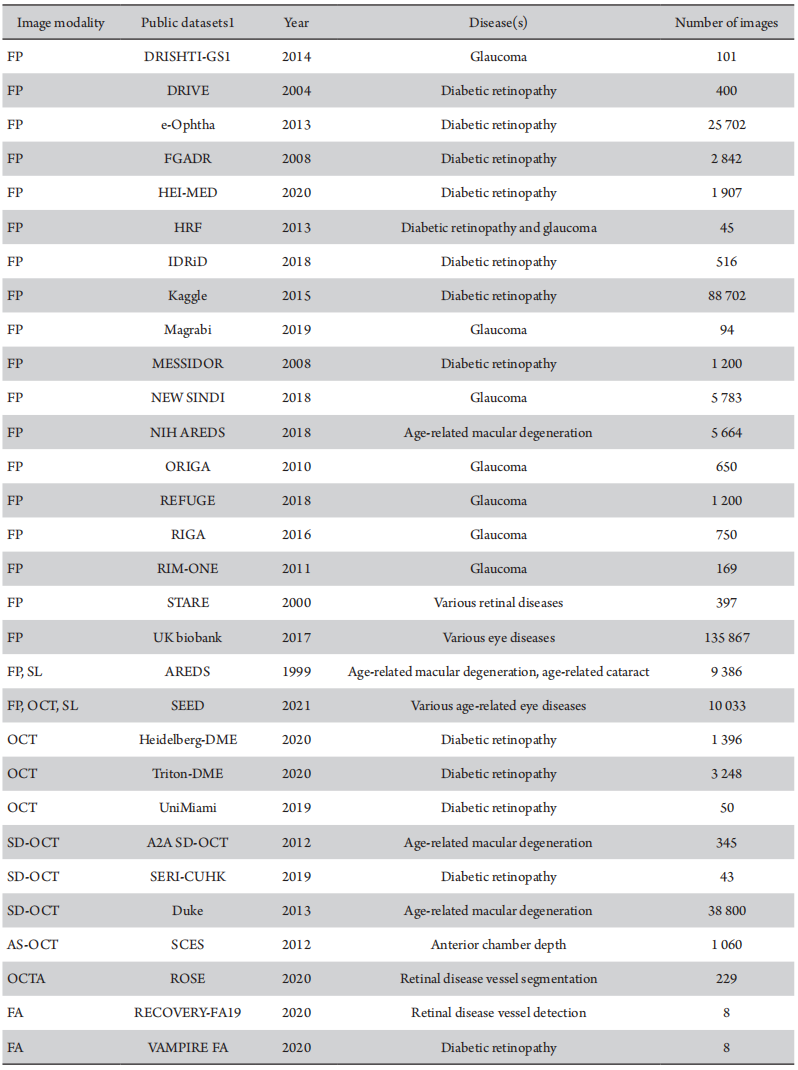
To provide a reference of publicly available datasets, the ocular disease image databases used in these 500 highest-impact articles were summarized with the pertinent information (Table 3 and Supplementary Table S2).
DL in ophthalmology is evolving rapidly and the status of the field is difficult to summarize comprehensively using conventional methods.[7–9] This study presents a comprehensive overview of all existing research using LLM-based methods combined with in-depth manual analysis to uncover trends in DL in ophthalmology from a large amount of publication data. The development of DL in ophthalmology is in sync with the overall progress in DL domain, relying heavily on the foundational support provided by the latter. In this study, we fine-tuned the BERT-based large-scale language model with manually annotated paper data to build an intelligent LLM capable of reviewing medical literature and extracting key information. Its accuracy was verified in extracting disease and image modality information, suggesting that incorporating an LLM into the literature analysis workflow could enhance the efficiency and productivity of researchers in the field. The number of articles and development trends of various eye diseases were categorized and summarized using the LLM–assisted approach.
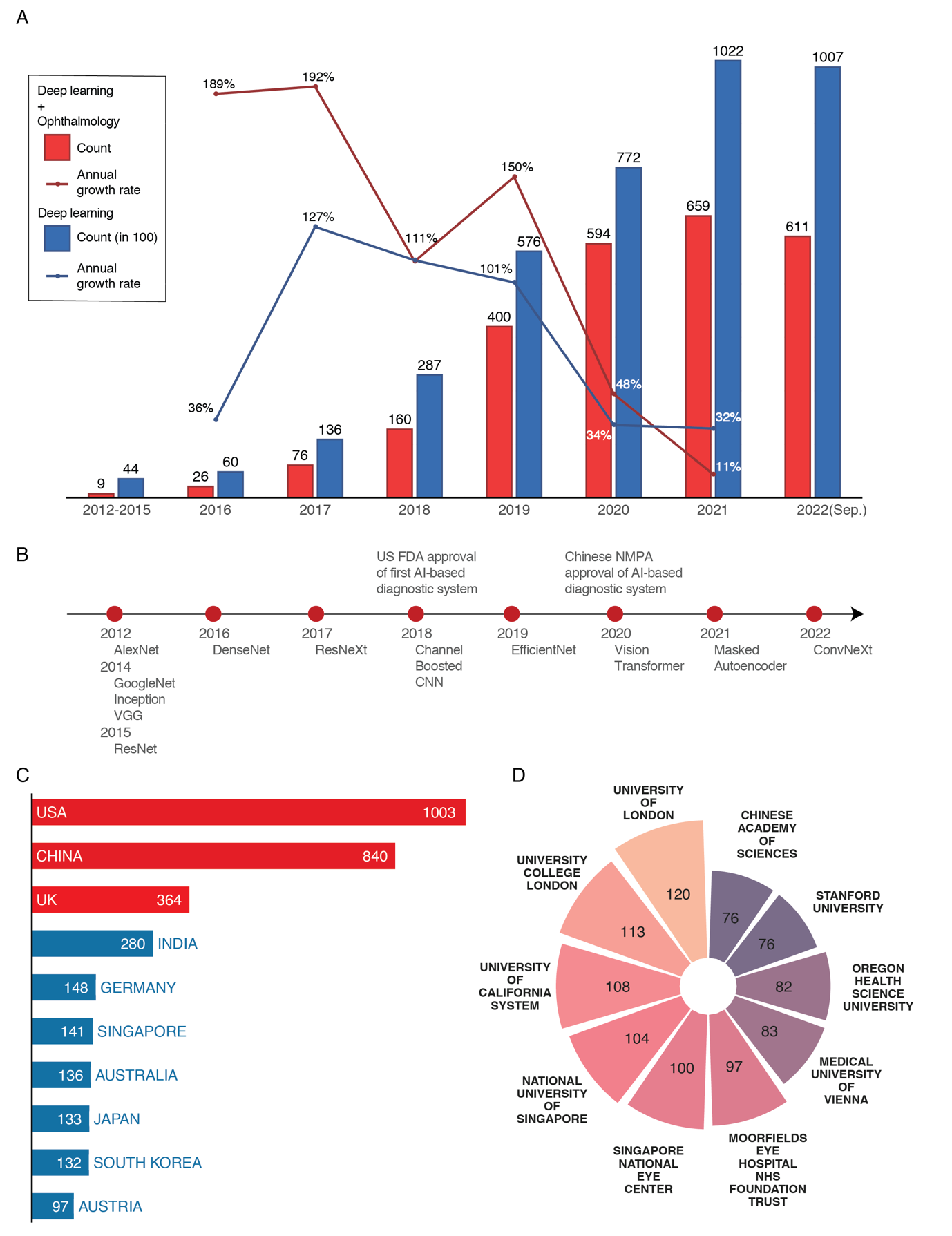
Advances in the DL field have accelerated DL research in ophthalmology. From 2012 to 2015, there were revolutionary developments in the DL field, including the construction of benchmark image datasets ImageNet,[16] COCO[17] and development of convolutional neural network architectures such as AlexNet[18] and ResNet.[19] The subsequent rapid growth of DL research in ophthalmology occurred as a result of these breakthroughs in DL advancing the state-of-the-art and laying the foundation for further research and development of DL in ophthalmology. Many of the leading ophthalmic deep learning studies have come from technologically advanced countries including the United States, China, the United Kingdom, and India. With major research contributions from these countries, deep learning-based diagnostic systems for ocular diseases have gained approval from global regulatory bodies. In 2018, the first U.S.license for such a system was issued,[20] followed by the Chinese license in 2020.[21] This widespread regulatory approval reflects the progress in deep learning research and validation of its ability to accurately detect and diagnose eye diseases, signaling a shift from laboratory studies to clinical applications.
Ophthalmic DL research has gradually transitioned from initial feasibility studies to real-world clinical applications. Early feasibility DL studies focused on DR based on FP, as the large patient population and extensive labeled image datasets enabled algorithm development and validation with minimal technical complexity.[22] By establishing capabilities and clinical value on a common ocular disease (e.g. DR) with abundant training data, researchers laid the groundwork to then investigate expanding DL to other ocular diseases.[1] The initial successes have led to further research broadening real-world deployment of DL models across diverse clinical applications, such as telemedicine screening and smartphone-based diagnostic tools at point-of-care.[23-24]
Moving beyond reliance on single data modalities like FP and OCT, there is instead increasing utilization of multimodal approaches that integrate information from diverse clinical exams and tests to enable more comprehensive and accurate diagnosis. This transformation is driven by complex ocular conditions like glaucoma that require assimilating data from basic ophthalmic exams, visual field testing, cup-to-disc ratios from fundus imaging, and optic nerve layer thickness from OCT to facilitate robust clinical judgments.[25]Moreover, combining other data sources like genomic tests with imaging data has been proven efficacy for predictive modeling in diseases like age-related macular degeneration.[26] As algorithms become more sophisticated, they can synergistically combine disparate inputs from various ophthalmic subspecialties, testing modalities, and data types. By amalgamating these diverse datasets, DL models can mimic multifaceted clinical decision making and enable more precise disease diagnosis and prognosis across ophthalmology.
Data quantity and quality are crucial in DL applications in ophthalmology. However, the necessary sample size is contingent upon the complexity of the disease and detection task, as well as the intricacy of the model. In an early experiment, the impact of dataset size on DL algorithm performance in detecting DR was analyzed.[3] The results revealed that peak performance was reached at approximately 60,000 images, suggesting that increasing the dataset size beyond this point did not improve algorithm performance. However, with advancements in DL algorithms, it is now unclear what amount of data is required for optimal performance. By investigating high-impact DL articles on DR detection, our findings indicate that the DL model’s performance decreased markedly from laboratory to clinical studies. This observation implies that the self-reported AUC and other evaluation criteria employed in these studies may not adequately represent the real-world performance of the DL models due to the reproducibility issues.[27] Alternatively, it could be that the data analyzed, extracted from previously published DR-related studies, lack broad applicability to other ocular diseases, which necessitates more rigorous experimental investigations in future studies. Additionally, we found that the proportion of studies using externally validated datasets and public datasets was not high (20–35%) which might be due to the difficulty in obtaining resources for external validation datasets and public datasets. The question about the sample size required for deep learning is the one without a standard answer. Determining the optimal sample size for DL studies is challenging due to the disease complexity, specific medical tasks, and the complexity of DL models.[28-29] Given that the results derived from different deep learning algorithms on various datasets cannot be directly compared, it is imperative to establish a unified and objective standard evaluation method. This will ensure a greater consistency in the assessment of model performance, thereby enhancing the reliability of the outcomes.
This study has several limitations. This study is based on previously published articles, and therefore may not capture emerging trends in research that has not yet been published. Additionally, the citation time-frame used in this study is restricted to high-impact articles published mainly before 2021, resulting in fewer articles from 2022 onward that were included in the refined analysis. To obtain more concrete conclusions, a more robust study with a larger sample size is warranted. Although we analyzed ophthalmology research trends, data modality, volume, and types of studies, some other aspects of deep learning in ophthalmology research were not addressed in this paper. These aspects include data privacy and security, interpretability and transparency of AI models, and regulation and standardization of AI in practical applications. There are some articles that are not indexed in PubMed and do not have MeSH words, which is mitigated by our thorough analysis of titles and abstracts, ensuring a comprehensive review that minimizes the impact of this limitation on our study's findings.
In conclusion, we showed that an LLM combined with in-depth manual analysis were capable of reviewing medical literature and extracting information. Using the LLM–assisted approach, we have identified trends and challenges among common ophthalmic DL research and further provided prospects for future applications. This includes the necessity of validating AI models via real world clinical setting, and creating standardized, public accessible datasets to enhance collaboration, benchmarking for DL applications. Additionally, this study offers a practical approach to comprehensively investigate current status and future trends in the field, making it a valuable reference for other researchers.
Correction notice
NoneAcknowledgement
NoneAuthor Contributions
(I) Conception and design: HTL,DRL,MJL,ZZL(II)Administrative support: HTL, DRL
(III) Provision of study materials or patients: HTL, DRL
(IV) Collection and assembly of data: MJL, WXZ, ZMZ, JYP
(V) Data analysis and interpretation: MJL, JYP, ZZL, LQZ
(VI) Manuscript writing: MJL, WXZ
(VII) Final approval of manuscript: All authors
Conflict of Interests
The authors disclose that the corresponding author, Haotian Lin, serves as Editor-in-Chief of Eye Science. To avoid any potential conflict of interest, the editorial handling of this manuscript was assigned to an independent editor, and the authors were not involved in the peer-review or decision-making process. All editorial procedures were conducted in accordance with COPE guidelines and the journal's standard policies.
The authors declare no other competing financial or non-financial interests relevant to this work