





Polypoid choroidal vasculopathy (PCV), characterized by subretinal orange-red nodular lesions on the fundus, recurrent submacular hemorrhage (SMH), and serous or hemorrhagic retinal pigment epithelial detachment (PED), is a risk factor for severe visual impairment in Asian populations. Large submacular hemorrhages may develop into dense breakthrough vitreous hemorrhages. Polypoid choroidal vasculopathy with breakthrough vitreous hemorrhage (PCVVH) was first reported by Kleiner in 1985, with 4.5-19.9% of patients with PCV running the risk of developing vitreous hemorrhage[1-3].To date, vitrectomy is the primary treatment method, although the prognosis of vitrectomy for PCVVH varies greatly. In addition, a consensus on the timing of this operation, namely the duration from vitreous hemorrhage to perform vitrectomy, as well as whether cataract surgery and intraocular lens implantation can improve the vision or not, is lacking. Furthermore, few relevant studies exist in the literature. Therefore, this study aimed to investigate the prognosis and risk factors for vitrectomy for massive vitreous hemorrhage (VH) secondary to polypoidal choroidal vasculopathy.
This was a retrospective study, and ethical approval was obtained from the Institutional Review Board of Zhongshan Ophthalmic Center. The clinical characteristics of 49 eyes of 48 patients with polypoidal choroidal vasculopathy and breakthrough vitreous hemorrhage treated with a 23-gauge pars plana vitrectomy(PPV) between January 2015 and December 2020 were evaluated.
The inclusion criteria were as follows: (1) diagnosis of PCV based on the results of fundus examination, B ultrasound, optical coherence tomography (OCT), fundus fluorescein angiography (FFA), and indocyanine green angiography (ICGA), both preoperatively and postoperatively, and the presence of orange red polypoidal lesions during the operation; (2) vision loss due to PCVVH was treated with 23-gauge PPV; and (3) the follow-up duration was at least six months.
Similarly, the exclusion criteria were as follows: (1) traumatic vitreous hemorrhage; (2) other ocular diseases that could affect visual acuity, such as primary glaucoma, optic neuritis, age-related macular degeneration(AMD), retinal vein occlusion, choroidal melanoma, and retinal vasculitis; and (3) a medical history of photodynamic therapy(PDT) or PPV.
All patients underwent a comprehensive eye examination, including best-corrected visual acuity (BCVA), slit-lamp microscopy, and B-scan before surgery. The patients also underwent 23-gauge PPV, including central and peripheral vitrectomies, performed by multiple skilled surgeons. We used a wide-angle viewing system for PPV and identified the peripheral retina during the peripheral vitrectomy. If subretinal hemorrhage was observed during surgery without retinal tears, this was not treated due to the significant damage caused by clearing subretinal hemorrhage. According to the surgeon’s judgment, silicone oil or C3F8 was used to fill the vitreous cavity. In cases with retinal tears or poor fundus conditions, silicone oil is generally chosen. Epiretinal membrane enucleation was performed if a macular epiretinal membrane was observed. The following data was collected from each of the patients enrolled : BCVA before and after surgery, duration of VH until surgery, hypertension, diabetes, anticoagulant use, smoking history, anti-vascular endothelial growth factor (VEGF) use, spectrum OCT (SD-OCT; Heidelberg, Germany), postoperative complications, and secondary surgery during follow-up.
BCVA was measured using an early treatment diabetic retinopathy study(EDTRS) chart and converted to the logarithm of the minimum angle of resolution (logMAR) scale for statistical analysis. No light perception was set at 3.5 logMAR, light perception was set at 3.2 logMAR, hand movement(HM) was set at 2.3 logMAR, and counting fingers (CF) was set at 1.85 logMAR, according to a previous method.[4]
A paired t-test was used to compare the visual improvement and differences between the BCVA of post-vitrectomy at three, six, and 12 months and at the final follow-up from the BCVA of baseline pre-vitrectomy. The BCVA of post-cataract surgery at one week and three months was compared to the BCVA of baseline pre-cataract surgery performed after vitrectomy. The prognostically relevant factors were grouped, and independent sample t-tests were used to compare the differences in BCVA at each time point. One-way analysis of variance (ANOVA) was used to compare the BCVA at baseline and post-vitrectomy at three, six, and 12 months and at a final follow-up between the age groups and BCVA groups. A value of P≤0.05 was considered statistically significant. Statistical analyses were performed using SPSS for Windows (version 17.0; SPSS Inc., Chicago, IL, USA).
Table 1 Clinical characteristics of patients with massive vitreous hemorrhage secondary to polypoidal choroidal vasculopathy
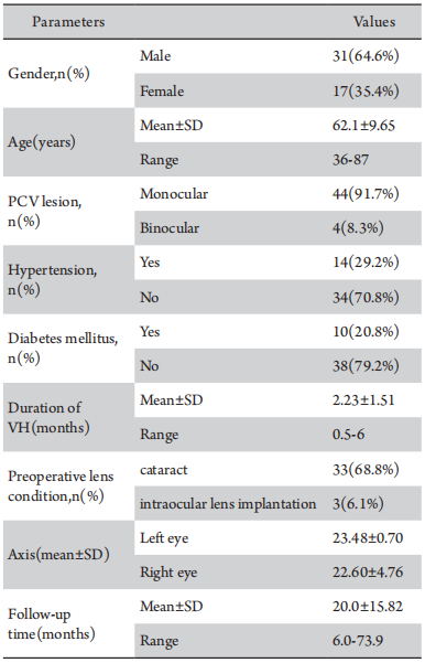
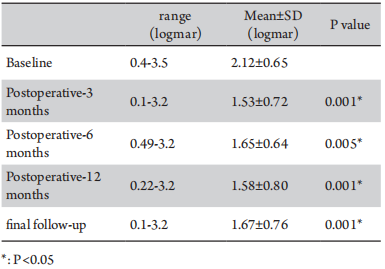
Table 2 BCVA at baseline and three, six, and 12 months final follow-up after vitrectomy
Eight eyes underwent vitrectomy combined with silicone oil filling, and the silicone oil was taken out at 11.99±6.25 months (range 3.8-25 months) after surgery. Four eyes were observed recurrent retinal detachment after removing silicone oil at 11.54±8.04 months(range 3.8-25 months).
Fourteen eyes (28.6%) were subjected to phacoemulsification and intraocular lens implantation in an average time of 10.16±5.14 months (range 1.97-21.43 months) after vitrectomy. Six eyes underwent vitrectomy combined with cataract surgery due to postoperative complications. Compared with the BCVA before cataract surgery, the final BCVA was stable or improved in 13 eyes, increased in 1 eye. BCVA at one week and three months post-cataract surgery was improved compared with BCVA before cataract surgery (P<0.05)(Table 3). Among these 14 eyes, the BCVA at the final follow-up was better than 1.3 logMAR only in six eyes (28.6%); the final follow-up BCVA of two eyes improved compared with the blind standard, and the other two eyes improved compared with the low visual acuity standard.
Table 3 BCVA before cataract surgery and 1 week and 6 months after cataract surgery
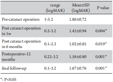
Table 4 Factors related to visual prognosis after PPV surgery
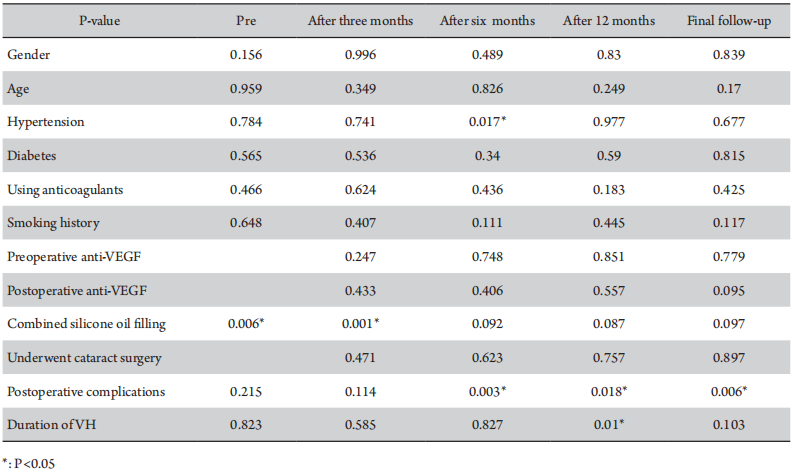
Patients were separated into three groups, 3.5-3.2 logMAR(Group A), 2.3-1.85 logMAR(Group B), and >1.85 logMAR(Group C), according to BCVA. Then, the BCVA of post-vitrectomy at three, six, and 12 months and at the final follow-up between the three groups was compared. Significant differences were observed in terms of the visual acuity between group A and group B at three, six, and 12 months and at the final follow-up, as well as between group A and group C(P<0.05). There was no statistically significant difference in visual acuity between group B and group C (P>0.05). Extremely poor preoperative vision, with or without light perception, is associated with poor vision after surgery. Therefore, when patients with PCVVH have extremely poor vision, it is necessary to consider whether vitrectomy should be performed.
There is no consensus on the timing of surgery for patients with PCVVH or whether anti-VEGF therapy is required before surgery in the clinical setting. Some surgeons tend to inject anti-VEGF into the vitreous after the onset of vitreous hemorrhage and then perform PPV surgery after the vitreous hemorrhage is slightly reduced, which could reduce the difficulty of the operation. Chen et al.[11] reported that anti-VEGF injection into the vitreous before PPV surgery could help reduce the frequency of anti-VEGF treatments and postoperative complications and improve short-term vision after surgery. Perioperative anti-VEGF could reduce the difficulty of surgery and reduce the occurrence of postoperative complications.[11] Previous animal experiments have shown that subretinal hemorrhage lasts for seven days, causing irreversible damage to the photoreceptor cells and retinal pigment epithelial(RPE) atrophy. Subsequently, local retinal necrosis occurs within 14 days, which allows red blood cell fragments to pass through the damaged retina, causing vitreous hemorrhage.[12-13] Kimura et al.[14] proposed an optimal operation time for SMH secondary to PCV of 7-10 days after the onset of SMH. On the one hand, a premature intervention can lead to recurrent bleeding after operation. However, on the other hand, if the intervention time is too late, this can cause irreversible damage to the retina, leading to poor postoperative vision. Nevertheless, the duration of VH in the enrolled patients ranged from half to six months. Therefore, we divided them into groups according to whether the duration of VH exceeded one month or not, and found that patients who underwent vitrectomy within one month of the onset of VH had better BCVA 12 months after vitrectomy than those who underwent vitrectomy one month later (P=0.015). There was no statistically significant relation between preoperative anti-VEGF levels and BCVA(P>0.05). If PPV surgery is performed early, the retina can easily be damaged owing to a thick subretinal hemorrhage. Blood cell fragments stay in the vitreous for a long time, which may cause irreversible toxic effects on the retina, and damage the retina and RPE layer. Thus, we propose that vitrectomy surgery should be performed within one month of the onset of massive vitreous hemorrhage in order to improve vision after surgery.
PPV is the primary treatment for PCVVH. Zhao et al.[15] retrospectively analyzed 103 eyes with PCVVH and found that BCVA was significantly better after PPV than before surgery. Lin et al.[16] studied 17 eyes with PCVVH that underwent vitrectomy, of which visual acuity improved in 16 eyes. Severe cataract and macular scarring cause poor visual acuity. In the present study, BCVA at three, six, and 12 months and at the last follow-up after vitrectomy improved compared to the average preoperative BCVA (P<0.05), consistent with the results of previous studies.[15-19] However, the visual acuity of patients with PCVVH varies considerably after vitrectomy, with most patients maintaining a low level of visual acuity. As shown in Figure 1, PCVVH is characterized by large polypoid lesions and strong activity. If the lesion does not involve the macula, the visual prognosis is better. In our study, the BCVA ranged from 0.1 logMAR to 3.2 logMAR at the final follow-up. The BCVA at the final follow-up was better than 1.3 logMAR only in 14 eyes (28.6%). Zhao et al.[16] found that the hemorrhagic retinal detachment, baseline central macular thickness, and best-corrected visual acuity were factors associated with final best-corrected visual acuity (P < 0.05) in PCV patients. Previous studies have shown that the incidence of secondary epiretinal membranes is 16.6% and that older individuals are more likely to develop an epiretinal membrane.[20] In this study, 58% of patients underwent epiretinal membrane enucleation, and the incidence of epiretinal membrane enucleation was much higher than that in the normal population. We speculate that this may be because PCV patients are mostly elderly individuals aged 50-70 years, with factors such as vitreous hemorrhage and retinal detachment exacerbating the progression of the epiretinal membrane.
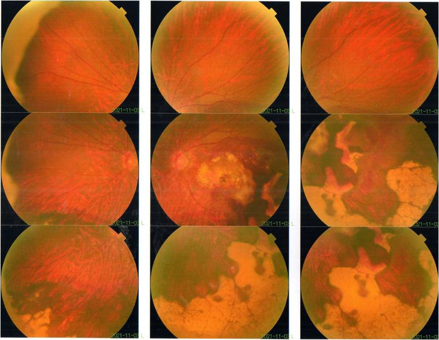
Figure 1 Fundus photography of the left eye: a large orange polypoid lesion at the posterior pole, the choroid in the inferior temporal and peripheral areas is chronic hemorrhagic foci
Patients with PCVVH complained of decreased and blurred vision due to cataract complications half a year after PPV. Phacoemulsification and intraocular lens implantation can be considered to improve vision if polypoid lesions are inactive. Based on our study results, improvements were observed atone week and three months post-cataract surgery compared to before surgery(P<0.05). However, vision prognosis after cataract surgery can vary significantly. In fact, long-term visual acuity in the 14 patient’s post-cataract surgery ranged from 0.1 logMAR to 2.3 logMAR. Compared to BCVA before cataract surgery, the final BCVA improved or stabilized in 13 eyes and increased in one eye. Among these 14 eyes, BCVA at the final follow-up was better than 1.3 logMAR only in six eyes (28.6%).
Common postoperative complications of PPV in patients with PCVVH include iatrogenic retinal tears, recurrent vitreous hemorrhage, complicated cataracts, hyphemia, secondary glaucoma, macular subretinal fibrosis, retinal detachment, and choroidal detachment.[17,21-22] Iatrogenic retinal tears are the most common surgical complication, with an incidence of approximately 17.8%, mostly observed during the induction of posterior vitreous detachment (PVD).[23] Studies have also reported sympathetic ophthalmia in the other eye, which is a rare postoperative complication.[23] Postoperative complications were observed in 10 eyes (20.4%) and included recurrent retinal detachment(18.4%), vitreous hemorrhage(6.1%), macular holes (2%), hyphemia(6.1%), and lens dislocation(4.1%). The peak period of postoperative complications was within one year after PPV, although one patient had recurrent retinal detachment more than two years after the first PPV. Notably, recurrent retinal detachment was observed in four eyes after silicone oil removal. Therefore, choosing the appropriate time to perform the SO removal surgery is a key factor in effectively reducing postoperative complications. Postoperative complications are another heavy blow to patients, with their vision often worsening as a result. Among patients with postoperative complications, BCVA was mostly less than 1.3 logMAR.
All patients included in this single-center study were from the Zhongshan Ophthalmology Centre of Sun Yat-Sen University. Since this is a retrospective study, we hope that a prospective longitudinal study will shed further light on the prognosis and factors influencing patients with PCVVH.
When PCV is secondary to VH, visual acuity drops sharply, for which vitrectomy is the primary clinical treatment. This study evaluates the visual prognosis of patients with PCVVH undergoing PPV surgery and shows that PPV can improve the short- and long-term visual acuity of patients with PCVVH. However, visual acuity after PPV was found to differ significantly, with only 28.6% of the patients showing a better long-term visual acuity than the non-blind standard. If polypoid lesions are not active, phacoemulsification and intraocular lens implantation can be considered to improve vision approximately half a year after PPV. Although some patients had a good visual acuity after cataract surgery, up to 57.1% of their vision either did not improve or deteriorated. Postoperative complications were observed in 10 eyes (20.4%). We also found that hypertension, vitrectomy combined with silicone oil filling, postoperative adverse events, and vitreous hemorrhage lasting >1 month were associated with a poor postoperative BCVA.
Correction notice
NoneAcknowledgement
NoneAuthor Contributions
(I) Conception and design: YL(II)Administrative support: TL,BQL,YL
(III) Provision of study materials or patients: TL,BQL,YL
(IV) Collection and assembly of data: CXC and JLC
(V) Data analysis and interpretation: CXC and QW
(VI) Manuscript writing:All authors
(VII) Final approval of manuscript: All authors