




Introduction
Retinopathy of prematurity (ROP) is an important cause
of blindness in children (1). It is a proliferative vascular
disorder of the retina that exclusively affects premature
infants. Infants who undergo proper screening and
treatment for ROP have improved long-term functional
and structural outcomes (2). The Early Treatment of
Retinopathy of Prematurity (ETROP) study defined highrisk
prethreshold patients as type 1 ROP (3). The authors
recommended type 1 ROP should be properly treated
by laser ablation of peripheral avascular retina to avoided
unfavorable structural outcomes such as retinal detachment
or macular ectopia (4). Although laser treatment can
effectively cause regression of type 1 ROP, some limitations
and adverse reactions exist. Some patients still progress to
unfavorable outcome despite repeated laser treatment (4).
Because of permanent destruction of a considerable portion
of the retina, visual field loss and high myopia may occur
after laser ablation (5,6).
Elevated intraocular vascular endothelial growth factor (VEGF) was found in patients with advanced ROP (7). VEGF can lead to abnormal angiogenesis and subsequent retinal neovascularization, which is known to play a significant role in the pathogenesis of ROP (8). Intravitreal injection of anti-VEGF agents such as bevacizumab, pegaptanib, or ranibizumab has become an adjuvant therapy for laser or monotherapy for severe ROP (9). Bevacizumab (Avastin™, Genentech Inc., South San Francisco, CA, USA) is a humanized monoclonal antibody to VEGF-A. Intravitreal bevacizumab (IVB) can result in a marked decrease of aqueous VEGF concentration in eyes with advanced ROP (10). The Bevacizumab Eliminates the Angiogenic Threat of Retinopathy of Prematurity (BEATROP) study found that IVB was effective in the treatment of severe ROP (11). Bevacizumab injection resulted in better outcome in zone I ROP and equal outcome in zone II ROP comparing to laser treatment (11-13). Significantly lower degree of myopia was found in eyes receiving IVB than in eye receiving laser, owing to more preservation of normal retinal tissue and better growth of eye globe (14-16). There were some disadvantages following bevacizumab administration, such as reactivation of ROP, tissue fibrosis, persistence of peripheral avascular area, and possibly affecting maturation of other organs due to lowering systemic VEGF (17-23).
The purpose of this study is to compare the structural outcome of IVB and laser treatment for type 1 ROP in Chinese infants after long-term follow-up.
Methods
After approval from the Institutional Review Board at
Far Eastern Memorial Hospital in Taipei, we conducted
a retrospective chart review of infants with type 1 ROP
receiving treatments from December 2002 to January
2016. The agreement of participation in the study
was not required for the participants because this is a
retrospective trial reviewing medical charts. ROP staging
was defined as International Classification of Retinopathy of
Prematurity (24). Type 1 ROP includes zone I, any stage
ROP with plus disease; zone I, stage 3 ROP without plus
disease; or zone II, stage 2 or 3 ROP with plus disease
according to criteria of ETROP study (3). Informed
consents agreeing to the treatment procedures were
obtained from family members of all patients.
From December 2002 to April 2009, infants with type 1 ROP were treated by diode laser photocoagulation (IRIS Medical Instruments Inc., Mountain View, CA, USA) for peripheral retinal avascular zone ablation by one ophthalmologist (Jia-Kang Wang). The laser settings were duration of 0.5 second, power of 0.3–0.7 W until appearance of creamy-white spots, and in nearly confluent pattern with burns placed half- to one-burn widths apart (25). Dexamethasone eye solution was given 4 times a day for 1 week after the procedure.
From May 2009 to January 2015, IVB was performed for patients with type 1 ROP by four ophthalmologists (JiaKang Wang, Yun-Ju Chen, Fang-Ting Chen, and YungRay Hsu). We used a 30-gauge needle for an injection of bevacizumab 0.625 mg (0.025 mL solution) through the pars plicata, aiming the needle directly toward the optic nerve in direction of visual axis. Norfloxacin eye solution was given 4 times a day for 1 week after the procedure.
The patients were closely followed until disappearance of retinal neovascularization in the laser group and regression of avascular zone in the bevacizumab group. Persistence or reactivation of ROP was allowed repeated treatments of laser procedures or bevacizumab injections. The unfavorable structural outcomes were recorded, such as posterior retinal fold involving the macula, a retinal detachment involving the macula, or retrolental tissue or ‘‘mass’’ obscuring the view of the posterior pole (3). The gestational age (GA), birth body weight (BBW), postmenstrual age (PMA) for intervention, and fundus findings from pre-treatment to the last follow-up were recorded. The numerical difference between laser and bevacizumab groups was compared by Student t-test. Fisher’s exact test was used for categorical comparison between groups. All P values were two sided and were considered statistically significant if P<0.05.
ROP, retinopathy of prematurity; IVB, intravitreal bevacizumab; SD, standard deviation.
From December 2002 to April 2009, infants with type 1 ROP were treated by diode laser photocoagulation (IRIS Medical Instruments Inc., Mountain View, CA, USA) for peripheral retinal avascular zone ablation by one ophthalmologist (Jia-Kang Wang). The laser settings were duration of 0.5 second, power of 0.3–0.7 W until appearance of creamy-white spots, and in nearly confluent pattern with burns placed half- to one-burn widths apart (25). Dexamethasone eye solution was given 4 times a day for 1 week after the procedure.
From May 2009 to January 2015, IVB was performed for patients with type 1 ROP by four ophthalmologists (JiaKang Wang, Yun-Ju Chen, Fang-Ting Chen, and YungRay Hsu). We used a 30-gauge needle for an injection of bevacizumab 0.625 mg (0.025 mL solution) through the pars plicata, aiming the needle directly toward the optic nerve in direction of visual axis. Norfloxacin eye solution was given 4 times a day for 1 week after the procedure.
The patients were closely followed until disappearance of retinal neovascularization in the laser group and regression of avascular zone in the bevacizumab group. Persistence or reactivation of ROP was allowed repeated treatments of laser procedures or bevacizumab injections. The unfavorable structural outcomes were recorded, such as posterior retinal fold involving the macula, a retinal detachment involving the macula, or retrolental tissue or ‘‘mass’’ obscuring the view of the posterior pole (3). The gestational age (GA), birth body weight (BBW), postmenstrual age (PMA) for intervention, and fundus findings from pre-treatment to the last follow-up were recorded. The numerical difference between laser and bevacizumab groups was compared by Student t-test. Fisher’s exact test was used for categorical comparison between groups. All P values were two sided and were considered statistically significant if P<0.05.
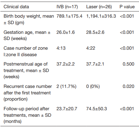
Table 1 Comparison of clinical data between patients with type 1 ROP treated by intravitreal bevacizumab or laser
ROP, retinopathy of prematurity; IVB, intravitreal bevacizumab; SD, standard deviation.
Results
We collected 43 patients (86 eyes) with type 1 ROP,
including 30 male and 13 female infants. Their mean GA
and BBW were 27.5±2.2 weeks and 1,034.0±334.9 gm.
Zone I and zone II disease were found in 8 (16 eyes) and
35 (70 eyes) patients respectively. The mean PMA for
treatment was 37.3±2.2 weeks. The mean follow-up period
was 54.4±48.1 months.
Laser treatment was administered in 26 patients (52 eyes), including 18 male and 8 female infants. Their mean GA and BBW were 28.5±2.6 weeks and 1,194.1±316.3 gm. Zone I and zone II disease were found in 4 (8 eyes) and 22 (44 eyes) patients respectively. Of patients with zone II disease, 2 (4 eyes) and 20 (40 eyes) patients were identified as posterior and anterior zone II disease. The mean PMA for treatment was 37.7±2.1 weeks. Complete regression of ROP was found in all patients in the laser group, including disappearance of plus sign (decreased vessel tortuosity), retinal avascular zone, and retinal neovascularization. There was no recurrence of ROP after single session of laser. Complications associated with laser treatment were not found, such as hyphema, vitreous hemorrhage, cataract, glaucoma and corneal damage. All cases of the laser group had favorable structural outcome after the mean follow-up of 74.5±50.3 months.
IVB was injected in 17 infants (34 eyes), including 12 male and 5 female infants. Their mean GA and BBW were 26.0±1.6 weeks and 789.1±175.4 gm. Zone I and zone II disease were found in 4 (8 eyes) and 13 (26 eyes) patients respectively. Of infants with zone II disease, anterior and posterior zone II disease were noted in 10 (20 eyes) and 3 (6 eyes) patients. The mean PMA for treatment was 37.2±2.2 weeks. Complete regression of ROP was found in 15 infants following the first IVB with continuation of normal vascularization toward the periphery of the retina, vanishing of plus and retinal neovascularization. Four eyes in two patients (2/17, 11.7%) had recurrence of ROP. The GA and BBW were 29 weeks and 760 gm in one case with ROP reactivation. The patient had anterior zone II and stage 3 plus disease and underwent the first bevacizumab therapy at PMA of 38 weeks. Retinal neovascularization and plus sign waned, but not accompanying with expansion of retinal vascular zone 1 week later. Second IVB was performed owing to reappearance of neovascularization and plus sign 8 weeks after the first treatment. Temporary regression of ROP was observed after the second injection. Laser salvage was administered 10 weeks after the second IVB due to ROP reactivation. The case had favorable structural outcome after 1-year follow-up. The GA and BBW were 24 weeks and 795 gm in the other case with recurrent ROP. The patient had zone I and stage 3 plus disease and underwent the first bevacizumab therapy at PMA of 33 weeks. Second IVB was performed owing to recurrence of ROP 4 weeks after the first treatment. Adjuvant laser therapy was administered 4 weeks after the second IVB due to ROP second reactivation. The case did not have progression to stage 4 or macular ectopia after 18-month follow-up. There were no ocular or systemic complications such as cataract, endophthalmitis, or delayed maturation of other vital organs in all patients receiving bevacizumab. All cases of the bevacizumab group had favorable structural outcome after the mean follow-up of 23.7±20.7 months.
The GA, BBW, and follow-up period were significantly less in the bevacizumab group than in the laser group (P<0.001) (Table 1). More zone I disease was found in the bevacizumab group than in the laser group (P<0.001). The PMA for management was comparable between both group (P=0.5). ROP reactivation was discovered significantly more in the bevacizumab group than in the laser group (P<0.001). No cases in both groups had unfavorable anatomical outcome at the final follow-up.
There were several reports comparing the anatomical results of laser and IVB treatment. A prospective study randomly conducted laser in one eye and IVB in the fellow eye in 12 infants with zone I type 1 ROP (18). The authors found all eyes treated with a bevacizumab were noted to have abnormalities at the periphery (large avascular area, abnormal branching, shunt) or the posterior pole (hyperfluorescent lesion, absence of foveal avascular zone). But these lesions were not observed in the majority of the lasered eyes after examination of fundus fluorescein angiography at age of 9 months (18). Higher chance of ROP reactivation was reported after bevacizumab monotherapy than after laser monotherapy in several prior studies. A prospective study performed IVB in one eye and laser therapy in contralateral eye of 14 patients with stage 3 posterior ROP (13). Three of 14 eyes (21.42%) had recurrence of ROP after single bevacizumab treatment, higher than one of 14 eyes (7.14%) with recurrence receiving conventional laser therapy (13). Another preceding retrospective comparative study demonstrated that more ROP recurred in 22 bevacizumabtreated eyes (14%) than 32 laser-treated eyes (3%) (21). We also discovered our patients receiving bevacizumab treatment had higher rate of ROP reactivation than those treated by laser. The different response between laser and bevacizumab for ROP reactivation can be hypothetically explained as follows: bevacizumab can temporarily inhibit intraocular VEGF, ROP may reactivate while vitreous VEGF levels elevate as anti-VEGF effect vanishing. Laser treatment destroys the avascular retinal tissue, which can permanently lower the intraocular VEGF (30). Close followup and possible repeated IVB or/and laser salvage should be addressed before bevacizumab management for type 1 ROP. Favorable structural outcome can be achieved in most of the patients with proliferative ROP treated either bevacizumab monotherapy or conventional laser according to prior reports (13,21,22). Huang CK and coauthors reported only one eye developed retinal detachment among 32 laser-treated eyes with type 1 ROP, while no unfavorable results found in 22 bevacizumab-treated eyes (21). Favorable outcomes were discovered in all eyes with neovascular ROP after laser or IVB in two prior reports (13,22). We had similar findings in this study, in which neither laser-treated eyes nor bevacizumab-treated eyes had unfavorable outcome.
In this case series, laser was performed in the earlier years in patients with ROP (from 2002 to 2009), whereas intraocular bevacizumab injected in the recent years (from 2009 to 2015). The fact can explain the earlier cases undergoing laser had longer follow-up period than cases receiving bevacizumab treatment lately. Because recent advances of pediatric care for prematurity, cases with smaller GA and lower BBW can survive. The rate of zone I disease, that is, vasculopathy occurring close to the optic nerve, because the eyes were extremely immature and associated younger age and lighter body weight at birth, was on the rise (2). The bevacizumab group in current years tends to have significantly less GA and BBW and more zone I disease than the laser group in the early years in the study. There were some limitations to our study. Some bias may occur because this was not a prospective or multicenter study and all subjects coming from the same institution. Our study composed of small case number that may underestimate or overestimate the results. However, we uniquely compared clinical effects of laser and IVB in Chinese infants with type 1 ROP after average follow-up more than 4 years.
In summary, laser therapy and IVB were both effective treatments for type 1 ROP. Single session of laser ablation on peripheral retinal avascular area in nearly confluent pattern was sufficient for complete regression of ROP. Single IVB was appropriate for managing most of cases with ROP in bevacizumab group, but a small proportion (nearly one tenth) of them had reactivation requiring repeated injections or/and laser salvage.
Laser treatment was administered in 26 patients (52 eyes), including 18 male and 8 female infants. Their mean GA and BBW were 28.5±2.6 weeks and 1,194.1±316.3 gm. Zone I and zone II disease were found in 4 (8 eyes) and 22 (44 eyes) patients respectively. Of patients with zone II disease, 2 (4 eyes) and 20 (40 eyes) patients were identified as posterior and anterior zone II disease. The mean PMA for treatment was 37.7±2.1 weeks. Complete regression of ROP was found in all patients in the laser group, including disappearance of plus sign (decreased vessel tortuosity), retinal avascular zone, and retinal neovascularization. There was no recurrence of ROP after single session of laser. Complications associated with laser treatment were not found, such as hyphema, vitreous hemorrhage, cataract, glaucoma and corneal damage. All cases of the laser group had favorable structural outcome after the mean follow-up of 74.5±50.3 months.
IVB was injected in 17 infants (34 eyes), including 12 male and 5 female infants. Their mean GA and BBW were 26.0±1.6 weeks and 789.1±175.4 gm. Zone I and zone II disease were found in 4 (8 eyes) and 13 (26 eyes) patients respectively. Of infants with zone II disease, anterior and posterior zone II disease were noted in 10 (20 eyes) and 3 (6 eyes) patients. The mean PMA for treatment was 37.2±2.2 weeks. Complete regression of ROP was found in 15 infants following the first IVB with continuation of normal vascularization toward the periphery of the retina, vanishing of plus and retinal neovascularization. Four eyes in two patients (2/17, 11.7%) had recurrence of ROP. The GA and BBW were 29 weeks and 760 gm in one case with ROP reactivation. The patient had anterior zone II and stage 3 plus disease and underwent the first bevacizumab therapy at PMA of 38 weeks. Retinal neovascularization and plus sign waned, but not accompanying with expansion of retinal vascular zone 1 week later. Second IVB was performed owing to reappearance of neovascularization and plus sign 8 weeks after the first treatment. Temporary regression of ROP was observed after the second injection. Laser salvage was administered 10 weeks after the second IVB due to ROP reactivation. The case had favorable structural outcome after 1-year follow-up. The GA and BBW were 24 weeks and 795 gm in the other case with recurrent ROP. The patient had zone I and stage 3 plus disease and underwent the first bevacizumab therapy at PMA of 33 weeks. Second IVB was performed owing to recurrence of ROP 4 weeks after the first treatment. Adjuvant laser therapy was administered 4 weeks after the second IVB due to ROP second reactivation. The case did not have progression to stage 4 or macular ectopia after 18-month follow-up. There were no ocular or systemic complications such as cataract, endophthalmitis, or delayed maturation of other vital organs in all patients receiving bevacizumab. All cases of the bevacizumab group had favorable structural outcome after the mean follow-up of 23.7±20.7 months.
The GA, BBW, and follow-up period were significantly less in the bevacizumab group than in the laser group (P<0.001) (Table 1). More zone I disease was found in the bevacizumab group than in the laser group (P<0.001). The PMA for management was comparable between both group (P=0.5). ROP reactivation was discovered significantly more in the bevacizumab group than in the laser group (P<0.001). No cases in both groups had unfavorable anatomical outcome at the final follow-up.
Discussion
Laser treatment is the standard of care for threshold or highrisk
prethreshold ROP. The ETROP study included 361 eyes
with type 1 ROP treated mostly by laser and rest of them by
cryotherapy (3). Retreatment was conducted in 13.9% of the
infants (3). After 6-year follow-up, 8.9% of the treated children
had unfavorable structural results (4). In the current study,
there was no one requiring laser retreatment and surgical
intervention in 26 patients undergoing primary laser ablative
therapy. The fair outcome is possibly caused from small
sample size, less proportion of zone I disease, and using nearly
confluent laser pattern. Small sample size in the study can lead
to statistical bias. Infants with zone I disease often had a poor
outcome. Unfavorable structural outcomes following laser
therapy were found in 22.2% of the high-risk prethreshold
eyes with zone I disease at age 2 years in the ETROP trial (3).
In a prior study, 64% of patients having eyes with anterior
zone I disease needed retinal detachment surgery following
laser therapy (26), and 100% of posterior zone I eyes requiring
surgical repair reported in another article (27). Different
mechanisms of zones I and II ROP have been speculated.
Zone I disease was thought to be correlated with aberrant
vasculogenesis, which was less dependent on VEGF-mediated
mechanisms, thus explaining its insensitivity to laser therapy,
while zone II disease was considered angiogenesis-dependent
and associated with VEGF; thus, laser ablation was effective (2).
Superior outcomes in this study was possibly because zone
I disease was found in 15.4% of the laser group in our case
series, less than 41.3% of the patient with zone I disease in
the ETROP study (3). Conventional laser treatment regimens
suggested a burn density spaced one to one and a half burn
widths apart (28,29). The prior study advocated that dense
laser spots with more complete destruction of the avascular
retina resulted in better anatomical outcome than sparse laser
application (25). We had fair clinical results in laser group by
applying nearly confluent laser photocoagulation with half- to
one-burn widths apart to avoid under-treatment, or ‘skip areas’
leading to persistent abnormal vascularity with progression to
retinal detachment.
In recent years, intravitreal anti-VEGF has become another option for treating advanced ROP. Bevacizumab injection initially serves as a supplemental therapeutic agent for severe laser-refractory ROP (1,20). IVB can also treat ROP when media opacities precluding laser photocoagulation such as vitreous hemorrhage, hyphema, and undilated pupils with tunica vasculosa lentis (2,20). Because the procedure of intravitreal injection was quick, bevacizumab therapy is better for the patient too sick for lengthy laser (19). Bevacizumab monotherapy has been a trend for treatment of ROP currently. The BEAT-ROP study collected 140 eyes with zone I or zone II posterior stage 3 plus disease receiving IVB management (11). Before 54 weeks’ PMA, 4% of the treated eyes needed repeated bevacizumab injection, and 4.3% of them having unfavorable anatomical outcome (11). A multicenter study recruited 162 eyes with prethreshold ROP treated by IVB (12). After mean 13.7-month follow-up, 1% of the treated children needed repeated bevacizumab injection due to persistence of plus disease, 9% of them requiring laser salvage therapy because of lack of response to bevacizumab, and 2% of them having unfavorable anatomical outcome (12). In this article, 11.7% of bevacizumab group received repetitive bevacizumab or laser salvage, which was comparable to the results in previous studies (from 4% to 10%) (11,12). All the infants of bevacizumab group had favorable outcome either after single injection of bevacizumab or/and additional laser treatment. The fair clinical result is possibly caused from small sample size, less proportion of zone I disease, and using laser salvage treatment. Zone I disease was found in 23.5% of the bevacizumab group in our case series, less than 44% of the patient with zone I disease in the BEAT-ROP study (11). A prior article described 17 eyes with recurrence of ROP after initial treatment with IVB monotherapy (17). Five eyes progress to retinal detachment without laser salvage, but the other 12 eyes had favorable outcome following adjuvant laser applied (17). Laser salvage can further inhibit VEGF level in the eye by destroying the avascular retina that is responsible for continued VEGF expression. Complete regression of ROP was found after performing laser salvage in two patients with ROP reactivation in our study.
In recent years, intravitreal anti-VEGF has become another option for treating advanced ROP. Bevacizumab injection initially serves as a supplemental therapeutic agent for severe laser-refractory ROP (1,20). IVB can also treat ROP when media opacities precluding laser photocoagulation such as vitreous hemorrhage, hyphema, and undilated pupils with tunica vasculosa lentis (2,20). Because the procedure of intravitreal injection was quick, bevacizumab therapy is better for the patient too sick for lengthy laser (19). Bevacizumab monotherapy has been a trend for treatment of ROP currently. The BEAT-ROP study collected 140 eyes with zone I or zone II posterior stage 3 plus disease receiving IVB management (11). Before 54 weeks’ PMA, 4% of the treated eyes needed repeated bevacizumab injection, and 4.3% of them having unfavorable anatomical outcome (11). A multicenter study recruited 162 eyes with prethreshold ROP treated by IVB (12). After mean 13.7-month follow-up, 1% of the treated children needed repeated bevacizumab injection due to persistence of plus disease, 9% of them requiring laser salvage therapy because of lack of response to bevacizumab, and 2% of them having unfavorable anatomical outcome (12). In this article, 11.7% of bevacizumab group received repetitive bevacizumab or laser salvage, which was comparable to the results in previous studies (from 4% to 10%) (11,12). All the infants of bevacizumab group had favorable outcome either after single injection of bevacizumab or/and additional laser treatment. The fair clinical result is possibly caused from small sample size, less proportion of zone I disease, and using laser salvage treatment. Zone I disease was found in 23.5% of the bevacizumab group in our case series, less than 44% of the patient with zone I disease in the BEAT-ROP study (11). A prior article described 17 eyes with recurrence of ROP after initial treatment with IVB monotherapy (17). Five eyes progress to retinal detachment without laser salvage, but the other 12 eyes had favorable outcome following adjuvant laser applied (17). Laser salvage can further inhibit VEGF level in the eye by destroying the avascular retina that is responsible for continued VEGF expression. Complete regression of ROP was found after performing laser salvage in two patients with ROP reactivation in our study.
There were several reports comparing the anatomical results of laser and IVB treatment. A prospective study randomly conducted laser in one eye and IVB in the fellow eye in 12 infants with zone I type 1 ROP (18). The authors found all eyes treated with a bevacizumab were noted to have abnormalities at the periphery (large avascular area, abnormal branching, shunt) or the posterior pole (hyperfluorescent lesion, absence of foveal avascular zone). But these lesions were not observed in the majority of the lasered eyes after examination of fundus fluorescein angiography at age of 9 months (18). Higher chance of ROP reactivation was reported after bevacizumab monotherapy than after laser monotherapy in several prior studies. A prospective study performed IVB in one eye and laser therapy in contralateral eye of 14 patients with stage 3 posterior ROP (13). Three of 14 eyes (21.42%) had recurrence of ROP after single bevacizumab treatment, higher than one of 14 eyes (7.14%) with recurrence receiving conventional laser therapy (13). Another preceding retrospective comparative study demonstrated that more ROP recurred in 22 bevacizumabtreated eyes (14%) than 32 laser-treated eyes (3%) (21). We also discovered our patients receiving bevacizumab treatment had higher rate of ROP reactivation than those treated by laser. The different response between laser and bevacizumab for ROP reactivation can be hypothetically explained as follows: bevacizumab can temporarily inhibit intraocular VEGF, ROP may reactivate while vitreous VEGF levels elevate as anti-VEGF effect vanishing. Laser treatment destroys the avascular retinal tissue, which can permanently lower the intraocular VEGF (30). Close followup and possible repeated IVB or/and laser salvage should be addressed before bevacizumab management for type 1 ROP. Favorable structural outcome can be achieved in most of the patients with proliferative ROP treated either bevacizumab monotherapy or conventional laser according to prior reports (13,21,22). Huang CK and coauthors reported only one eye developed retinal detachment among 32 laser-treated eyes with type 1 ROP, while no unfavorable results found in 22 bevacizumab-treated eyes (21). Favorable outcomes were discovered in all eyes with neovascular ROP after laser or IVB in two prior reports (13,22). We had similar findings in this study, in which neither laser-treated eyes nor bevacizumab-treated eyes had unfavorable outcome.
In this case series, laser was performed in the earlier years in patients with ROP (from 2002 to 2009), whereas intraocular bevacizumab injected in the recent years (from 2009 to 2015). The fact can explain the earlier cases undergoing laser had longer follow-up period than cases receiving bevacizumab treatment lately. Because recent advances of pediatric care for prematurity, cases with smaller GA and lower BBW can survive. The rate of zone I disease, that is, vasculopathy occurring close to the optic nerve, because the eyes were extremely immature and associated younger age and lighter body weight at birth, was on the rise (2). The bevacizumab group in current years tends to have significantly less GA and BBW and more zone I disease than the laser group in the early years in the study. There were some limitations to our study. Some bias may occur because this was not a prospective or multicenter study and all subjects coming from the same institution. Our study composed of small case number that may underestimate or overestimate the results. However, we uniquely compared clinical effects of laser and IVB in Chinese infants with type 1 ROP after average follow-up more than 4 years.
In summary, laser therapy and IVB were both effective treatments for type 1 ROP. Single session of laser ablation on peripheral retinal avascular area in nearly confluent pattern was sufficient for complete regression of ROP. Single IVB was appropriate for managing most of cases with ROP in bevacizumab group, but a small proportion (nearly one tenth) of them had reactivation requiring repeated injections or/and laser salvage.