





HIGHLIGHTS
•Anterior scleral thickness is affected by age, daily variations, and ocular diseases, making it crucial for assessing eye health.
•AS-0CT provides a non-invasive method to measure anterior scleral thickness as an indicator of ocular health, which is an area with limited systemic reviews and significant research potential.
•AS-0CT may enhance clinical evaluation and research on ocular biomechanics, aiding in disease monitoring and progression understanding.
INTRODUCTION
The sclera is a crucial component of the eye. Forms around 85% of the outer tunic of the human eyeball. It is a connective tissue that consists of irregularly arranged lamellae of collagen fibrils interspersed with proteoglycans and glycoproteins, serving as a protective layer that maintains the shape of the eyeball and provides an attachment point for the extraocular muscles.[1] It can also influence the biomechanical characteristics of many intraocular tissues, such as the cornea. A variety of ocular disorders affect the sclera, such as scleritis, myopia, and central serous chorioretinopathy (CSC).[2-4] The scleral thickness may change with different ocular conditions. Nowadays, limited imaging techniques are available for comprehensively analyzing the whole structure of the sclera. Ultrasound biomicroscopy and anterior segment optical coherence tomography (AS-OCT) can be used to visualize the anterior segment, including the anterior sclera.[5] At present, OCT can also visualize the sclera of the posterior pole in highly myopic eyes.The advent of AS-OCT has revolutionized the evaluation of anterior ocular structures, including the anterior sclera.[6] AS-OCT offers high-resolution, non-invasive imaging that allows for precise measurement of anterior scleral thickness in vivo. Initially, the ASOCT was developed for the assessment of the cornea and anterior chamber. AS-OCT has expanded its utility to include detailed visualization of the sclera, providing clinicians with valuable insights into the structural integrity and pathology of the eye. The growing body of research utilizing AS-OCT underscores its importance in advancing our understanding of ocular biomechanics and pathology. Recently, researchers have utilized AS.OCT to investigate the anterior scleral thickness not only in healthy eyes, but also in certain ocular disorders such as keratoconus, myopia, CSC and others.[3-4,7] Given the critical role of the sclera in maintaining the structural integrity of the globe and regulating choroidal venous flow and intraocular pressure, changes in scleral structure can serve as indicators of susceptibility to various ocular diseases.[8-9] These changes are often reflected in scleral thickness measurements. Compared to ultrasound biomicroscopy, AS-OCT, with its superior axial resolution of 10 um, offers a more effective and precise tool for evaluating these conditions.[4,10]
The purpose of this systematic review is to identify and summarize the current literature regarding the anterior scleral thickness measurement in different ocular conditions. By systematically evaluating the available evidence, we aim to better understand the anterior scleral changes in certain ocular diseases and to suggest future directions of research.
METHODS
Search strategy
A comprehensive literature search was conducted across electronic databases, including PubMed, Scopus and Embase, to identify studies on measurement of anterior scleral thickness using AS-OCT across different ocular conditions. The search strategy employed a combination of Medical Subject Headings (MeSH) terms and keywords related to "scleral thickness", "optical coherence tomography" and "anterior segment OCT" The search was limited to articles published in English and included studies from the inception of the databases until January 2024. Bibliographies of selected articles were also reviewed to identify any additional relevant studies.
The inclusion criteria and exclusion criteria
The inclusion criteria were as follows: 1) use of AS-OCT to measure anterior scleral thickness; 2) quantitative measurements of anterior scleral thickness; and 3) full-text available.The exclusion criteria were as follows: 1) involved animal subjects or cadaveric eyes; 2) used imaging techniques other than AS-OCT; 3) no information about anterior scleral thickness; 3) case reports, reviews, editorials, or conference abstracts without full text.
Data extraction and risk of bias assessment
Data extraction was performed independently by two authors (Lihui Meng and Qianyi Yu) using a standardized data extraction form, The extracted data included study characteristics (author, year of publication, country), population characteristics (age, sex, ocular condition), AS-OCT parameters (device used), outcomes (mean anterior scleral thickness, standard deviation, measurement locations) and main study conclusions. Discrepancies between reviewers were resolved through discussion or consultation with the corresponding author (Youxin Chen).For risk of bias assessment, we used the Newcastle-Ottawa Scale (NOS) and the JBl Critical Appraisal Checklist for analytical cross-sectional studies to assess(as shown in supplementary file 1 and 2). The NOS included the following items: 1) the representativeness of the sample; 2) risk of bias in the measurement of exposures; 3) risk of bias in the measurement of outcomes; 4) risk of bias due to confounding factors. And the JBI Checklist included eight items including inclusion criteria, study subjects, exposure, objective, confounding factors, strategies to deal with confounding factors, outcome measurement and statistical analysis.
Data synthesis
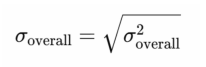
We did not perform any meta-analysis because of the heterogeneity of these studies. In our study, heterogeneity primarily arose from differences in the sample characteristics (such as age, gender, and comorbidities), variations in study equipment (different AS-OCT devices), and inconsistencies in measurement locations. Additionally, the analytical techniques employed in the included studies varied, which could have influenced the comparability of results. Given these factors, the variability between studies was substantial. and a meta-analysis would not have been appropriate. We summarized anterior scleral thickness in different ocular conditions in different studies and their main findings. Besides, we selected studies which have same location measurement in healthy eyes and CSC subjects, calculating the overall mean values (μoverall) and SD (σoverall) via the following formulas:
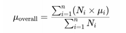
Ni is the sample size of subgroup i.
μi is the mean value of subgroup i.
n is the total number of subgroups.
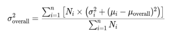
oi is the sample size of subgroup i.
RESULTS
The screening process was presented in Figure 1. We potentially publications initially identified 745 potentially publications related to the anterior scleral thickness using AS-OCT. After removing the duplicates and reviewing the titles and abstracts, 78 full-text articles were assessed for eligibility. Eventually, 32studies were included in this systematic review.
Main characteristics of included studies
Except healthy subjects, this review included 11 different types of ocular disorders or conditions, including myopia, keratoconus, exotropia, retinal vein occlusion, CSC, scleritis or episcleritis, glaucoma, fuchs endothelial dystrophy, nanophthalmos, cycloplegia and intravitreal injections. Researchers investigated the associations between anterior scleral thickness with age, measurement locations, diurnal variations, different disorders and local therapies (Table 1).Table 1. Summary of main characteristics of incuded studies.
|
|
Publication year |
Nationality |
Disease |
Sample size |
Age(years) |
Gender(M:F) |
Study design |
OCT device |
Study findings |
||||||||||
|
Zinkernagel et al |
2015 |
Switzerland |
Repeated Intravitreal Injections |
35 (70 eyes) |
76 (range 61-87)^ |
15:20 |
Retrospective interventional cross-sectional study |
SD-AS OCT (Heidelberg) |
Intravitreal injections may lead to thinner sclera when applied repeatedly in the same quadrant. |
||||||||||
|
Pekel et al |
2015 |
Turkey |
High Myopia |
62 (62 eyes) |
(30.5±13.2)vs(30.9±10.6)* |
20:42 |
Cross-sectional comparative study |
SD-AS OCT (Heidelberg) |
The thickness of anterior wall structures of myopia patients is not statistically different with healthy controls. |
||||||||||
|
Schlatter et al |
2015 |
Switzerland |
Keratoconus |
55 (62 eyes) |
(30.8±8.5)vs(30.7±6.3) |
42:13 |
Comparative case-control study |
SD-AS OCT (Heidelberg) |
The anterior scleral stroma thickness in keratoconus patients is similar to that in healthy controls. |
||||||||||
|
Ebneter et al |
2015 |
Switzerland |
No |
53 (53 eyes) |
48.6 (median: 47, range 18-92) |
25:28 |
Observational case series |
SD-AS OCT (Heidelberg) |
The anterior scleral thickness increases with age and varies significantly between quadrants. |
||||||||||
|
Buckhurst et al |
2015 |
United Kingdom |
No |
74 (74 eyes) |
27.7±5.3 |
28:46 |
Cross-sectional study |
Visante AS-OCT (Zeiss) |
Significant variations in AST occur depending on meridian and distance from the SS. |
||||||||||
|
Read et al |
2016 |
Australia |
No |
19 (19 eyes) |
21.5±2.3 |
9:10 |
Longitudinal study |
SD-AS OCT (Heidelberg) |
This study provides the first evidence of diurnal variations occurring in the thickness of the anterior sclera and conjunctiva. |
||||||||||
|
Woodman-Pieterse et al |
2018 |
Australia |
Myopia and emmetropes |
40 (40 eyes) |
21±2 |
20:20 |
Longitudinal study |
SD-AS OCT (Heidelberg) |
There is significant thinning of the anterior sclera during accommodation. These changes were more prominent in myopes. |
||||||||||
|
Ha et al |
2019 |
Korea |
Exotropia |
76 (76 eyes) |
11.6±6.6 |
30:46 |
Retrospective chart review |
Cirrus high-definition OCT (Zeiss) |
The scleral thickness at 1.0-0.5 mm anterior to OMR insertion site was thicker that than at the OMR insertion site. |
||||||||||
|
Adiyeke et al |
2020 |
Turkey |
CRVO |
67 (134 eyes) |
(62.2±11.6)vs(57.9±13.8) |
32:35 |
Case-control study |
SD-AS OCT (Heidelberg) |
Thicknesses of sclera and lamina cribrosa are increased in the CRVO, which may play a role in the pathogenesis of the disease. |
||||||||||
|
Dhakal et al |
2020 |
India |
Myopia |
95 (95 eyes) |
23.7±3.9 |
37:58 |
Cross-sectional study |
SS-AS OCT (Topcon) |
The relative significant thinning of the anterior sclera along the inferior meridian with increasing degree of myopia compared with the other three meridians. |
||||||||||
|
Kaya et al |
2020 |
Turkey |
Systemic Lupus Erythematosus |
91 (91 eyes) |
(37.9±12.5)vs(38.45±13.76) |
12:79 |
Cross-sectional study |
SD-AS OCT (Heidelberg) |
Scleral thickness is thicker in SLE patients compared to healthy controls. |
||||||||||
|
Imanaga et al |
2021 |
Japan |
Central Serous Chorioretinopathy |
87 (100 eyes) |
(48.0±10.7)vs(49.0±7.9) |
67:20 |
Retrospective comparative study |
SD-AS OCT (Heidelberg) |
Scleral thickness is greater in CSC eyes than in control, and thick sclera may have a role in the pathogenesis of CSC. |
||||||||||
|
Hau et al |
2021 |
Singapore |
Scleritis or Episcleritis |
37 (37 eyes) |
(42±10.0)vs(40±8.7) |
19:18 |
Prospective study |
AS OCT (Optovue) |
The degree of vascularity and tissue thickness were different between episcleritis, scleritis and controls. |
||||||||||
|
Sung et al |
2021 |
Korea |
Myopia |
79 (79 eyes) |
27.03±2.70 |
51:28 |
Cross-sectional study |
SD-AS OCT (Heidelberg) |
Scleral thicknesses are influenced significantly by axial length, central corneal thickness, intraocular pressure, and Bruch’s membrane opening area in myopic eyes. |
||||||||||
|
Niyazmand et al |
2021 |
Australia |
Myopia |
45 (45 eyes) |
24.8±4.6 |
20:25 |
Cross-sectional study |
SD-AS OCT (Heidelberg) |
The biomechanical forces acting on the eye led to nasal anterior scleral thickening and forward movement of the nasal scleral surface. |
||||||||||
|
Lee et al |
2021 |
Korea |
Central Serous Chorioretinopathy |
30 (30 eyes) |
(50.27±14.42)vs(54.73±11.82) |
24:6 |
Prospective case control study |
SD-AS OCT (Heidelberg) |
Scleral thickness of the study group were significantly greater than those of the control group. Choroidal thickness was positively correlated with scleral thickness. |
||||||||||
|
Park et al |
2021 |
Korea |
Primary Open‑Angle Glaucoma |
54 (54 eyes) |
51.35±13.27 |
NA |
Prospective observational study |
SD-AS OCT (Heidelberg) |
There were no significant changes in AST after using the DTFC drugs, the AST showed a significant reduction after using PG analogues. These might be related with the increased uveoscleral outflow. |
||||||||||
|
Fernández-Vigo et al |
2021 |
Spain |
No |
596 (596 eyes) |
42.6±17.2 |
258:338 |
Cross-sectional study |
SS-AS OCT (Topcon) |
The CTT dimensions were then correlated with age, sex, refractive error and AST. |
||||||||||
|
Imanaga et al |
2022 |
Japan |
Central Serous Chorioretinopathy |
158 (158 eyes) |
(50.2±10.9)vs(55.1±12.7) |
130:28 |
Retrospective cross-sectional study |
SD-AS OCT (Heidelberg) |
A thick choroid and thick sclera appeared to be related to the presence of LOF in CSC. |
||||||||||
|
Wang et al |
2022 |
Canada |
Repeated Intravitreal Injections |
79 (158 eyes) |
73 (range 34–94) |
30:49 |
Cross-sectional study |
NA |
Compared to injecting naive eyes, multiple intravitreal injections at the repeated scleral quadrant results in scleral thinning. |
||||||||||
|
Burguera-Giménez et al |
2022 |
Spain |
No |
50 (50 eyes) |
29.02±9.58 |
14:36 |
Cross-sectional prospective nonrandomized study |
Casia 2 (AS SS-OCT; Tomey) |
Scleral thickness is associated with the biomechanical corneal response metrics. Significant meridional variances in scleral thickness occur and prove that AST is associated with AL, CCT and IOP. |
||||||||||
|
Li et al |
2023 |
China |
High Myopia |
76 (76 eyes) |
(34.8±13.3)vs(39.1±12.5) |
32:44 |
Cross-sectional study |
Casia SS-1000 (AS SS-OCT; Tomey) |
AST and SS length were not significantly different between high myopia and control groups. |
||||||||||
|
Zhou et al |
2023 |
China |
Myopia |
93 (93 eyes) |
30.2±8.8 |
37:56 |
Cross-sectional study |
CIRRUS HD-OCT 5000 (Zeiss) |
The AST is negatively correlated with AL and positively correlated with age. Compared with emmetropic eyes, the AST is thinner in highly myopic eyes. |
||||||||||
|
Mohapatra et al |
2022 |
India |
Central Serous Chorioretinopathy |
100 (100 eyes) |
(39.72±8.4)vs(36.16±8.4) |
92:8 |
Prospective case-control study |
CIRRUS HD-OCT 5000 (Zeiss) |
There is a significant increase in posterior scleral thickness in patients with CSCR while AST is not affected. |
||||||||||
|
Korkmaz et al |
2023 |
Turkey |
Fuchs endothelial dystrophy |
62 (62 eyes) |
(62.5±13.2)vs(64±8.1) |
10:52 |
Prospective cross-sectional study |
SS-AS OCT (Topcon) |
In patients with FED, scleral thickness was significantly higher. |
||||||||||
|
Aichi et al |
2023 |
Japan |
Central Serous Chorioretinopathy |
115 (230 eyes) |
50.7±12.1 |
99:16 |
Retrospective comparative study |
SD-AS OCT (Heidelberg) |
The affected and fellow eyes showed no significant difference in scleral thickness. |
||||||||||
|
Fernández-Vigo et al |
2023 |
Spain |
No |
113 (113 eyes) |
(38.7±12.3)vs(41.8±11.7) |
50:63 |
Cross-sectional study |
SS-AS OCT (Topcon) |
CTT and AST measurements were thicker in the temporal quadrant of Hispanic patients compared to Caucasians. |
||||||||||
|
Yildiz et al |
2023 |
Turkey |
Keratoconus |
71 (71 eyes) |
(25.3±4.9)vs(26.5±2.3) |
45:26 |
Cross-sectional study |
SS-AS OCT (Topcon) |
Structural features of the cornea, sclera, and lamina cribrosa with similar collagen content may be similarly affected in patients with keratoconus. |
||||||||||
|
Fernández-Vigo et al |
2023 |
Spain |
Nanophthalmos |
58 (106 eyes) |
NA |
NA |
Cross-sectional comparative study |
SD-AS OCT (Heidelberg)&SS-AS OCT (Zeiss) |
Significant anatomical differences are found in nanophthalmic eyes, but no relevant differences in the AST were observed. |
||||||||||
|
Korkmaz et al |
2023 |
Turkey |
Cycloplegia |
25 (25 eyes) |
30.6±12.4 |
8:17 |
Prospective cross-sectional study |
SS-AS OCT (Topcon) |
After cycloplegia, there was a significant thinning of AST posterior to SS and a slight increase in AST at the SS. |
||||||||||
|
Burguera-Giménez et al |
2023 |
Spain |
Keratoconus |
111 (111 eyes) |
(32.3±12.0)vs(29.0±9.6) |
47:64 |
Prospective non-randomized case-control study |
Casia 2 (AS SS-OCT; Tomey) |
KC eyes presented significant thickness variations among eccentricities over the paracentral sclera. |
||||||||||
|
Teeuw et al |
2023 |
Netherland |
No |
107 (214 eyes) |
51.6±18.5 |
48:59 |
Comprehensive prospective imaging study |
SD-AS OCT (Heidelberg) |
The mean AST of the inferior quadrant was the |
||||||||||
*(Age of patient group)vs(Age of control group)
SD-AS OCT Spectral domain anterior segment optical coherence tomography
AST Anterior scleral thickness
SS Scleral spur
OMR Original medial rectus
CRVO Central retinal vein occlusion
CSC Central Serous Chorioretinopathy
SLE Systemic lupus erythematosus
DTFC Dorzolamide/timolol fixed combination
PG Prostaglandin
CTT Conjunctival and Tenon’s capsule thickness
AL Axial length
IOP Intraocular pressure
LOF Loculation of fluid
FED Fuchs endothelial dystrophy
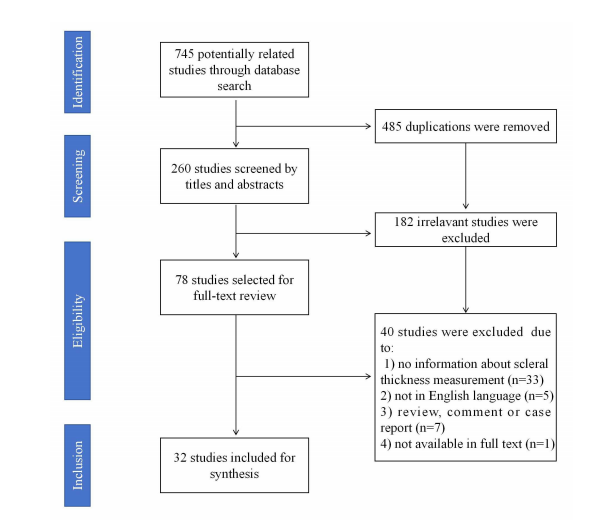
Figure 1 Screening process of the included studies
Risk of bias assessment
Supplementary file 1. Risk of bias assessments for the 32 included studies (NOS)
|
First author |
Publication year |
Selection |
Comparability |
Exposure |
Score |
|
Zinkernagel |
2015 |
3 |
1 |
3 |
|
|
Pekel |
2015 |
3 |
1 |
2 |
6 |
|
Schlatter |
2015 |
3 |
1 |
3 |
7 |
|
Ebneter |
2015 |
3 |
1 |
2 |
6 |
|
Buckhurst |
2015 |
4 |
1 |
2 |
7 |
|
Read |
2016 |
2 |
1 |
2 |
5 |
|
Woodman-Pieterse |
2018 |
3 |
1 |
2 |
6 |
|
Ha |
2019 |
3 |
1 |
2 |
6 |
|
Adiyeke |
2020 |
3 |
1 |
3 |
7 |
|
Dhakal |
2020 |
3 |
1 |
2 |
6 |
|
Kaya |
2020 |
4 |
1 |
1 |
6 |
|
Imanaga |
2021 |
4 |
1 |
2 |
7 |
|
Hau |
2021 |
2 |
1 |
3 |
6 |
|
Sung |
2021 |
4 |
1 |
1 |
6 |
|
Niyazmand |
2021 |
2 |
1 |
2 |
5 |
|
Lee |
2021 |
2 |
1 |
3 |
6 |
|
Park |
2021 |
3 |
1 |
3 |
7 |
|
Fernández-Vigo |
2021 |
3 |
2 |
2 |
7 |
|
Imanaga |
2022 |
4 |
2 |
2 |
8 |
|
Wang |
2022 |
4 |
1 |
2 |
7 |
|
Burguera-Giménez |
2022 |
2 |
1 |
3 |
6 |
|
Li |
2023 |
4 |
1 |
2 |
7 |
|
Zhou |
2023 |
4 |
1 |
2 |
7 |
|
Mohapatra |
2022 |
4 |
1 |
3 |
8 |
|
Korkmaz |
2023 |
3 |
1 |
2 |
6 |
|
Aichi |
2023 |
4 |
1 |
3 |
8 |
|
Fernández-Vigo |
2023 |
4 |
1 |
2 |
7 |
|
Yildiz |
2023 |
3 |
1 |
2 |
6 |
|
Fernández-Vigo |
2023 |
3 |
1 |
2 |
6 |
|
Korkmaz |
2023 |
2 |
1 |
3 |
6 |
|
Burguera-Giménez |
2023 |
3 |
1 |
3 |
7 |
|
Teeuw |
2023 |
4 |
1 |
2 |
7 |
Anterior scleral thickness in healthy eyes
Table 2. Anterior scleral thickness of healthy eyes (included in the calculation)
|
First author |
Publication year |
Nationality |
Sample size (Healthy) |
Age (Healthy) |
Gender (M:F,healthy) |
OCT device |
0mm-SS-T (μm) |
1mm-SS-T (μm) |
2mm-SS-T (μm) |
3mm-SS-T (μm) |
0mm-SS-N (μm) |
1mm-SS-N (μm) |
2mm-SS-N (μm) |
3mm-SS-N (μm) |
|
Buckhurst |
2015 |
United Kingdom |
74 (74 eyes) |
27.7±5.3 |
28:46 |
Visante AS-OCT (Zeiss) |
731±64 |
666±57 |
659±61 |
673±65 |
698±63 |
674±63 |
690±62 |
712±57 |
|
Adiyeke |
2020 |
Turkey |
35 (70 eyes) |
57.9±13.8 |
18:17 |
SD-AS OCT (Heidelberg) |
702.0±30.8 |
659.0±28.4 |
649.0±22.2 |
655.0±24.9 |
665.3±24.2 |
651.9±21.7 |
655.9±26.7 |
682.8±21.9 |
|
Fernández-Vigo |
2021 |
Spain |
596 (596 eyes) |
42.6±17.2 |
258:338 |
SS-AS OCT (Topcon) |
556.5±60.7 |
522.4±65.7 |
513.3±67.3 |
548.8±71.9 |
546.8±63.4 |
558.4±71.5 |
574.4±71.6 |
590.1±76.6 |
|
Burguera-Giménez |
2022 |
Spain |
50 (50 eyes) |
29.02±9.58 |
14:36 |
Casia 2 (AS SS-OCT; Tomey) |
|
522±65 |
497±63 |
513±71 |
|
525±51 |
531±58 |
532±65 |
|
Li |
2023 |
China |
42 (42 eyes) |
39.1±12.5 |
18:24 |
SS-OCT |
767.48±104.28 |
558.43±87.99 |
560.29±84.29 |
589.29±91.62 |
690.76±68.69 |
524.98±59.00 |
554.88±47.07 |
564.26±55.06 |
|
Fernández-Vigo |
2023 |
Spain |
53 (53 eyes) Hispanic |
38.7±12.3 |
24:29 |
SS-AS OCT (Topcon) |
|
536.1±77.9 |
559.8±80.8 |
591.6±83.0 |
|
589.3±68.3 |
611.3±78.8 |
613.0±75.0 |
|
60 (60 eyes) Caucasian |
41.8±11.7 |
26:34 |
SS-AS OCT (Topcon) |
|
535.5±56.0 |
520.7±50.1 |
558.9±54.7 |
|
565.8±75.1 |
577.5±74.5 |
589.8±76.4 |
|||
|
Fernández-Vigo |
2023 |
Spain |
30 (60 eyes) |
NA |
NA |
SS-AS OCT (Zeiss) |
|
670±100 |
704±89 |
691±118 |
|
687±98 |
740±94 |
767±120 |
|
30 (60 eyes) |
NA |
NA |
SD-AS OCT (Heidelberg) |
|
691±99 |
666±87 |
691±74 |
|
682±99 |
707±84 |
719±69 |
|||
|
Korkmaz |
2023 |
Turkey |
25 (25 eyes) |
30.6±12.4 |
8:17 |
Casia 2 (AS SS-OCT; Tomey) |
718.4±40.1 |
522.5±24.7 |
527.2±39.9 |
|
697.5±46.0 |
512.3±34.4 |
529.6±34.2 |
|
|
Burguera-Giménez |
2023 |
Spain |
50 (50 eyes) |
29.0±9.6 |
14:36 |
SD-AS OCT (Heidelberg) |
|
522±65 |
497±63 |
513±71 |
|
525±51 |
531±58 |
532±65 |
1 mm-SS-T: Anterior scleral thickness at a temporal distance of 1mm from the scleral spur
1 mm-SS-N: Anterior scleral thickness at a nasal distance of 1mm from the scleral spur
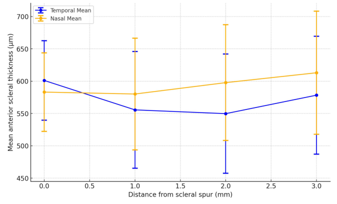
Figure 2 The overall mean temporal/nasal anterior scleral thickness in healthy eyes
Table 3. Anterior scleral thickness of eyes in two genders
|
First author |
Publication year |
Nationality |
Disease |
Sample size |
Age |
Gender |
0~6mm-SS-S (μm) |
0~6mm-SS-T (μm) |
0~6mm-SS-I (μm) |
0~6mm-SS-N (μm) |
0~6mm-SS-4q (μm) |
0~2mm-LB-4q (μm) |
|
Zhou |
2023 |
China |
Myopia |
93 (93 eyes) |
30.2±8.8 |
Male (n=37) |
495±42 |
603±48 |
575±62 |
569±67 |
559±50 |
|
|
Female (n=56) |
489±34 |
589±46 |
573±59 |
562±55 |
552±38 |
|
||||||
|
Teeuw |
2023 |
Netherland |
No |
107 (214 eyes) |
51.6±18.5 |
Male (n=48) |
|
|
|
|
|
552±57 |
|
Female (n=59) |
|
|
|
|
|
524±44 |
0~6 mm-SS-S: Average anterior scleral thickness at a superior distance of 0~6 mm from the scleral spur.
0~6 mm-SS-T: Average anterior scleral thickness at a temporal distance of 0~6 mm from the scleral spur.
0~6 mm-SS-I: Average anterior scleral thickness at a inferior distance of 0~6 mm from the scleral spur.
0~6 mm-SS-N: Average anterior scleral thickness at a nasal distance of 0~6 mm from the scleral spur.
0~6 mm-SS-4q: Average anterior scleral thickness of 4 quadrant at a distance of 0~6 mm from the scleral spur.
0~2 mm-LB-4q: Average anterior scleral thickness of 4 quadrant at a distance of 0~2 mm from the limbus.
Anterior scleral thickness in different ocularc onditions
Scleritis and episcleritis
Inflammatory conditions like scleritis and episcleritis show significant changes in scleral structure. Hau et al found the degree of vascularity and thickness of sclera and episclera were different between episcleritis, scleritis and controls using AS-OCT or AS-OCTA.[16] AS-OCT may potentially be a useful tool in evaluating patients with scleral inflammation. And several studies have investigated the conjunctival and scleral complex thickness in scleral inflammatory diseases.[2] We did not include them since they had no information about isolated anterior scleral thickness.
Myopia
50% studies proposed that myopic eyes often exhibited thinner sclera when compared to emmetropic and hypertropic eyes. For example, Dhakal et al found that the relative thinning of the anterior sclera along the inferior meridian with increasing myopia degree compared to other meridians (r = 0.27; P = 0.008).[17] The anterior scleral thickness can also be influenced by axial length, corneal thickness, intraocular pressure and even Bruch’s membrane opening area[18] This thinning is associated with the elongation of the eye in myopia, contributing to the biomechanical instability of the sclera. However, the other 50% of publications did not find significant differences. For example, Pekel et al and Li et al found that the thickness of anterior wall structures of myopic eyes has no statistical difference with healthy eyes.[3,19]
Keratoconus
In Keratoconus, anterior scleral thickness tends to be abnormal. Yildiz et al. found that structural features of the cornea, sclera, and lamina cribrosa might be similarly affected in patients with keratoconus due to similar collagen content of them.[7] Burguera-Giménez et al. reported that keratoconus eyes presented significant thickness variations among eccentricities over the paracentral scleral. The anterior scleral thickness significantly varied with scleral eccentricity over the temporal meridian (P = 0.009) in healthy controls, whereas in KC eyes, this variation was over the nasal (P = 0.001), temporal (P = 0.029) and inferior (P = 0.006) meridians.[20] However, Schlatter et al reported that the anterior scleral stroma thickness in keratoconus eyes is similar to that in healthy subjects.[20]
Central serous chorioretinopathy
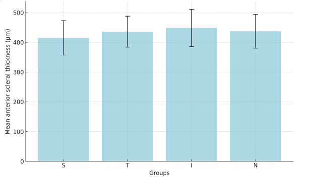
In CSC (Table 4 and Figure 3), 60% of the studies was found that anterior scleral thickness was greater than controls and thought that thick sclera might have a role in the pathogenesis of CSC.[4,21] For example, lmanaga et al found that anterior scleral thickness was significantly greater in CSC eyes than in normal control eyes at the superior ( 429.4±50.3 ) μm vs. ( 395.2±55.4) μm; P =0.005, temporal ( 447.7±45.7 ) μm vs. ( 396.5±64.1 ) μm; P <0.001, inferior ( 455.7±81.2 ) μm vs. ( 437.8±46.9 ) μm; P = 0.022, and nasal ( 454.9±44.7 ) μm vs. ( 416.6±51.2 ) μm; P = 0.001 points. 4 Besides, the anterior scleral thickness may also have a positive association with choroidal thickness. Lee et al found a positive association between subfoveal choroidal thickness and the sub-lateral rectus muscle scleral thickness (r = 0.394, P = 0.031).[21] However, Mohapatra et al found that there is a significant increase in posterior scleral thickness in CSC but no significant differences in anterior scleral thickness. [22] Aichi et al also found no significant differences between CSC and controls.[23]
Other ocular conditions
Some local therapies of the eye may lead to scleral changes. For example, intravitreal injections may lead to thinner sclera if repeating in the same quadrant,[24-25] Zinkemagel et al.[25] reported that eyes with more than 30injections had thinner average anterior scleral thickness in the inferotemporal quadrant ( 568.46±66 ) μm than the fellow eyes ( 590.6±75 ) μm, P = 0.003. Wang et al.[24] reported that injected eyes had a mean anterior scleral thickness of ( 588±95 ) μm versus ( 618±85 ) μm in fellow naive eyes ( P < 0.001). The anterior scleral thickness showed a significant reduction after using Prostaglandin analogues ( P < 0.05), which might be associated with the increase uveoscleral outflow.[26] And Korkmaz found that after cyeloplegia, there was a significant thinning of anterior scleral thickness posterior to scleral spur in nasal and temporal quadrants ( P < 0.05) and a slight increase in anterior scleral thickness at the scleral spur but no significant differences.[27]
In retinal vascular diseases, such as retinal vein occlusion, Adiyeke et al.[28] found that thickness of sclera in CRVO eyes were significantly increased at scleral spur in all quadrants (P < 0.05). Also, in other corneal diseases, such as Fuchs endothelial dystrophy, sclera was significantly thicker at 6 mm posterior to the scleral spur in all quadrants (P < 0.05).[29]
Noteworthy, AS-OCT has been applied in some systemic diseases’ researches. For example, Kaya et a investigated the anterior scleral thickness in patients with systemic lupus erythematosus (SLE), and found that inSLE patients, the anterior sclera thickness is thicker at all distances compared with controls (P < 0.05).[30] This broadens the application scope of AS-OCT.
Table 4. Anterior scleral thickness of eyes with central serious chorioretinopathy (included in the calculation)
|
First author |
Publication year |
Nationality |
Disease |
Sample size (CSC) |
Age (CSC) |
Gender (M:F, CSC) |
6mm-SS-S (μm) |
6mm-SS-T (μm) |
6mm-SS-I (μm) |
6mm-SS-N (μm) |
||
|
Imanaga |
2021 |
Japan |
Central Serous Chorioretinopathy |
40 (47 eyes) |
48.0±10.7 |
33:7 |
429.4±50.3 |
447.7±45.7 |
455.7±81.2 |
454.9±44.7 |
||
|
Imanaga |
2022 |
Japan |
Central Serous Chorioretinopathy (loculation of fluid (LOF)) |
98 (98 eyes) |
NA |
86:12 |
426.2±63.0 |
445.7±55.9 |
459.2±62.3 |
445.4±59.3 |
||
|
Central Serous Chorioretinopathy (non-loculation of fluid (non-LOF)) |
60 (60 eyes) |
NA |
44:16 |
395.1±52.1 |
414.9±47.4 |
428.8±49.6 |
414.3±55.9 |
|||||
|
Aichi |
2023 |
Japan |
Central Serous Chorioretinopathy |
115 (230 eyes) |
46.4±18.3 |
54:46 |
410.3±58.4 |
434.3±52.9 |
447.9±59.5 |
434.9±59.1 |
||
CSC : Central Serous Chorioretinopathy.
6mm-SS-S: Anterior scleral thickness at a superior distance of 6mm from the scleral spur.
6mm-SS-T: Anterior scleral thickness at a temporal distance of 6mm from the scleral spur.
6mm-SS-I: Anterior scleral thickness at a inferior distance of 6mm from the scleral spur.
6mm-SS-N: Anterior scleral thickness at a nasal distance of 6mm from the scleral spur.
DISCUSSION
In this systematic review, we provided a comprehensive overview of the current understanding of anterior scleral thickness across various ocular conditions using AS-OCT. These studies collectively examined11 ocular disorders as well as healthy eyes. Variations in anterior scleral thickness can indicate the presented progression of ocular conditions. For instance. Thinner sclera in myopic eyes suggests biomechanical alterations, while increased thickness in conditions like active scleritis may reflects inflammatory responses. Additionally, age and diurnal variations further impact scleral measurements, offering insights into their dynamic nature. The precision of AS-OCT in detecting these subtle changes enhances its utility in monitoring ocular health, potentially aiding early diagnosis and tracking disease progression in clinical settings. The findings underscore the importance of anterior scleral thickness to be a potential biomarker in ocular health.
It is regarded that the anterior scleral thickness is positively with age, This might be due to age-related changes in scleral collagen and biomechanics.[11,13] The observed regional variations in anterior scleral thickness such as the thicker inferior quadrant compared to other quadrants, suggested that these variations could be linked to the different mechanical stresses exerted on the sclera by ocular movements and external forces.[13] The diurnal variations reported by Read et al indicated that the sclera was not static throughout the day, which might be due to physiological changes in ocular blood flow, intraocular pressure and other metrics fluctuating with the circadian rhythm,[14] The ethnic differences revealed by Femández. Vigo et al., with Hispanic patients showing higher temporal quadrant thickness compared to Caucasians, suggest that genetic or environmental factors may also influence scleral structure.[31] This finding may explain the phenomenon to some extent that different ethics have quite different epidemiology in certain ocular disorders. Males had thicker anterior scleral thickness compared to females, but the significance of this phenomenon is not known and could perhaps represent an anthropological difference between two genders.[2]
The sclera has tight associations with other ocular structures, such as the cornea, choroid and ciliary muscle. The anterior scleral thickness is associated with the biomechanical corneal response metrics, Although the measurement profiles did not differ between keratoconus and control groups in some studies, the inferior-superior asymmetry differences demonstrated scleral changes over the vertical meridian in keratoconus, which need further investigation.[7,20,32] According to Korkmaz study, inhibition of forward inward movement of the ciliary body by cycloplegia can affect anterior scleral thickness via causing a change in the mechanical force of the ciliary muscle on the sclera.[27] In CSC, the association between choroidal changes and anterior scleral thickness has been observed.[21]
The association between myopia and reduced anterior scleral thickness is well-documented in the reviewed studies, The thinning of the sclera, particularly along certain meridians, reflects the biomechanical alterations associated with axial elongation in myopic eyes. This thinning compromises the structural integrity of the sclera, potentially exacerbating myopic progression and increasing the risk of myopia-related complications.[33] However, the inconsistencies reported by Pekel et al. suggest that anterior scleral thickness changes in myopia may not be uniform and could depend on additional factors such as the degree of myopia, axial length, and individual variations in scleral composition.[3,34]
In inflammatory conditions like scleritis and episcleritis, the significant thickening of the sclera observed in the reviewed studies underscores the utility of AS-OCT in diagnosing and monitoring these conditions.AS-OCT was instrumental in identifying necrosis and scleral nodules in subclinical cases, and different subtypes of scleritis indicate different pathogenesis. Moreover, complete resolution of scleral inflammation can be followed by OCT.[35] The ability of AS-OCT to detect subtle changes in scleral structure makes it a valuable tool in distinguishing between different types of scleral inflammation and in assessing the effectiveness of therapeutic interventions.
The findings related to CSC suggest a potential role for inereased anterior scleral thickness in the pathogenesis of the disease, possibly due to its association with choroidal thickness. However, the lack of consensus, as seen in the studies by Mohapatra et al. and Aichi et al. indicates that further investigation was required to elucidate the exact relationship between anterior scleral thickness and CSC.[22-23,36] The potential influence of other factors, such as intraocular pressure and choroidal circulation, should also be considered in future studies.
The impact of local therapies, such as intravitreal injections and the use of prostaglandin analogues, cycloplegia on anterior scleral thickness further broadens the clinical relevance of anterior scleral thickness measurements. The observed thinning of the sclera following repeated injections or chronic medication use suggests that these interventions may have unintended effects on scleral integrity, which could influence treatment outcomes, particularly in patients requiring long-term therapy, This may remind clinicians to monitor anterior scleral thickness in patients undergoing such treatments to prevent adverse effects.
The variations in anterior scleral thickness observed in conditions like retinal vein occlusion, Fuchs endothelial dystrophy, and systemic diseases like SLE suggest that scleral changes are not confined to ocular pathologies but may also reflect systemic health. The thicker sclera in SLE patients, as reported by Kaya et al, implies that systemic inflammatory or autoimmune processes could impact scleral structure, providing a potential link between systemic health and ocular manifestations.[30] This finding opens new avenues for research into the role of anterior scleral thickness as a marker for systemic disease.
Strengths of our systematic review include that this topic was the first time to report. And we followed the strict protocol in the synthesis process. However, there was significant variability in the standards used across studies, particularly in relation to diagnostic tools and measurement techniques. For example, AS-OCT equipment from different manufacturers can yield slightly varying results due to differences in resolution scanning protocols, or imaging software. Additionally, measurement techniques are often not uniformly applied across studies, which can lead to inconsistencies in the data. Standardization of these tools and methods would not only enhance the accuracy and consistency of measurements but also facilitate better comparability across studies. Establishing standardized protocols for AS-OCT imaging, measurement analysis, and reporting could significantly improve the reproducibility of research findings and lead to more robust conclusions in future studies. Our analysis was limited due to thehigh risk of bias and heterogeneity of included studies, which preclude the further statistical analysis and pooling data for meta-analysis. Specifically, variations in patient characteristics (e.g., age, gender, comorbidities and ethnicities) and study equipment (e.g., SD-OCT vs. SS-OCT) could introduce bias or lead to divergent findings. To better understand the robustness of the conclusions, it would be useful to evaluate how such heterogeneity might affect the outcomes with large sample size in the future.
To advance future research in this area, several critical steps should be taken to tackle the deficiencies identified in the current literature. These steps include 1) measuring same locations in cohort studies; 2)conducting research in a large sample size and maximizing the representativeness; 3) use design such as matching, and analytic strategies (multivariable adjustment, propensity score matching, or inverse probability weighting)to control for potential confounding factors; 4) conducting subgroup analyses or sensitivity tests to assess the consistency of results across different settings or groups; 5) promoting well-designed studies to investigate anterior scleral thickness in differentiate group, gender group and different ocular conditions.
In conclusion, this systematic review underscores the significant role of anterior scleral thickness in ocular health and disease, as well as the growing importance of AS-OCT in clinical practice. Continued research into anterior scleral thickness, with a focus on standardization, longitudinal analysis, and multimodal imaging, will further advance our understanding of scleral biomechanics and its implications for ocular and systemic health.
Correction notice
NoneAcknowledgement
NoneAuthor Contributions
(I)Conception and design: Lihui Meng and Qianyi Yu(II)Administrative support: Jingyuan Yang and YouxinChen
(III)Provision of study materials or patients: Allauthors
(IV)Collection and assembly of data: Lihui Meng andQianyi Yu
(V)Data analysis and interpretation: Lihui Meng and Qianyi Yu
(VI)Manuscript writing: All authors
(VII)Final approval of manuscript: All authors