





Introduction
Rubeosis iridis, also known as iris neovascularization, is
caused by ocular ischemia in patients with proliferative
diabetic retinopathy(1-3). Intravitreal anti-vascular
endothelial growth factor (anti-VEGF), such as bevacizumab,
ranibizumab, and pegaptanib, were reported to be benefi cial
for anterior segment neovascularization(4-7). Aflibercept,
a new anti-VEGF, was not described for treating rubeosis
iridis after reviewing the literature.
Case presentation
A 61-year-old man had diabetes mellitus under insulin control
for 10 years. HbA1c level was between 7 to 8.5 mg/dL for
these 3 years. He did not have other systemic disease.
Pan-retinal argon laser photocoagulation was performed
for proliferative diabetic retinopathy 3 years ago in both
eyes. He underwent pars plana vitrectomy and silicone
oil infusion for vitreous hemorrhage and tractional
retinal detachment 2 years ago in the left. Neovascular
glaucoma occurred subsequently, which caused phthisis
bulbi and vision without light perception. The patient
complained of right progressive blurred vision for 1 week
3 months ago. His corrected visual acuity was 20/30 in
the right eye. Intraocular pressure and anterior segment
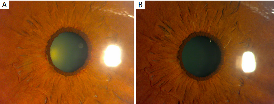
were normal, except neovascularization around pupillary
margin (Figure 1A). Mild nuclear sclerosis of lens was
found. Neovascularization of angle was not demonstrated
under gonioscopic examination. Spectral-domain optical
coherence tomography revealed normal macular structure.
Fundus examination showed scanty vitreous hemorrhage
and laser spots scattering on mid-peripheral retina, without
accompanying preretinal fibrosis. Iris neovascularization
secondary to proliferative diabetic retinopathy was diagnosed.
After full explanation of benefi t, risk, and off -label usage, the patient signed the informed consent. Single intravitreal aflibercept (2 mg/0.05 mL) was injected. Rubeosis iridis disappeared on the next day (Figure 1B). One week later, scattered retinal laser photocoagulation was added on the peripheral retina.
Vitreous hemorrhage gradually vanished without increasing preretinal fibrosis. Neovascularization of angle and iris was not found, along with improved corrected visual acuity to 20/25 3 months following the injection.
After full explanation of benefi t, risk, and off -label usage, the patient signed the informed consent. Single intravitreal aflibercept (2 mg/0.05 mL) was injected. Rubeosis iridis disappeared on the next day (Figure 1B). One week later, scattered retinal laser photocoagulation was added on the peripheral retina.
Vitreous hemorrhage gradually vanished without increasing preretinal fibrosis. Neovascularization of angle and iris was not found, along with improved corrected visual acuity to 20/25 3 months following the injection.
Figure 1 (A) Neovascularization found on the pupillary margin, especially on the superior, temporal, and nasal-inferior part in the right eye;
(B) disappearance of neovascularization of iris on the next day following intravitreal aflibercept.
Discussion
In eyes with proliferative diabetic retinopathy, the elevated
level of VEGF is found in response to retinal hypoxia (1).
Higher VEGF concentration in vitreous cavity and aqueous
humor is strongly associated with development of retinal
and iris neovascularization (2,3). Rubeosis iridis may cause
hyphema, which obstructing the visual axis and impairing
vision. Iris neovascularization may extend to anterior
chamber angle, resulting in neovascular glaucoma and
subsequent optic nerve damage associated with visual field
and acuity defect.
Pan-retinal laser photocoagulation can lower the severity of retinal ischemia, as the standard of care for management of rubeosis iridis secondary to proliferative diabetic retinopathy. Intravitreal injection anti-VEGF can serve as an adjunct or main treatment for anterior segment neovascularization. Bevacizumab is a full-length recombinant humanized monoclonal antibody directed against all isoforms of VEGF-A, the most important VEGF for pathologic angiogenesis. Bevacizumab used intravitreally resulted in regression of rubeosis iridis within several days(5). Ranibizumab is an antigen-binding fragment of monoclonal antibody against all isoforms of VEGF-A. Intravitreal ranibizumab was described to cause receding of iris neovascularization secondary to diabetic retinopathy(6). Pegaptanib is RNA aptamer to inhibit one isoform of VEGF-A, VEGF 165. Disappearance of iris neovascularization was also reported after intravitreal pegaptanib in diabetic patients(7). However, intravitreal anti-VEGF agents have temporary effects for controlling rubeosis iridis(8).Iris neovascularization may recur 2 to 4 months after anti-VEGF injection. Besides, anti-VEGF can cause tractional retinal detachment in cases with preexisting preretinal fibrosis(8).
Aflibercept (Eylea™, Regeneron Pharmaceuticals, Inc., and Bayer Pharma AG, Berlin, Germany) is a decoy receptor fusion protein, composed of the second domain of human VEGF receptor 1 and the third domain of VEGF receptor 2, which are fused to the Fc domain of human IgG1 (9). Aflibercept can downregulate both VEGF-A, VEGF-B, and placental growth factor, which are synergistic for pathologic angiogenesis. The binding affinity of VEGF for this drug is higher than for ranibizumab and bevacizumab(10). Aflibercept displays a prolonged VEGF inhibition in comparison with the other VEGF-antagonists ranibizumab and bevacizumab in retinal pigment epithelium/choroid organ cultures(11). For diabetic patients, aflibercept have been proven to reduce macular edema and to restore visual acuity (12). Aflibercept was not described for treating rubeosis iridis secondary to diabetic retinopathy previously.
In our case with proliferative diabetic retinopathy, iris neovascularization can occur after incomplete pan-retinal laser photocoagulation. Intravitreal afl ibercept caused rapid regression of iris neovascularization, preventing further deterioration to neovascular glaucoma or hyphema. Adding scattering retinal laser photocoagulation was helpful to avoid recurrence of rubeosis iridis. Because there was no pre-existing preretinal fibrosis, aflibercept did not cause subsequent tractional retinal detachment in this diabetic patient.
In summary, we described a case with iris neovascularization secondary to proliferative diabetic retinopathy responding rapidly to intravitreal aflibercept. Aflibercept may serve as another anti-VEGF for treating rubeosis iridis.