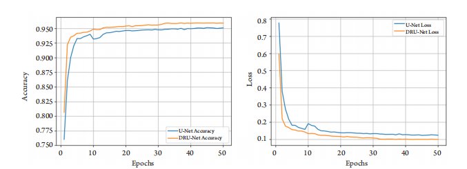
Figure 1 The network of optimized DRUNET, which follows the infrastructure of DRUNET. Output in down-sampling path is upsampled to the same scale of original images. Deconvolution with stride of 1, 2, 4 are respectively used in the three module outputs in down-sampling path, incorporating multi-scale feature extraction, the feature maps will reach output layer after being concatenated in channels
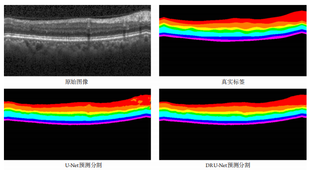
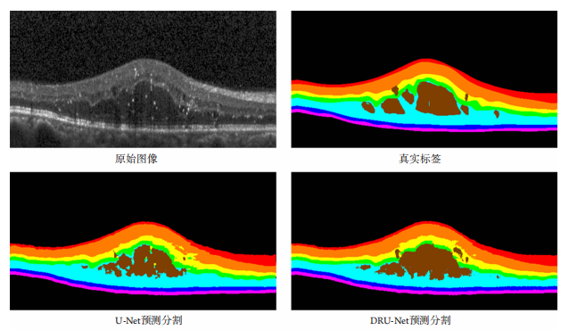
Figure 4 Comparison of segmentation results on samples with effusion, U-Net shows many cavities in effusion area, while the segmentation result of DRUNet is closer than true label
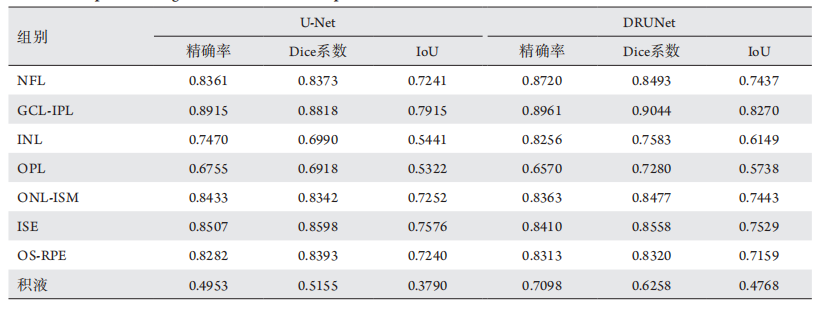
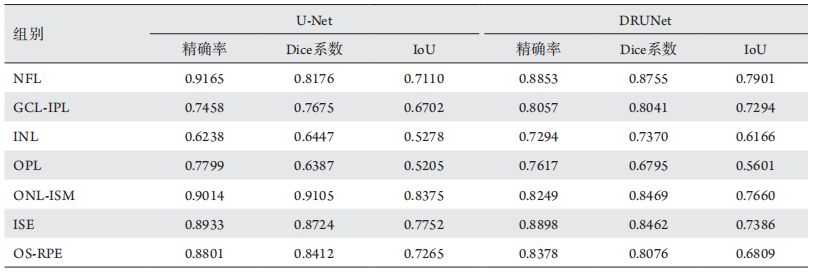
表 1 不带积液样本的分割结果统计
Table 1 Comparison of segmentation results on samples without effffusion
1. Clevert DA, Unterthiner T, Hochreiter S. Fast and accurate deep
network learning by exponential linear units (ELUs)[C]//International
Conference on Learning Representations, 2016: 1-14.
2. Ioffe S, Szegedy C. Batch normalization: Accelerating deep network
training by reducing internal covariate shift[C]//Proceedings of the
32nd International Conference on Machine Learning, 2015: 448-456.
3. Chiu SJ, Allingham MJ, Mettu PS, et al. Kernel regression-based
segmentation of optical coherence tomography images with diabetic
macular edema[ J]. Biomedical Optics Express, 2015, 6(4): 1172-1194.
4. Liu Y, Cheng MM, Hu X, et al. Richer convolutional features for edge
detection[ J]. IEEE Transactions on Pattern Analysis and Machine
Intelligence, 2019, 41(8): 1939-1946.
5. He K, Zhang X, Ren S, et al. Deep residual learning for image
recognition[C]//IEEE Conference on Computer Vision and Pattern
Recognition (CVPR), 2016: 770-778.
6. Yu F, Koltun V. Multi-scale contex t aggregation by dilated
convolutions[ J]. arXiv preprint arXiv: 1511.07122,2015.
7. Devalla SK, Renukanand PK, Sreedhar BK, et al. DRUNET: a dilated-
residual U-Net deep learning network to segment optic nerve head
tissues in optical coherence tomography images[ J]. Biomedical Optics
Express, 2018, 9(7): 3244-3265.
8. Lang A, Carass A, Hauser M, et al. Retinal layer segmentation of
macular OCT images using boundary classification[ J]. Biomedical
Optics Express, 2013, 4(7): 1133-1152.
9. 许毓鹏. 视网膜血管性疾病光学相干断层成像图像的自动分层
研究[ J]. 上海交通大学学报: 医学版, 2019, 39(6): 613-621.
XU Yupeng. Automatic layer segmentation of optical coherence
tomography images in retinal vascular diseases[ J]. Journal of Shanghai
Jiaotong University. Medical Science, 2019, 39(6): 613-621.
10. 杨云. 集成支持向量机在OCT血管内斑块分割中的应用与研
究[ J]. 计算机应用与软件, 2019, 36(4): 103-107.
YANG Yun. Application and research of adaboost-SVM in OCT
intravascular patch segmentation[ J]. Computer Applications and
Software, 2019, 36(4): 103-107.
11. Vermeer KA , van der Schoot J, Lemij HG, et al. Automated
segmentation by pixel classification of retinal layers in ophthalmic OCT
images[ J]. Biomedical Optics Express, 2011, 2(6): 1743-1756.
12. Lang A, Carass A, Sotirchos E, et al. Segmentation of retinal OCT
images using a random forest classifier[C]//Medical Imaging 2013:
Image Processing. International Society for Optics and Photonics,
2013: 145-155.
13. Fang L, Li S, Cunefare D, et al. Segmentation based sparse
reconstruction of optical coherence tomography images[ J]. IEEE Trans
Med Imaging, 2017, 36(2): 407-421.
14. 贺琪欲. 基于光学相干层析成像的视网膜图像自动分层方
法[ J]. 光学学报, 2016, 36(10): 309-318.
HE Qiyu. Automated retinal layer segmentation based on optical
coherence tomographic images[ J]. Acta Optica Sinica, 2016, 36(10):
309-318.
15. 蔡怀宇. 眼科光学相干层析成像的图像处理方法[ J]. 中国光学,
2019, 12(4): 731-740.
CAI Huaiyu. Image processing method for ophthalmic optical
coherence tomography[ J]. Chinese Optics, 2019, 12(4): 731-740.
16. Rogers W, 祝庆麟. 计算机辅助医学诊断文献回顾[ J]. 国外医
学·生物医学工程分册, 1981(4): 26-31.
ROGERS W, ZHU Qinglin. Review of computer aided medical
diagnosis[ J]. Journal of Biomedical Engineering Foreign Medical
Sciences Biomedical Engineering, 1981(4): 26-33.
18. Chakravarty A , Sivaswamy J. A super vised joint multi-layer
segmentation framework for retinal optical coherence tomography
images using conditional random field[ J]. Comput Methods Programs
Biomed, 2018, 165: 235-250.
19. 滕岩, 刘英伟, 杨明明, 等. 糖尿病视网膜病变患者全视网膜激
光光凝术后黄斑区功能与形态变化[ J]. 中华眼底病杂志, 2010,
26(2): 120-123.
TENG Yan, LIU Yingwei, YANG Mingming, et al. The functional and
morphological changes of macular after panretinal photocoagulation
in the patients with diabetic retinopathy[ J]. Chinese Journal of Ocular
Fundus Diseases, 2010, 26(2): 120-123.
20. 吴彩云. 高度近视黄斑病变的形态学分类及其影响因素[D]. 兰
州: 兰州大学, 2014.
WU Caiyun. The morphological classification of myopia maculopathy
and influence factors[D]. Lanzhou: Lanzhou University, 2014.
'%20fill='white'%20fill-opacity='0.01'/%3e%3cmask%20id='mask0_3477_29692'%20style='mask-type:luminance'%20maskUnits='userSpaceOnUse'%20x='0'%20y='0'%20width='16'%20height='16'%3e%3crect%20id='&%23232;&%23146;&%23153;&%23231;&%23137;&%23136;_2'%20x='16'%20width='16'%20height='16'%20transform='rotate(90%2016%200)'%20fill='white'/%3e%3c/mask%3e%3cg%20mask='url(%23mask0_3477_29692)'%3e%3cpath%20id='&%23232;&%23183;&%23175;&%23229;&%23190;&%23132;'%20d='M14%205L8%2011L2%205'%20stroke='%23333333'%20stroke-width='1.5'%20stroke-linecap='round'%20stroke-linejoin='round'/%3e%3c/g%3e%3c/g%3e%3c/svg%3e)