微血管成像模块获取的结膜血管网络图像和动态血流视频,需进一步应用成像算法处理,以提取定量的血流形态学和血流动力学功能参数。图像处理软件的开发已在之前的文献中叙述[31-32]。对于结膜血管网络图像,首先,通过自适应直方图均衡化来提高图像质量,然后通过减去背景图像来进一步增强图像质量;然后,将增强后的图像转换为二值图像,利用骨架化算法得到网络骨架;最后,对血管骨架图像进行分析,计算分形维数复度,提取反应血管网复杂度的分形维数单分形值(Dbox)和反应血管网密度的多重分形(D0)值,用于评价血管密度和复杂性。对于结膜血流影像,首先采用基于球结膜时空图像的运动校正算法,对眼球运动进行补偿,得到配准图像。在对血管图像进行增强和分割后,利用多帧图像的均值,通过计算血管信号轮廓的半高宽(full width at half maxima,FWHM)来测量血管直径(D,μm)。通过分析红细胞的运动,定量结膜血流动力学参数,直接测出轴向血流速度(Va,mm/s),以此计算出截面血流速度,截面血流速度乘以血管横截面积(假设血管为圆形截面,由血管直径计算得出)获得血流量(Q,pl/s)[48]。
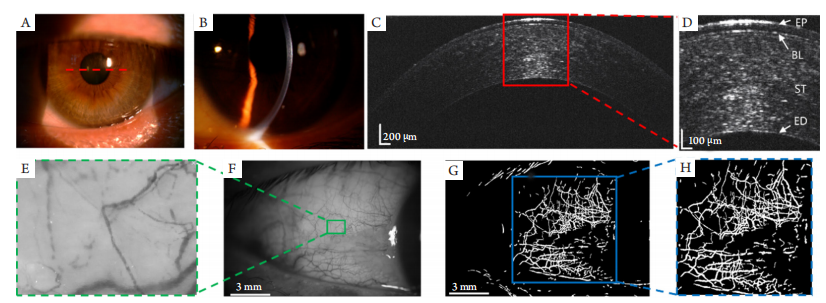
(A, B) Slit-lamp corneal imaging of healthy subjects, the red dashed line indicates the OCT scanning position. (C, D) OCT image of the healthy cornea and the zoom-in area showing clear corneal layer structures. EP: epithelial layer; BL: Bowman’s layer; ST: stroma; ED: endothelial layer. (E) Screenshots of eye microvascular dynamics in the selected area of conjunctival microvascular network. (F) The bulbar conjunctival vessel network image of the healthy subject. (G, H) The segmented vessel network image and the skeletonized vessel network image of the selected region.
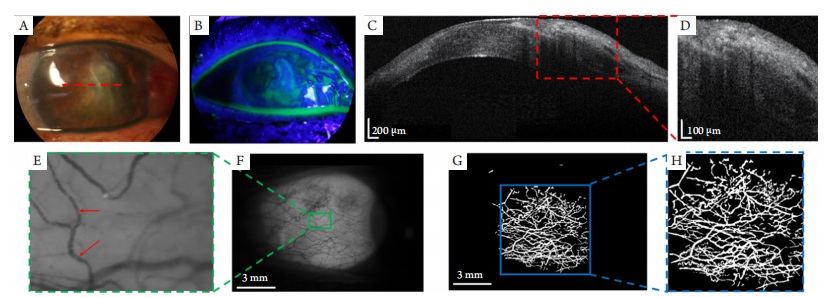
图3 角膜炎患者多模态成像
Figure 3 Experimental results of the keratitis patient
(A) Appearance image of the keratitis patient’s eye; the red dashed line indicates the OCT scanning position; (B) Fluorescein stain image of the keratitis patient; (C, D) OCT image of the inflamed cornea and the zoom-in area showing the highly reflective, inflamed lesion.(E) Screenshots of eye microvascular dynamics in the selected area of conjunctival microvascular network; (F) The bulbar conjunctival vessel network image of the keratitis patient; (G, H) Results of skeletonization of conjunctival microvascular network.
表1 血流形态学及动力学参数对比
Table 1 Comparison of the quantitative vessel parameters
1. Ting DSW, Pasquale LR, Peng L, et al. Artificial intelligence and
deep learning in ophthalmology[ J]. Br J Ophthalmol, 2019, 103(2):
167-175.
2. Hogarty DT, Mackey DA, Hewitt AW. Current state and future
prospects of artificial intelligence in ophthalmology: a review[ J]. Clin
Exp Ophthalmol, 2019, 47(1): 128-139.
3. Koutsiaris AG, Tachmitzi SV, Batis N, et al. Volume flow and wall shear
stress quantification in the human conjunctival capillaries and post-
capillary venules in vivo[ J]. Biorheology, 2007, 44(5/6): 375-386.
4. Atchison DA, Smith G. Optics of the human eye[M]. Boston:
Butterworth-Heinemann, 2000.
5. American National Standards Institute. American National Standard for
Ophthalmics—Light Hazard Protection for Ophthalmic Instruments,
ANSI Z80.36-2016[R]. New York, NY, USA: American National
Standards Institute, 2016.
6. Savini G, Barboni P, Carbonelli M, et al. Repeatability of automatic
measurements by a new Scheimpflug camera combined with Placido
topography[ J]. J Cataract Refract Surg, 2011, 37(10): 1809-1816.
7. R abinowitz YS, Li X , Canedo ALC, et al. Optical coherence
tomography combined with videokeratography to differentiate mild
keratoconus subtypes[ J]. J Refract Surg, 2014, 30(2): 80-87.
8. Schiano-Lomoriello D, Bono V, Abicca I, et al. Repeatability of anterior
segment measurements by optical coherence tomography combined
with Placido disk corneal topography in eyes with keratoconus[ J]. Sci
Rep, 2020, 10(1): 1124.
9. Mueller M, Schulz-Wackerbarth C, Steven P, et al. Slit-lamp-adapted
fourier-domain OCT for anterior and posterior segments: preliminary
results and comparison to time-domain OCT[ J]. Curr Eye Res, 2010,
35(8): 722-732.
10. Stehouwer M, Verbraak FD, De Vries H, et al. Fourier domain optical
coherence tomography integrated into a slit lamp; a novel technique
combining anterior and posterior segment OCT[ J]. Eye, 2010, 24(6):
980-984.
11. Zawadzki RJ, Zhang P, Zam A, et al. Adaptive-optics SLO imaging
combined with widefield OCT and SLO enables precise 3D localization
of fluorescent cells in the mouse retina[ J]. Biomed Opt Express, 2015,6(6): 2191-2210.
12. Felberer F, Kroisamer JS, Baumann B, et al. Adaptive optics SLO/
OCT for 3D imaging of human photoreceptors in vivo[ J]. Biomed Opt
Express, 2014, 5(2): 439-456.
13. Song W, Wei Q, Liu T, et al. Integrating photoacoustic ophthalmoscopy
with scanning laser ophthalmoscopy, optical coherence tomography,
and fluorescein angiography for a multimodal retinal imaging
platform[ J]. J Biomed Opt, 2012, 17(6): 061206.
14. Malone JD, El-Haddad MT, Bozic I, et al. Simultaneous multimodal
ophthalmic imaging using swept-source spectrally encoded scanning
laser ophthalmoscopy and optical coherence tomography[ J]. Biomed
Opt Express, 2016, 8(1): 193-206.
15. Mujat M, Ferguson RD, Patel AH, et al. High resolution multimodal
clinical ophthalmic imaging system[ J]. Opt Express, 2010, 18(11):
11607-11621.
16. Chen W, Deng Y, Jiang H, et al. Microvascular abnormalities in dry eye
patients[ J]. Microvasc Res, 2018, 118: 155-161.
17. Cheung ATW, Hu BS, Wong SA, et al. Microvascular abnormalities
in the bulbar conjunctiva of contact lens users[ J]. Clin Hemorheol
Microcirc, 2012, 51(1): 77-86.
18. Xu Z, Jiang H, Tao A, et al. Measurement variability of the bulbar
conjunctival microvasculature in healthy subjects using functional slit
lamp biomicroscopy (FSLB)[ J]. Microvasc Res, 2015, 101: 15-19.
19. Wang L, Yuan J, Jiang H, et al. Vessel sampling and blood flow velocity
distribution with vessel diameter for characterizing the human bulbar
conjunctival microvasculature[ J]. Eye Contact Lens, 2016, 42(2): 135.
20. Jiang H, Zhong J, DeBuc DC, et al. Functional slit lamp biomicroscopy
for imaging bulbar conjunctival microvasculature in contact lens
wearers[ J]. Microvasc Res, 2014, 92: 62-71.
21. Lee WD, Devarajan K, Chua J, et al. Optical coherence tomography
angiography for the anterior segment[ J]. Eye Vis (Lond), 2019, 6(1): 4.
22. Br unner M, Romano V, Steger B, et al. Imaging of corneal
neovascularization: optical coherence tomography angiography and
fluorescence angiography[ J]. Invest Ophthalmol Vis Sci, 2018, 59(3):
1263-1269.
23. Shousha MA, Karp CL, Perez VL, et al. Diagnosis and management
of conjunctival and corneal intraepithelial neoplasia using ultra high-
resolution optical coherence tomography[ J]. Ophthalmology, 2011,
118(8): 1531-1537.
24. Pantalon A, Pfister M, Aranha dos Santos V, et al. Ultrahigh‐resolution
anterior segment optical coherence tomography for analysis of corneal
microarchitecture during wound healing[ J]. Acta Ophthalmol, 2019,
97(5): e761-e771.
25. Christopoulos V, Kagemann L, Wollstein G, et al. In vivo corneal high-
speed, ultra–high-resolution optical coherence tomography[ J]. Arch
Ophthalmol, 2007, 125(8): 1027-1035.
26. Bizheva K, Tan B, MacLelan B, et al. Sub-micrometer axial resolution
OCT for in-vivo imaging of the cellular structure of healthy and
keratoconic human corneas[ J]. Biomed Opt Express, 2017, 8(2):
800-812.
27. Du C, Shen M, Li M, et al. Anterior segment biometry during
accommodation imaged with ultralong scan depth optical coherence
tomography[ J]. Ophthalmology, 2012, 119(12): 2479-2485.
28. Werkmeister RM, Alex A, Kaya S, et al. Measurement of tear film
thickness using ultrahigh-resolution optical coherence tomography[ J].
Invest Ophthalmol Vis Sci, 2013, 54(8): 5578-5583.
29. Fujimoto JG, Drexler W. Optical coherence tomography: technology
and applications[M]. New York: Springer Science & Business Media,
2008.
31. Nanji AA, Sayyad FE, Galor A, et al. High-resolution optical coherence
tomography as an adjunctive tool in the diagnosis of corneal and
conjunctival pathology[ J]. Ocul Surf, 2015, 13(3): 226-235.
32. Ang M, Baskaran M, Werkmeister RM, et al. Anterior segment optical
coherence tomography[ J]. Prog Retin Eye Res, 2018, 66: 132-156.
33. Xiao P, Mazlin V, Grieve K, et al. In vivo high-resolution human retinal
imaging with wavefront-correctionless full-field OCT[ J]. Optica, 2018,
5(4): 409-412.
34. Mazlin V, Xiao P, Dalimier E, et al. In vivo high resolution human
corneal imaging using full-field optical coherence tomography[ J].
Biomed Opt Express, 2018, 9(2): 557-568.
35. Drexler W, Liu M, Kumar A, et al. Optical coherence tomography
today: speed, contrast, and multimodality[ J]. J. Biomed Opt, 2014,
19(7): 071412.
36. Adhi M, Duker JS. Optical coherence tomography–current and future
applications[ J]. Curr Opin Ophthalmol, 2013, 24(3): 213.
37. Huang D, Swanson EA , Lin CP, et a l . Optical coherence
tomography[ J]. Science, 1991, 254(5035): 1178-1181.
38. Wilson G, Ren H, Laurent J. Corneal epithelial fluorescein staining[ J].
J Am Optom Assoc, 1995, 66(7): 435.
39. Oliphant H, Kennedy A, Comyn O, et al. Commercial slit lamp anterior
segment photography versus digital compact camera mounted on a
standard slit lamp with an adapter[ J]. Curr Eye Res, 2018, 43(10):
1290-1294.
40. Stanzel TP, Devarajan K, Lwin NC, et al. Comparison of optical
coherence tomography angiography to indocyanine green angiography
and slit lamp photography for corneal vascularization in an animal
model[ J]. Sci Rep, 2018, 8(1): 11493.
41. Yuan J, Jiang H, Mao X, et al. Slit-lamp photography and videography
with high magnifications[ J]. Eye Contact Lens, 2015, 41(6): 391.
42. van den Berg TJTP. Intraocular light scatter, reflections, fluorescence
and absorption: what we see in the slit lamp[ J]. Ophthalmic Physiol
Opt, 2018, 38(1): 6-25.
43. Mantopoulos D, Cruzat A, Hamrah P. In vivo imaging of corneal
inflammation: new tools for clinical practice and research[ J]. Semin
Ophthalmol, 2010, 25(5/6): 178-185.
44. Oatts JT, Keenan JD, Mannis T, et al. Multimodal assessment of corneal
thinning using optical coherence tomography, scheimpflug imaging,
pachymetry and slit lamp examination[ J]. Cornea, 2017, 36(4): 425.
45. Mondino BJ. Inflammatory diseases of the peripheral cornea[ J].
Ophthalmology, 1988, 95: 463-472.
46. Abdulkhaleq LA, Assi MA, Abdullah R, et al. The crucial roles of
inflammatory mediators in inflammation: A review[ J]. Veterinary
World, 2018, 11(5): 627.
'%20fill='white'%20fill-opacity='0.01'/%3e%3cmask%20id='mask0_3477_29692'%20style='mask-type:luminance'%20maskUnits='userSpaceOnUse'%20x='0'%20y='0'%20width='16'%20height='16'%3e%3crect%20id='&%23232;&%23146;&%23153;&%23231;&%23137;&%23136;_2'%20x='16'%20width='16'%20height='16'%20transform='rotate(90%2016%200)'%20fill='white'/%3e%3c/mask%3e%3cg%20mask='url(%23mask0_3477_29692)'%3e%3cpath%20id='&%23232;&%23183;&%23175;&%23229;&%23190;&%23132;'%20d='M14%205L8%2011L2%205'%20stroke='%23333333'%20stroke-width='1.5'%20stroke-linecap='round'%20stroke-linejoin='round'/%3e%3c/g%3e%3c/g%3e%3c/svg%3e)