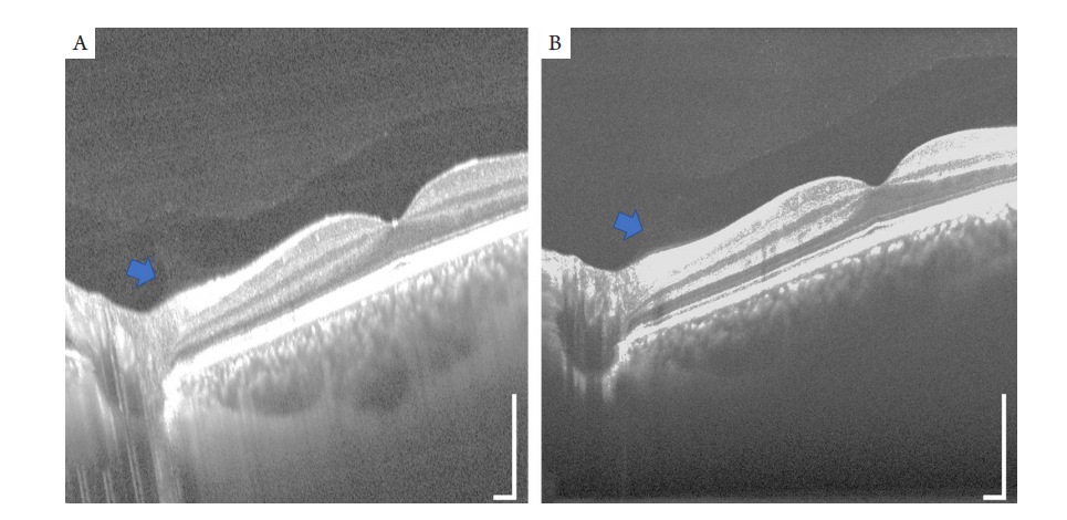
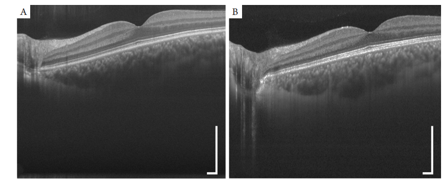
(A) The retina OCT image from a custom-built SD-OCT system (36 kHz, 850 nm SD-OCT system); (B) the retina OCT image from a custom-built SS-OCT (200 kHz, 1 060 nm SS-OCT system). Scale bar: 400 μm.
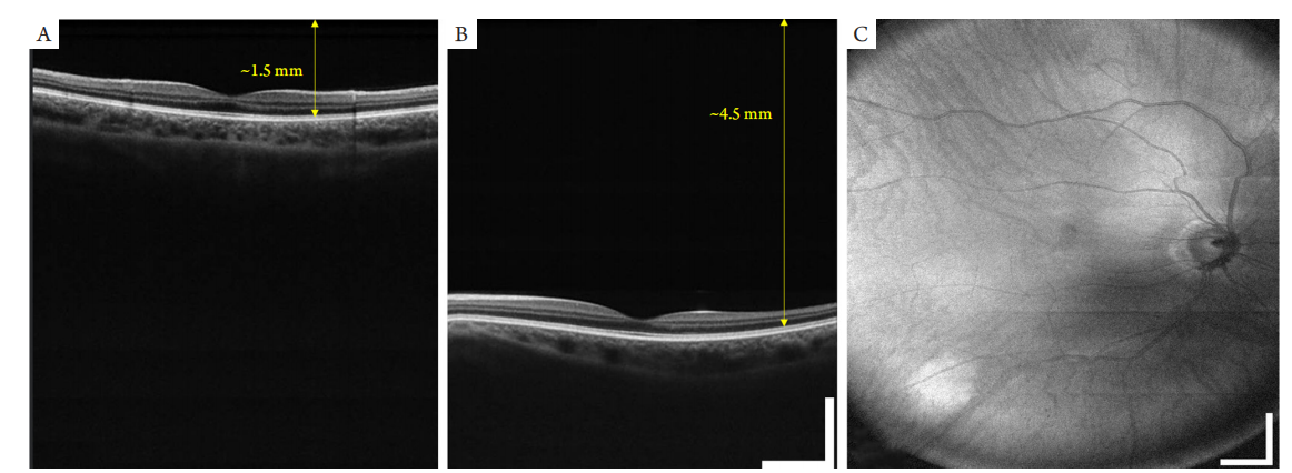
(A,B) OCT images with 6 mm imaging depth. (C) Wide-field SS-OCT results. Images were acquired using a custom-built 200 kHz,1060 nm SS-OCT. Scale bar: 1 mm.
(A) SS-OCT imaging results (with a custom-built 200 kHz, 1 060 nm SS-OCT); (B) SD-OCT imaging results (with a custom-built 36 kHz, 850 nm SD-OCT). Scale bar: 400 μm.
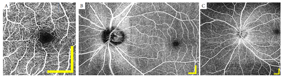
图7 SS-OCTA成像结果
Figure 7 SS-OCTA results
(A) 3 mm × 3 mm 高清OCTA成像,(B) 7 mm × 12 mm的大视场OCTA图像,(C) 10 mm × 10 mm的大视场OCTA图像(使用自研的200 kHz,1 060 nm SS-OCT系统)。比例尺:1 mm。
(A) 3 mm × 3 mm OCTA; (B) 7 mm × 12 mm OCTA; (C) 10 mm × 10 mm OCTA. Images were acquired using a custom-built 200 kHz, 1 060 nm SS-OCT. Scale bar: 1 mm.
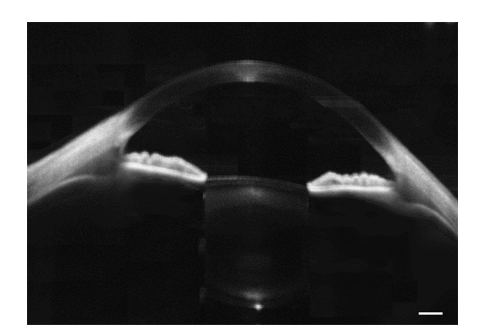
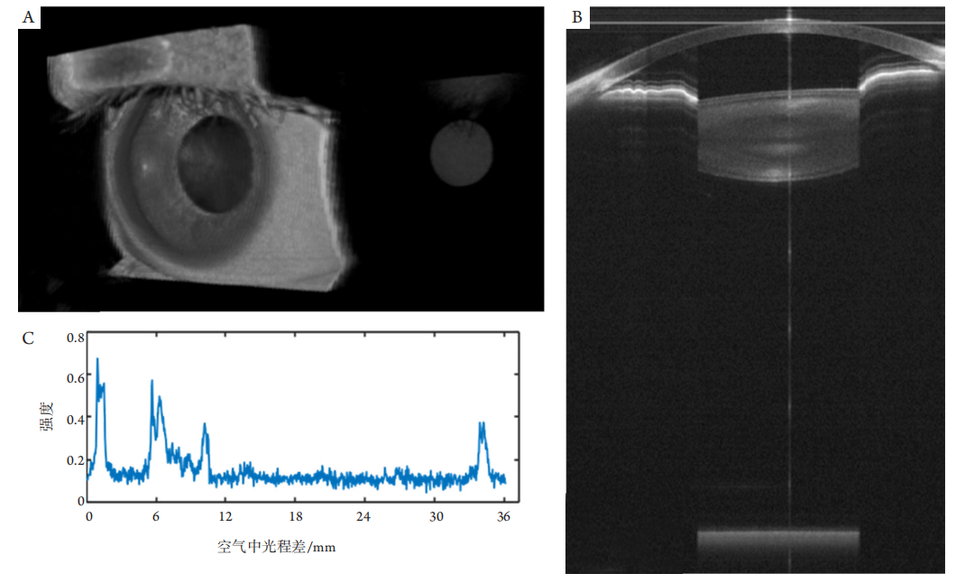
Figure 8 SS-OCT imaging of anterior segment, from the cornea to the posterior surface of the lens (with a custom-built 50 kHz, 1 310 nm SS-OCT). Scale bar: 400 μm
(A) The 3D rendering of the 3D OCT images of the whole eye. (B) One single OCT image of the whole eye. (C) The axial profile of human eye acquired with SS-OCT. Images were acquired using a custom-built 10 kHz, 1 060 nm SS-OCT system. Scale bar: 1 mm.
1. Choma MA, Sarunic MV, Yang CH, et al. Sensitivity advantage of swept
source and Fourier domain optical coherence tomography[ J]. Opt
Express, 2003, 11(18): 2183-2189.
2. de Boer JF, Cense B, Park BH, et al. Improved signal-to-noise ratio
in spectral-domain compared with time-domain optical coherence
tomography[ J]. Opt Lett, 2003, 28: 2067-2069.
3. Leitgeb R, Hitzenberger CK, Fercher AF, et al. Performance of fourier
domain vs. time domain optical coherence tomography[ J]. Opt
Express, 2003, 11: 889-894.
4. Wojtkowski M, Leitgeb R, Kowalczyk A, et al. In vivo human retinal
imaging by Fourier domain optical coherence tomography[ J]. J
Biomed Opt, 2002, 7(3): 457-463.
5. Alibhai AY, Or C, Witkin AJ. Swept source optical coherence
tomography: a review[ J]. Curr Ophthalmol Rep, 2018, 6(1): 7-16.
6. Potsaid B, Jayaraman V, Fujimoto JG, et al. MEMS tunable VCSEL light
source for ultrahigh speed 60kHz-1MHz axial scan rate and long range
centimeter class OCT imaging[M]//Izatt JA, Fujimoto JG, Tuchin VV,
ed al. Optical coherence tomography and coherence domain optical
methods in biomedicine Xvi. Vol 8213. Bellingham: Spie-Int Soc
Optical Engineering, 2012.
7. Drexler W, Fujimoto JG. Optical coherence tomography[C].
Nondestructive Material Testing Using OCT, 2015.
8. Jirauschek C, Huber R. Wavelength shifting of intra-cavity photons:
adiabatic wavelength tuning in rapidly wavelength-swept lasers[ J].
Biomed Opt Express, 2015, 6(7): 2448-2465.
9. Pfeiffer T, Draxinger W, Grill C, et al. Long-range live 3D-OCT at
different spectral zoom levels[C]. SPIE Proceedings (Optical Society
of America, 2017), paper 104160L.
10. Choma MA, Hsu K , Izatt JA. Swept source optical coherence
tomography using an all-fiber 1300-nm ring laser source[ J]. J Biomed
Opt, 2005, 10(4): 6.
11. Jacques SL. Optical properties of biological tissues: a review[ J]. Phys
Med Biol, 2013, 58(11): R37.
12. Marschall S, Klein T, Wieser W, et al. FDML swept source at 1060 nm
using a tapered amplifier[M]//Izatt JA, Fujimoto JG, Tuchin VV, ed al.
Optical coherence tomography and coherence domain optical methods
in biomedicine Xiv. Vol 7554. Bellingham: Spie-Int Soc Optical
Engineering, 2010.
13. Pavlin C, Christopehr D, Burns P. High frequency doppler ultrasound
examination of blood flow in the anterior segment of the eye[ J]. Am J
Ophthalmol, 1998, 126: 597-600.
14. Klein T, Wieser W, Reznicek L, et al. Multi-MHz retinal OCT[ J].
Biomed Opt Express, 2013, 4(10): 1890-1908.
15. Dastiridou A, Bousquet E, Kuehlewein L, et al. Choroidal imaging
with swept-source optical coherence tomography in patients with
birdshot chorioretinopathy: choroidal reflectivity and thickness[ J].
Ophthalmology, 2017, 124(8): 1186-1195.
16. Migacz JV, Gorczynska I, Azimipour M, et al. Megahertz-rate optical
coherence tomography angiography improves the contrast of the
choriocapillaris and choroid in human retinal imaging[ J]. Biomed Opt
Express, 2019, 10(1): 50-65.
17. Farazdaghi MK, Ebrahimi KB. Role of the choroid in age-related
macular degeneration: a current review[ J]. J Ophthalmic Vis Res, 2019,
14(1): 78.
18. Keenan TD, Klein B, Agrón E, et al. Choroidal thickness and vascularity
vary with disease severity and subretinal drusenoid deposit presence
in nonadvanced age-related macular degeneration[ J]. Retina, 2020,
40(4): 632-642.
19. Michalewska Z, Swept-source O. Taking imaging deeper and wider[ J].
Retina Today, 2014, 11: 12.
20. Kumar P, Chawla R , Balakrishnan J, et al. ‘Solitary Idiopathic
Choroiditis’ or a tumour of scleral origin: a case report based
hypothesis[ J]. Med Hypotheses, 2020, 139: 109695.
21. Shinohara K, Moriyama M, Shimada N, et al. Characteristics of
peripapillary staphylomas associated with high myopia determined by
swept-source optical coherence tomography[ J]. Am J Ophthalmol,
2016, 169: 138-144.
22. Kim YC, Koo YH, Jung KI, et al. Impact of posterior sclera on
glaucoma progression in treated myopic normal-tension glaucoma
using reconstructed optical coherence tomographic images[ J]. Invest
Ophthalmol Vis Sci, 2019, 60(6): 2198-2207.
23. Wu J, Gerendas BS, Waldstein SM, et al. Stable registration of
pathological 3D-OCT scans using retinal vessels[C]. In Proceedings of
the Ophthalmic Medical Image Analysis First International Workshop,
Boston, MA, USA, 14 September 2014:1-8.
24. Liu G, Yang J, Wang J, et al. Extended axial imaging range, widefield
swept source optical coherence tomography angiography[ J]. Journal of Biophotonics, 2017, 10(11SI): 1464-1472.
25. Campbell JP, Nudleman E, Yang J, et al. Handheld optical coherence
tomography angiography and ultra-wide-field optical coherence
tomography in retinopathy of prematurity[ J]. JAMA Ophthalmol,
2017, 135(9): 977-981.
26. The International Committee for the Classification of the Late
Stages of Retinopathy of Prematurity. An international classification
of retinopathy of prematurity. II. The classification of retinal
detachment[ J]. Arch Ophthalmol, 1987, 105(7): 906-912.
27. Russell JF, Flynn HW Jr, Sridhar J, et al. Distribution of diabetic
neovascularization on ultra-widefield fluorescein angiography and on
simulated widefield OCT angiography[ J]. Am J Ophthalmol, 2019,
207: 110-120.
28. Schaal KB, Munk MR, Wyssmueller I, et al. Vascular abnormalities
in diabetic retinopathy assessed with swept-source optical coherence
tomography angiography widefield imaging[ J]. Retina, 2019, 39(1):
79-87.
29. Wolff B, Matet A, Vasseur V, et al. En face OCT imaging for the
diagnosis of outer retinal tubulations in age-related macular
degeneration[ J]. J Ophthalmol, 2012, 2012: 542417.
30. Zhang Q, Lee CS, Chao J, et al. Wide-field optical coherence
tomography based microangiography for retinal imaging[ J]. Sci Rep,
2016, 6(1): 1-10.
31. Mwanza JC, Budenz DL. New developments in optical coherence
tomography imaging for glaucoma[ J]. Curr Opin Ophthalmol, 2018,
29(2): 121-129.
32. Bekkers A, Borren N, Ederveen V, et al. Microvascular damage assessed
by optical coherence tomography angiography for glaucoma diagnosis:
a systematic review of the most discriminative regions[ J]. Acta
Ophthalmologica, 2020, 98(6): 537-558.
33. Liu JJ, Witkin AJ, Adhi M, et al. Enhanced vitreous imaging in healthy
eyes using swept source optical coherence tomography[ J]. PLoS One,
2014, 9(7): e102950.
34. Spaide RF. Visualization of the posterior vitreous with dynamic
focusing and windowed averaging swept source optical coherence
tomography[ J]. Am J Ophthalmol, 2014, 158(6): 1267-1274.
35. Itakura H, Kishi S, Li D, et al. Observation of posterior precortical
vitreous pocket using swept-source optical coherence tomography[ J].
Invest Ophthalmol Vis Sci, 2013, 54(5): 3102-3107.
36. Hua R, Ning H. Modified enhanced vitreous imaging modality of
spectral domain optic coherence tomography[ J]. Eye (Lond), 2021,
35(1): 351-352.
37. Liu G, Tan O, Gao SS, et al. Postprocessing algorithms to minimize
fixed-pattern artifact and reduce trigger jitter in swept source optical
coherence tomography[ J]. Optics Express, 2015, 23(8): 9824-9834.
38. Choi W, Mohler KJ, Potsaid B, et al. Choriocapillaris and choroidal
microvasculature imaging with ultrahigh speed OCT angiography[ J].
PLoS One, 2013, 8(12): e81499.
39. Choi W, Moult EM, Waheed NK, et al. Ultrahigh-speed, swept-source
optical coherence tomography angiography in nonexudative age-related
macular degeneration with geographic atrophy[ J]. Ophthalmology,
2015, 122(12): 2532-2544.
40. Miller AR, Roisman L, Zhang Q, et al. Comparison between spectral-
domain and swept-source optical coherence tomography angiographic
imaging of choroidal neovascularization[ J]. Invest Ophthalmol Vis Sci,
2017, 58(3): 1499-1505.
41. Zang P, Liu G, Zhang M, et al. Automated motion correction using
parallel-strip registration for wide-field en face OCT angiogram[ J].
Biomed Opt Express, 2016, 7(7): 2823-2836.
42. Naseripour M, Falavarjani KG, Mirshahi R, et al. Optical coherence
tomography angiography (OCTA) applications in ocular oncology[ J].
Eye (Lond), 2020, 34(9): 1535-1545.
43. Venkateswaran N, Galor A , Wang J, et al. Optical coherence
tomography for ocular surface and corneal diseases: a review[ J]. Eye
Vis (Lond), 2018, 5:13.
44. Maslin JS, Barkana Y, Dorairaj S. Anterior segment imaging in
glaucoma: an updated review[ J]. Indian J Ophthalmol, 2015, 63(8):
630-640.
45. Ang M, Baskaran M, Werkmeister RM, et al. Anterior segment optical
coherence tomography[ J]. Prog Retin Eye Res, 2018, 66: 132-156.
46. McNabb RP, Polans J, Keller B, et al. Wide-field whole eye OCT system
with demonstration of quantitative retinal curvature estimation[ J].
Biomed Opt Express, 2019, 10(1): 338-355.
47. Mak H, Xu G, Leung CKS. Imaging the iris with swept-source optical
coherence tomography: relationship between iris volume and primary
angle closure[ J]. Ophthalmology, 2013, 120(12): 2517-2524.
48. Karnowski K, Kaluzny BJ, Szkulmowski M, et al. Corneal topography
with high-speed swept source OCT in clinical examination[ J]. Biomed
Opt Express, 2011, 2(9): 2709-2720.
49. Mazlin V. Full-field optical coherence tomography for non-contact
cellular-level resolution in vivo human cornea imaging[D]. PSL
Research University, 2019.
50. Zvietcovich F, Nair A, Singh M, et al. Dynamic optical coherence
elastography of the anterior eye: understanding the biomechanics of
the limbus[ J]. Invest Ophthalmol Vis Sci, 2020, 61(13): 7-7.
51. Poddar R, Migacz JV, Schwartz DM, et al. Challenges and advantages in
wide-field optical coherence tomography angiography imaging of the
human retinal and choroidal vasculature at 1.7-MHz A-scan rate[ J]. J Biomed Opt, 2017, 22(10): 106018.
52. Wang Z, Potsaid B, Chen L, et al. Cubic meter volume optical
coherence tomography[ J]. Optica, 2016, 3(12): 1496-1503.
53. Huang J, Chen H, Li Y, et al. Comprehensive comparison of axial length
measurement with three swept-source OCT-based biometers and
partial coherence interferometry[ J]. J Refractive Surg, 2019, 35(2):
115-120.
54. Grulkowski I, Liu JJ, Potsaid B, et al. Retinal, anterior segment and full
eye imaging using ultrahigh speed swept source OCT with vertical-
cavity surface emitting lasers[ J]. Biomed Opt Express, 2012, 3(11):
2733-2751.
55. Grulkowski I, Manzanera S, Cwiklinski L, et al. Swept source optical
coherence tomography and tunable lens technology for comprehensive
imaging and biometry of the whole eye[ J]. Optica, 2018, 5(1): 52-59.
56. Brás JE, Sickenberger W, Hirnschall N, et al. Cataract quantification
using swept-source optical coherence tomography[ J]. J Cataract
Refract Surg, 2018, 44(12): 1478-1481.
'%20fill='white'%20fill-opacity='0.01'/%3e%3cmask%20id='mask0_3477_29692'%20style='mask-type:luminance'%20maskUnits='userSpaceOnUse'%20x='0'%20y='0'%20width='16'%20height='16'%3e%3crect%20id='&%23232;&%23146;&%23153;&%23231;&%23137;&%23136;_2'%20x='16'%20width='16'%20height='16'%20transform='rotate(90%2016%200)'%20fill='white'/%3e%3c/mask%3e%3cg%20mask='url(%23mask0_3477_29692)'%3e%3cpath%20id='&%23232;&%23183;&%23175;&%23229;&%23190;&%23132;'%20d='M14%205L8%2011L2%205'%20stroke='%23333333'%20stroke-width='1.5'%20stroke-linecap='round'%20stroke-linejoin='round'/%3e%3c/g%3e%3c/g%3e%3c/svg%3e)