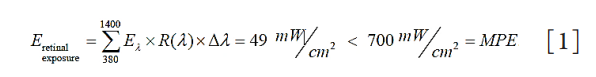
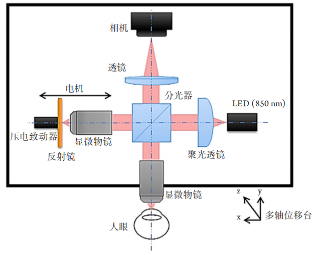
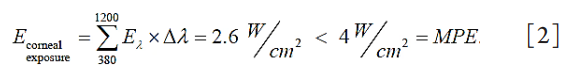
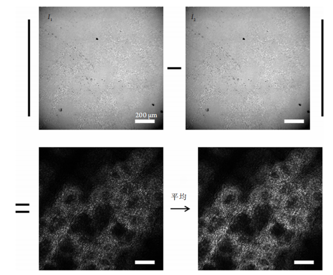
为验证FFOCT系统在活体人眼角膜成像中的质量,我们使用设备对1位27岁健康男性志愿受试者的角膜进行了活体角膜FFOCT成像实验。受试者自愿签署知情同意书,所有程序符合赫尔辛基宣言(1983年)。本研究内容已获得法国人事保护委员会CPP(Comité de Protection de Personnes) Sud-Est III de Bron和国家药品和健康产品安全局ANSM(Agence Nationale de Sécurité du Médicament et des Produits de Santé)批准(批准号:2019-A00942-55)。FFOCT成像实验前,对受试者进行了包括裂隙灯观察、眼压测量等常规的眼科检查。FFOCT成像时,受试者将下颌固定于颌托上,上额紧贴额托,非成像眼注视视标,保持稳定。通过调节设备高度及成像位置,分别对受试者角膜中央各深度进行FFOCT图像采集。相机采集频率设定为550 fps,每个成像深度通过双相位调制模式采集10张FFOCT图像。完成采集后,10张FFOCT图像通过ImageJ的图像配准插件[19]进行眼动导致的平面抖动配准、叠加平均后获得最终的活体角膜FFOCT图像。为了和所采集FFOCT影像作对比,我们利用接触式IVCM设备(HRT II with Rostock cornea module; Heidelberg Engineering,GmbH)采集了受试者角膜中央不同深度的IVCM影像。
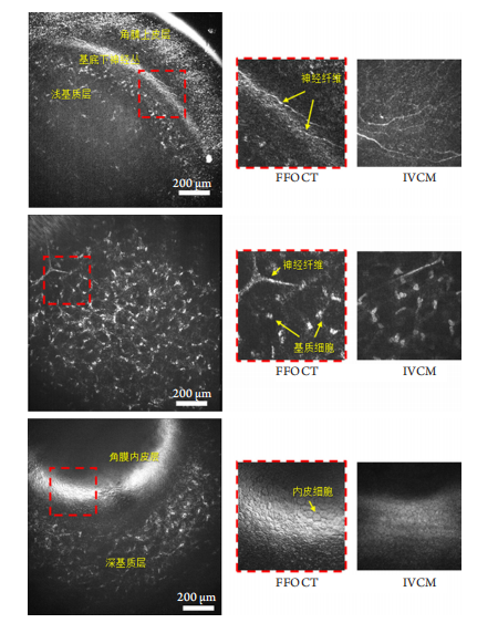
(A) FFOCT image of anterior cornea; (B) FFOCT image of middle cornea; (C) FFOCT image of posterior cornea. Field of view of FFOCT: 1.26 mm×1.26 mm; field of view of IVCM: 400 μm×400 μm.
1. Template matching and slice alignment—ImageJ Plugins[EB/OL].
[2016-06-15]. https://sites.google.com/site/qingzongtseng/template-
matching-ij-plugin.
2. Xiao P, Fink M, Boccara AC. Full-field spatially incoherent illumination
interferometry: a spatial resolution almost insensitive to aberrations[ J].
Opt Lett, 2016, 41(17): 3920-3923.
3. Ghouali W, Grieve K, Bellefqih S, et al. Full-field optical coherence
tomography of human donor and pathological corneas[ J]. Curr Eye
Res, 2015, 40(5): 526-534.
4. Dubois A. Handbook of full-field optical coherence microscopy:
Technology and applications[M]. Singapore: CRC Press, 2016.
5. Beaurepaire E, Boccara AC, Lebec M, et al. Full-field optical coherence
microscopy[ J]. Opt Lett, 1998, 23(4): 244-246.
6. Dorairaj SK, Stewart MW. Clinical applications of AS-OCT for corneal
disease[ J]. Focus, 2014, 102(3):195-207.
7. Yadav R, Kottaiyan R, Ahmad K, et al. Epithelium and Bowman's
layer thickness and light scatter in keratoconic cornea evaluated using
ultrahigh resolution optical coherence tomography[ J]. J Biomed Opt,
2012, 17(11): 116010.
8. Bizheva K, Tan B, MacLelan B, et al. Sub-micrometer axial resolution
OCT for in-vivo imaging of the cellular structure of healthy and
keratoconic human corneas[ J]. Biomed Optics Express, 2017, 8(2):
800-812.
9. Wang J, Abou Shousha M, Perez VL, et al. Ultra-high resolution optical
coherence tomography for imaging the anterior segment of the eye[ J].
Ophthalmic Surg Lasers Imaging, 2011, 42(4): S15-S27.
10. Ramos JL, Li Y, Huang D. Clinical and research applications of
anterior segment optical coherence tomography–a review[ J]. Clin Exp
Ophthalmol, 2009, 37(1): 81-89.
11. Drexler W, Fujimoto JG. Optical coherence tomography: technology
and applications[M]. Springer Science & Business Media, 2008.
12. Huang D, Swanson EA , Lin CP, et al . Optical coherence
tomography[ J]. Science, 1991, 254(5035): 1178-1181.
13. Guthoff RF, Zhivov A, Stachs O. In vivo confocal microscopy, an inner
vision of the cornea–a major review[ J]. Clin Exp Ophthalmol, 2009,
37(1): 100-117.
14. Hau SC, Dart JK, Vesaluoma M, et al. Diagnostic accuracy of microbial
keratitis with in vivo scanning laser confocal microscopy[ J]. Br J
Ophthalmol, 2010, 94(8): 982-987.
15. Villani E, Baudouin C, Efron N, et al. In vivo confocal microscopy of
the ocular surface: from bench to bedside[ J]. Curr Eye Res, 2014,
39(3): 213-231.
16. Ye Y, Jiang H, Zhang H, et al. Resolution of slit-lamp microscopy
photography using various cameras[ J]. Eye Contact Lens, 2013, 39(3):
205-213.
17. Krachmer JH, Mannis MJ, Holland EJ. Cornea[M]. Oxford: Gulf
Professional Publishing, 2005.
18. Pascolini D, Mariotti SP. Global estimates of visual impairment:
2010[ J]. Br J Ophthalmol, 2012, 96(5): 614-618.
19. Oliva MS, Schottman T, Gulati M. Turning the tide of corneal
blindness[ J]. Indian J Ophthalmol, 2012, 60(5): 423.
'%20fill='white'%20fill-opacity='0.01'/%3e%3cmask%20id='mask0_3477_29692'%20style='mask-type:luminance'%20maskUnits='userSpaceOnUse'%20x='0'%20y='0'%20width='16'%20height='16'%3e%3crect%20id='&%23232;&%23146;&%23153;&%23231;&%23137;&%23136;_2'%20x='16'%20width='16'%20height='16'%20transform='rotate(90%2016%200)'%20fill='white'/%3e%3c/mask%3e%3cg%20mask='url(%23mask0_3477_29692)'%3e%3cpath%20id='&%23232;&%23183;&%23175;&%23229;&%23190;&%23132;'%20d='M14%205L8%2011L2%205'%20stroke='%23333333'%20stroke-width='1.5'%20stroke-linecap='round'%20stroke-linejoin='round'/%3e%3c/g%3e%3c/g%3e%3c/svg%3e)