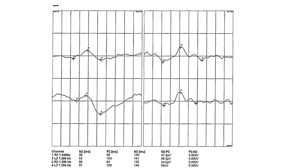
Figure 1 FVEP: indicated that the peak time of the P2 wave in the left eye was slightly delayed relative to the right eye, and the amplitude of the right eye was lower than that in the left eye
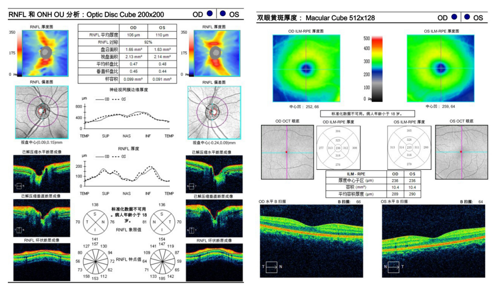
图 2 双眼视盘 + 黄斑区 OCT:未见明显异常
Figure 2 OCT of the optic disc and macula of both eyes showed no obvious abnormality
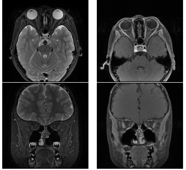
图 3 眼眶 MRI 平扫 + 增强:双眼未见视神经长 T2 及 T1 增强信号
Figure 3 Orbital MRI scan with enhanced enhancement showed that there was no long T2 and T1 enhanced signal of the optic nerve in both eyes
2 讨 论
非器质性视力下降也称为心因性或功能性视力下降,除视力下降外,还可伴有视野缺损,多由于精神心理疾病导致的转换障碍引起,也有部分患者为诈病以获取利益[1-2]。患者主诉视力下降或伴有其他视觉功能障碍,但客观检查均未见任何能够解释症状的器质性疾病[3]。非器质性视力下降患者最常见的视野缺损类型表现为向心性缩小,其中三叶草改变最具特征性[4]。使用动态视野检查可以发现此类患者的视野范围随着检查的进行而逐渐缩小,需要注意与视网膜疾病引起的向心性视野缩小相鉴别[5]。OCT 能够准确测量视盘周围神经纤维厚度以及黄斑区神经节细胞厚度,利用 OCT 对可疑非器质性视力下降患者进行随访,可以进一步明确是否存在视神经损害的证据[6]。
1. 河北省卫健委青年科技课题 (20170942; 20170948)。 This work was supported by the Youth Science and Technology Project of
Hebei Provincial Health and Family Planning Commission (20170942; 20170948) .
参考文献
1. Leavitt JA. Diagnosis and management of functional visual deficits[ J].
Curr Treat Options Neurol, 2006, 8(1): 45-51.
2. Zinkernagel SM, Mojon DS. Distance doubling visual acuity test:
a reliable test for nonorganic visual loss[ J]. Graefes Arch Clin Exp
Ophthalmol, 2009, 247(6): 855-858.
3. 孙平, 冯超逸, 孙兴怀, 等. 儿童非器质性视力下降临床特征分
析[ J]. 中国眼耳鼻喉科杂志, 2022, 22(1): 31-35.
SUN P, FENG CY, SUN XH, et al.Clinical characteristics of nonorganic
visual loss in a cohort of pediatric patients[ J]. Chin J Ophthalmol
Otorhinolaryngol, 2022, 22(1): 31-35.
4. Mu?oz-Hernández AM, Santos-Bueso E, Sáenz-Francés F, et al.
Nonorganic visual loss and associated psychopathology in children[ J].
Eur J Ophthalmol, 2012, 22(2): 269-273.
5. Karagiannis D, Kontadakis G, Brouzas D, et al. Nonorganic visual loss
in a child due to school bullying[ J]. Am J Ophthalmol Case Rep, 2016,
5: 90-91.
6. Taich A, Crowe S, Kosmorsky GS, et al. Prevalence of psychosocial
disturbances in children with nonorganic visual loss[ J]. J AAPOS,
2004, 8(5): 457-461.
7. Scarpina F, Melzi L, Castelnuovo G, et al. Explicit and implicit
components of the emotional processing in non-organic vision loss:
behavioral evidence about the role of fear in functional blindness[ J].
Front Psychol, 2018, 9: 494.
8. 娄华东, 徐鑫彦. 相对性瞳孔传入障碍及其在眼科的应用[ J]. 临
床眼科杂志, 2016, 24(2): 185-188.
LOU HD, XU XY. Quantitative relative afferent pupillary defect
examination and its role in ophthalmology[ J]. J Clin Ophthalmol,
2016, 24(2): 185-188.
9. 田国红, 王敏. 视神经病变与视网膜病变的鉴别要点[ J]. 中国眼
耳鼻喉科杂志, 2014, 14(3): 160-164.
TIAN GH, WANG M. Optic neuropathy versus retinal disease——
tips of differential diagnosis[ J]. Chin J Ophthalmol Otorhinolaryngol,
2014, 14(3): 160-164.
10. Melnick M D, Tadin D, Huxlin K R. Relearning to see in cortical
blindness[ J]. Neuroscientist, 2016, 22(2): 199-212.
12. Gise RA, Heidary G. Update on pediatric optic neuritis[ J]. Curr Neurol
Neurosci Rep, 2020, 20(3): 4.
13. Somers A, Casteels K, Van Roie E, et al. Non-organic visual loss
in children: prospective and retrospective analysis of associated
psychosocial problems and stress factors[ J]. Acta Ophthalmol, 2016,
94(5): e312-e316.
14. Savino P, Daneshmeyer H. Color atlas and synopsis of clinical
ophthalmology-Wills Eye Institute-Neuro-ophthalmology[M]. 2nd Ed.
New York: Lippincott Williams & Wilkins, 2012: 212-215.
15. Kevin R, Sitko, MD, et al. Pitfalls in the use of stereoacuity in the
diagnosis of nonorganic visual loss[ J]. Ophthalmology, 2016, 123(1):
198-202.
16. 田国红, 彭静婷, 张晓君. 非器质性视力下降的临床特征分
析[ J]. 中华眼底病杂志, 2010, 26(4): 379-380.
TIAN Guohong, PENG Jingting, ZHANG Xiaojun. Analysis of clinical
features of non-organic visual acuity loss[ J]. Chin J Ocular Fundus Dis,
2010,26(4): 379-380.
8. Kevin R, Sitko, MD, et
17. 王倩, 姜利斌. 光相干断层扫描在非青光眼性视神经病变中的
应用[ J]. 中华眼底病杂志, 2013, 29(3): 330-334.
WANG Q, JIANG LB. Application of optical coherence tomography in non-glaucoma optic neuropathy[ J]. Chin J Ocular Fundus Dis,
2013(3): 330-334.
18. 项剑, 王旭, 于丽丽, 等. 法医学视野客观评定范式研究——以视
网膜、视神经及高位视路损伤致视野缺损为例[ J]. 中国法医
学杂志, 2018, 33(4): 355-360.
XIANG J, WANG X, YU LL, et al. Exploration of visual field evaluation
methods in forensic science: an analysis of classical cases of visual field
defects caused by injury to the retina, optic nerve, and higher visual
pathway[ J]. Chin J Forensic Med, 2018, 33(4): 355-360.
19. Bruce B B, Newman N J. Functional visual loss[ J]. Neurol Clin, 2010,
28(3): 789-802.
20. 田国红. 非器质性视力下降的诊疗要点[ J]. 中国眼耳鼻喉科杂
志, 2016, 16(1): 68-70.
TIAN Guohong. The main points of diagnosis and treatment of
nonorganic vision loss[ J]. Chin J Ophthal Otorhinolaryngol, 2016,
16(1): 68-70.
21. Wandling L J G, Wandling G R Jr, Marshall M F, et al. Truth-telling
and deception in the management of nonorganic vision loss[ J]. Can J
Ophthalmol, 2016, 51(5): 390-392.
22. 刘琪, 徐柒华, 廖洪斐. 眼外伤后对侧健眼非器质性视力下降的
临床观察[ J]. 中华眼外伤职业眼病杂志, 2020, 42(7): 500-503.
LIU Q, XU QH, LIAO HF. Clinical observation on non-organic visual
acuity reduction of contralateral healthy eye after ocular trauma[ J].
Chin J Ocular trauma Occupation Eye Dis,2020,42(7):500-503.
'%20fill='white'%20fill-opacity='0.01'/%3e%3cmask%20id='mask0_3477_29692'%20style='mask-type:luminance'%20maskUnits='userSpaceOnUse'%20x='0'%20y='0'%20width='16'%20height='16'%3e%3crect%20id='&%23232;&%23146;&%23153;&%23231;&%23137;&%23136;_2'%20x='16'%20width='16'%20height='16'%20transform='rotate(90%2016%200)'%20fill='white'/%3e%3c/mask%3e%3cg%20mask='url(%23mask0_3477_29692)'%3e%3cpath%20id='&%23232;&%23183;&%23175;&%23229;&%23190;&%23132;'%20d='M14%205L8%2011L2%205'%20stroke='%23333333'%20stroke-width='1.5'%20stroke-linecap='round'%20stroke-linejoin='round'/%3e%3c/g%3e%3c/g%3e%3c/svg%3e)