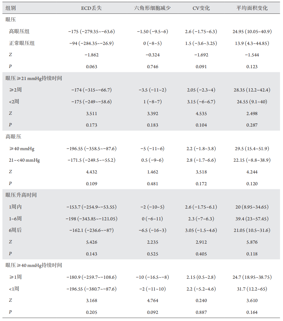
Table 1 Data of patients in the high intraocular pressure group and the normal intraocular pressure group
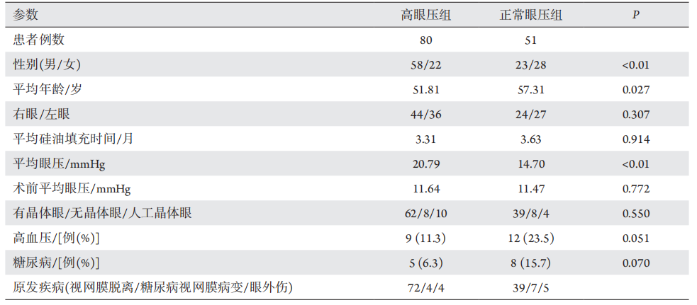
图1 高眼压组和正常眼压组填充硅油前后的ECD对比
Figure 1 ECD comparison of high intraocular pressure group and normal intraocular pressure group before and after silicone oil filling
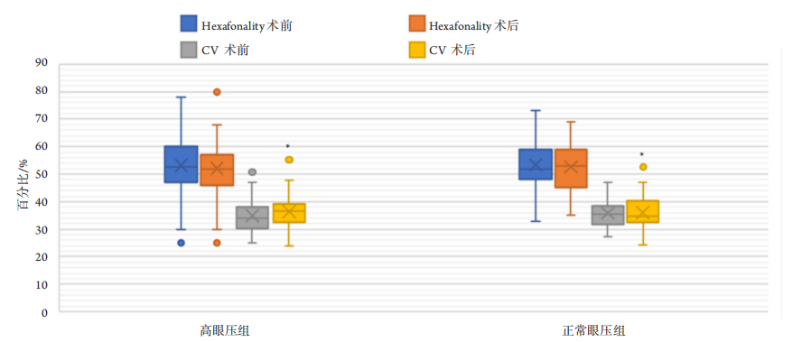
与术前比较,*P<0.05。
Compared with preoperation, *P<0.05.
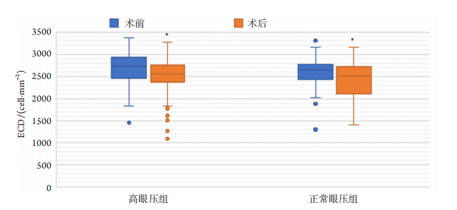
图2高眼压组和正常眼压组填充硅油前后的Hexafonality、CV对比
Figure 2 Comparison of Hexafonality and CV between high intraocular pressure group and normal intraocular pressure group before and after silicone oil filling
与术前比较,*P<0.05。
Compared with preoperation, *P<0.05.
图3 高眼压组和正常眼压组填充硅油前后的平均细胞面积对比
Figure 3 Comparison of Average size of high intraocular pressure group and normal intraocular pressure group before and after silicone oil filling
Table 2 Change of ECD loss, hexagonal cell changes, CV changes, and average endothelial cell area changes between different groups in the high intraocular pressure group [median (P25, P75)]
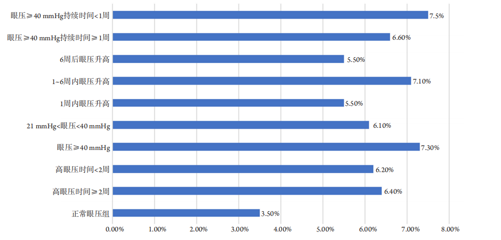
图4 在不同的眼压变化下,角膜内皮细胞的丢失率
figure 4 Loss rate of corneal endothelial cells at difffferent intraocular pressures
1. 2019 年度广东省基础与应用基础研究基金联合基金重点项目 (2019B1515120011)。 This work was supported by the
2019 Guangdong Basic and Applied Basic Research Foundation, China (2019B1515120011).
参考文献
1. Borislav D. Cataract after silicone oil implantation[ J]. Doc Ophthalmol,
1993, 83(1): 79-82.
2. Budenz DL, Taba KE, Feuer WJ, et al. Surgical management of
secondary glaucoma after pars plana vitrectomy and silicone oil
injection for complex retinal detachment[ J]. Ophthalmology, 2001,
108(9): 1628-1632.
3. Friberg TR, Guibord NM, et al. Corneal endothelial cell loss after
multiple vitreoretinal procedures and the use of silicone oil[ J].
Ophthalmic Surg Lasers, 1999, 30(7): 528-534.
4. Scott IU, Flynn HW Jr, Murray TG, et al. Outcomes of complex retinal
detachment repair using 1000- vs 5000-centistoke silicone oil[ J]. Arch
Ophthalmol, 2005, 123(4): 473-478.
5. Goezinne F, Nuijts RM, Liem AT, et al. Corneal endothelial cell density
after vitrectomy with silicone oil for complex retinal detachments[ J].
Retina, 2014, 34(2): 228-236.
6. Bikbova G, Oshitari T, Tawada A, et al. Corneal changes in diabetes
mellitus[ J]. Curr Diabetes Rev, 2012, 8(4): 294-302.
7. Friberg TR, Doran DL, Lazenby FL, et al. The effect of vitreous and
retinal surgery on corneal endothelial cell density[ J]. Ophthalmology,
1984, 91(10): 1166-1169.
8. Melamed S, Ben-Sira I, Ben-Shaul Y, et al. Corneal endothelial changes under induced intraocular pressure elevation: a scanning and
transmission electron microscopic study in rabbits[ J]. Br J Ophthalmol,
1980, 64(3): 164-169.
9. Gagnon MM, Boisjoly HM, Brunette I, et al. Corneal endothelial cell
density in glaucoma[ J]. Cornea, 1997, 16(3): 314-318.
10. Cho SW, Kim JM, Choi CY, et al. Changes in corneal endothelial
cell density in patients with normal-tension glaucoma[ J]. Jpn J
Ophthalmol, 2009, 53(6): 569-573.
11. Sihota R, Lakshmaiah NC, Titiyal JS, et al. Corneal endothelial status
in the subtypes of primary angle closure glaucoma[ J]. Clin Exp
Ophthalmol, 2003, 31(6): 492-495.
12. Guo T, Guo L, Fan Y, et al. Aqueous humor levels of TGFβ2 and SFRP1
in different types of glaucoma[ J]. BMC Ophthalmol, 2019, 19(1): 170.
13. Hu DN, Ritch R, et al. Hepatocyte growth factor is increased in the
aqueous humor of glaucomatous eyes[ J]. J Glaucoma, 2001, 10(3):
152-157.
14. Grus FH, Joachim SC, Sandmann S, et al. Transthyretin and complex
protein pattern in aqueous humor of patients with primary open-angle
glaucoma[ J]. Mol Vis, 2008, 14: 1437-1445.
15. Tham CC, Kwong YY, Lai JS, et al. Effect of a previous acute angle
closure attack on the corneal endothelial cell density in chronic angle
closure glaucoma patients[ J]. J Glaucoma, 2006, 15(6): 482-485.
16. Iwata K, Haruta M, Uehara K, et al. Influence of postoperative lens
status on intraocular pressure and corneal endothelium following
vitrectomy with silicone oil tamponade[ J]. Nippon Ganka Gakkai
Zasshi, 2013, 117(2): 95-101.
'%20fill='white'%20fill-opacity='0.01'/%3e%3cmask%20id='mask0_3477_29692'%20style='mask-type:luminance'%20maskUnits='userSpaceOnUse'%20x='0'%20y='0'%20width='16'%20height='16'%3e%3crect%20id='&%23232;&%23146;&%23153;&%23231;&%23137;&%23136;_2'%20x='16'%20width='16'%20height='16'%20transform='rotate(90%2016%200)'%20fill='white'/%3e%3c/mask%3e%3cg%20mask='url(%23mask0_3477_29692)'%3e%3cpath%20id='&%23232;&%23183;&%23175;&%23229;&%23190;&%23132;'%20d='M14%205L8%2011L2%205'%20stroke='%23333333'%20stroke-width='1.5'%20stroke-linecap='round'%20stroke-linejoin='round'/%3e%3c/g%3e%3c/g%3e%3c/svg%3e)