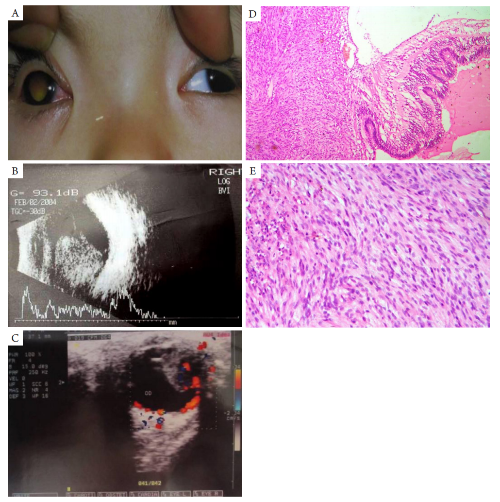
Figure 2 A 2-year-old female patient with chief complaint of right eye leukocoria. vision examination was noncooperation,intraocular pressure was T+1, mixed conjunctival congestion, transparent cornea and lens, normal anterior chamber depth,aqueous flare (?), pupil diameter was 4 mm, loss of pupillary reflex
(A) Yellow-white pupillary reflex could be found in the vitreous cavity. (B) Eye solid masses by B-ultrasonic examination. (C) Blood flow signal on color doppler. The clinical diagnosis was RB. (D) Tumor aroused from transitional zone of pars plana adjacent to ora serrata (HE,×40). (E) Cells became fusiform in shape and lose apical-basal polarity. Lots of mitotic nucleus could be found high power microscopic (HE,×100). Immunohistochemical staining for NSE, S100, CD117, Vimentin, GFAP, Keratin (+), and for SMA, BCL-2, Actin, Desmin, NF, SY(?). Pathological diagnosis: tumor originating from ciliary body interstitial cells.
2.2.3 与眼内炎症混淆
临床将RB误诊为眼内炎2例,患者病理特点为肿瘤小,坏死明显,沿视网膜表面和视网膜内生长。1例已明显侵犯虹膜,玻璃体腔内肿瘤沿锯齿缘附近视网膜表面生长约0.5 mm ×2 mm大小(图3)。另1例前节未见炎症反应,但玻璃体腔可见大量白色混浊和钙化。眼内炎误诊为RB共3例,本组特点病史时间较长,1例真菌性眼内炎病史1年,陈旧性穿通伤病史5年,剩余1例眼内非特异性炎症病史较短只有1个月,但有明显钙化。本组均存在眼后节无法视及的情况,2例是因为前房出血,1例因晶状体后增殖膜。本组患者年龄差异没有统计学意义(t=1.315,P=0.281)。
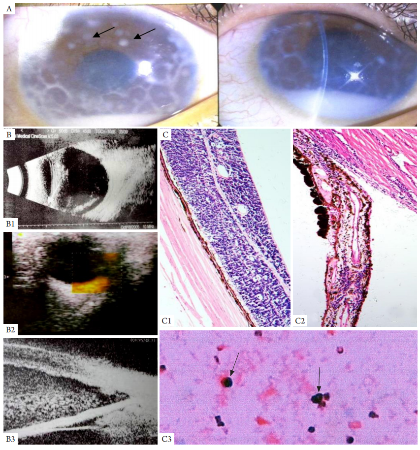
图3 患者,女,9岁。2005年因左眼红痛伴视力下降3个月为主诉入院
Figure 3 A 9-year-old female patient with chief complaint of ocular redness, pain and visual impairment on left eye about 3 months
The clinical diagnosis was uveitis. Visual acuity was 0.1 and unable corrected, intraocular pressure was 37 mmHg. Tumor cells floated in the anterior chamber (A, black arrow). (B1) High density fibrosis by B-ultrasonic examination. (B2) None blood flow signal on color doppler.(B3) Serious anterior chamber exudations on UBM. The results of bacterial culture and medical sensitivity experiments were negative. Visual acuity improved to 0.3 with douche of anterior chamber, patient discharged with improvement. But one month later, patients had been re-admitted to the hospital for relapse of focus. RB cells were found in aqueous humor after paracentesis (shown as arrows, C3: aqueous humor smear; HE, ×200), and the patient was diagnosed with RB and treated by enucleation. Postoperative pathological section shown the tumor cells were growth along the retina (C1: HE, ×100; B1), and obviously infiltrating iris and choroid (C2: HE, ×100; B3).
1. 林壮玲, 张平. 视网膜母细胞瘤诊疗的研究进展[ J]. 眼科学报,
2020, 35(4): 271-278.
LIN ZL, ZHANG P. Research progress on the diagnosis
and treatment of retinoblastoma[ J]. Yan Ke Xue Bao, 2020, 35(4):
271-278.
2. 吴中耀, 易玉珍, 吴德正, 等. 视网膜母细胞瘤的A和B型超声
诊断——113例超声探查结果的初步分析[ J]. 眼科学报, 1985,
1(1): 59-63.
WU ZY, YI YZ, WU DZ, et al. A- and B-scan in
diagnosis of retinoblastoma—a preliminary analysis of ultrasonic
examination in 113 patients[ J]. Yan Ke Xue Bao, 1985, 1(1): 59-63.
3. 吴中耀, 侯光辉. 52例视网膜母细胞瘤的CT扫描[ J]. 眼科学报,
1993, 9(2): 97-100.
WU ZY, HOU GH. CT findings in 52 cases of
retinoblastoma[ J]. Yan Ke Xue Bao, 1993, 9(2): 97-100.
4. 顾华丽, 王一卓, 黄东生, 等. CT显示瘤体内无钙化的儿童视网
膜母细胞瘤20例临床分析[ J]. 中华实用儿科临床杂志, 2017,
32(3): 187-190.
GU H, WANG YZ, HUANG DS, et al. Clinical analysis
of 20 cases of retinoblastoma without calcification on CT scan[ J].
Journal of Applied Clinical Pediatrics, 2017, 32(3): 187-190.
5. 何为民, 夏瑞南, 黄薇, 等. 359例眼内肿瘤的临床病理和误诊分
析[ J]. 中华眼底病 2002, 18(1): 28-30.
HE WM, XIA RN, HUANG W, et al. Clinical histopathological
and misdiagnostic analysis of 359 cases of intraocular tumors[ J].
Chinese Journal of Ocular Fundus Diseases, 2002, 18(1): 28-30.
6. Huang S, Rutar T, Bloomer M, et al. Analysis of clinical misdiagnoses in
children treated with enucleation[ J]. Arch Ophthalmol, 2010, 128(8):
1009-1013.
7. Howard GM, Ellsworth RM. Differential diagnosis of retinoblastoma.
A statistical survey of 500 children. I. Relative frequency of the lesions
which simulate retinoblastoma[ J]. Am J Ophthalmol, 1965, 60(4):
610-618.
8. Chawla B, Khurana S, Sen S, et al. Clinical misdiagnosis of
retinoblastoma in Indian children[ J]. Br J Ophthalmol, 2014, 98(4):
488-493.
9. Robertson DM, Campbell RJ. Analysis of misdiagnosed retinoblastoma
in a series of 726 enucleated eyes[ J]. Mod Probl Ophthalmol, 1977, 18:
156-159.
10. Margo CE, Zimmerman LE. Retinoblastoma: the accuracy of clinical
diagnosis in children treated by enucleation[ J]. J Pediatr ophthalmol
Strabismus, 1983, 20(6): 227-229.
11. Gerrish A, Stone E, Clokie S, et al. Non-invasive diagnosis of
retinoblastoma using cell-free DNA from aqueous humour[ J]. Br J
Ophthalmol, 2019, 103(5): 721-724.
12. Kogan L, Boniuk M. Causes for enucleation in childhood with special
reference to pseudogliomas and unsuspected retinoblastomas[ J]. Int
Ophthalmol Clin, 1962, 2: 507-524.
14. Sanders TE. Pseudoglioma: a clinicopathologic study of fifteen
cases[ J]. Trans Am Ophthalmol Soc, 1950, 48: 575-614.
15. 刘羽阳, 李琬悦, 张家墅, 等. 神经纤维瘤病Ⅱ型误诊为双侧视网
膜母细胞瘤一例[ J]. 中华耳科学杂志, 2020, 18(1): 45-48.
LIU YY, LI WY, ZHANG JS, et al. One case of bilateral
retinoblastoma was wrongly diagnosed as neurofibromatosis type 2[ J].
Chinese Journal of Otology, 2020, 18(1): 45-48.
16. 周思睿, 闵晓雪, 陶韵涵, 等. 66例视网膜母细胞瘤患儿临床资料
分析[ J]. 中华眼底病杂志, 2020, 36(1): 42-45.
ZHOU SR, MIN XX, TAO YH, et al. Clinical analysis of
66 patients of retinoblastoma[ J]. Chinese Journal of Ocular Fundus
Diseases, 2020, 36(1): 42-45.
'%20fill='white'%20fill-opacity='0.01'/%3e%3cmask%20id='mask0_3477_29692'%20style='mask-type:luminance'%20maskUnits='userSpaceOnUse'%20x='0'%20y='0'%20width='16'%20height='16'%3e%3crect%20id='&%23232;&%23146;&%23153;&%23231;&%23137;&%23136;_2'%20x='16'%20width='16'%20height='16'%20transform='rotate(90%2016%200)'%20fill='white'/%3e%3c/mask%3e%3cg%20mask='url(%23mask0_3477_29692)'%3e%3cpath%20id='&%23232;&%23183;&%23175;&%23229;&%23190;&%23132;'%20d='M14%205L8%2011L2%205'%20stroke='%23333333'%20stroke-width='1.5'%20stroke-linecap='round'%20stroke-linejoin='round'/%3e%3c/g%3e%3c/g%3e%3c/svg%3e)