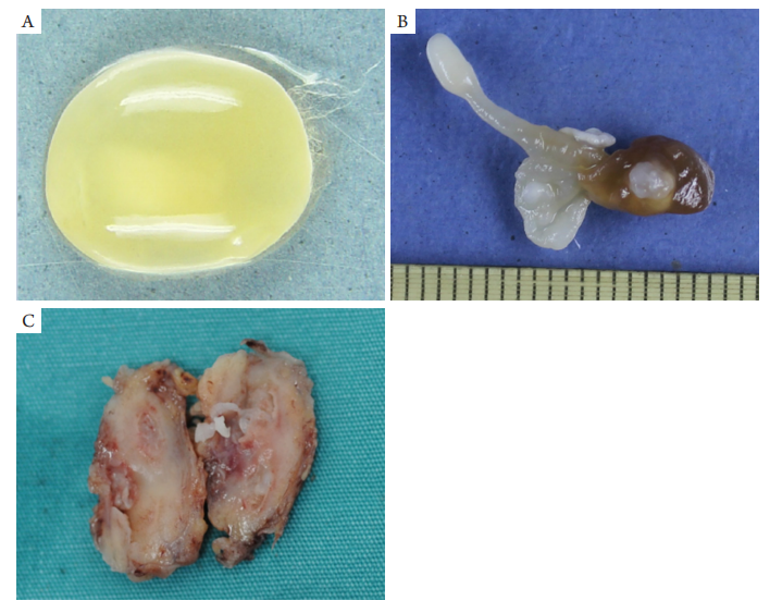
4例大体标本为虫体,其中3例为灰白色或淡黄色卵圆形囊性虫体,分别取自颞上方球结膜下、视盘颞下区视网膜下与下直肌,大小6 mm ×2 mm × 2 mm~10 mm × 6 mm × 4 mm,表面光滑,部分半透明,可透见白色头节(图1A),囊内充满液体;1例为不规则形状虫体,取自内直肌及周围,大小9 mm × 3 mm × 3 mm,前部细长可见白色头节,后部有囊样和囊皮样组织(图1B);仔细查找头节内有4个吸盘、1个顶突和2圈小钩。1例为眼球摘除术标本,切开眼球见视网膜完全性脱离,视网膜下为凝胶状黄绿色胶冻物,下方近中央处有一卵圆形白色物,大小约3 mm × 3 mm × 2 mm。2例取自眶前部,其中1例为黄白色无包膜组织,切面实性,黄白色,均质,边缘可见可疑囊性小结节;另1例为灰红色组织,切面实性,灰红色,质偏嫩,灶区见囊性病变,内见乳白色条索状虫体,当时未注意是虫体及查找头节(图1C)。
图1 眼囊尾蚴病大体表现
Figure 1 The gross presentation of specimen of ocular cysticercosis
(A) The bladderworm was light yellow, ovoid-shaped and translucent vesicle-like structures and the white scolex was visible; (B) The bladderworm was irregular shape; (C) The milky-white striped bladderworm was seen in the section of grayish-red specimen.
(A) Outer surface of cyst wall is distorted and wavy, and there is a “ridge-like” protrusion structure in the internal cavity space of the bladderworm (HE, ×100); (B) High magnification shows a large number of light blue calcareous corpuscles in the “ridge-like” protrusion structure of the internal cavity space of the bladderworm (HE, ×200); (C) The body wall of the bladderworm is divided into 3 parts from outside to inside: cortex, nuclear layer and stromal layer. The stromal layer contains a large number of light blue calcareous corpuscles(HE, ×100); (D) The suckers (short arrow) and small hooks (long arrow) are seen in the scolex (HE, ×200); (E) Intravitreal necrotic cysticercus causes granulomatous retinitis, preretinal inflammatory fibrous membrane, and tractional retinal detachment (HE, ×10);(F) The bladderworm is necrotic and disintegrating. Numerous blueviolet ovoid-shaped calcareous corpuscles are found in necroses whose surroundings are granulomatous inflammation with eosinophils (HE, ×200); (G) High magnification shows dark blue calcareous corpuscles in the bladderworm stroma (HE, ×400); (H) A large number of lymphocytes, plasma cells and eosinophils are infiltrated in tissues surrounding bladderworm and eosinophilic microabscess are formed (HE, ×40); (I) High magnification shows a small amount of dark blue calcareous corpuscle and smooth muscle in the bladderworm stroma (HE, ×200).
1. Rizvi SA, Saleh AM, Frimpong H, et al. Neurocysticercosis: A case
report and brief review[ J]. Asian Pac J Trop Med, 2016, 9(1): 100-102.
2. 李焕璋, 臧新中, 钱门宝, 等. 囊尾蚴病流行现况及研究进展[ J].
中国血吸虫病防治杂志, 2018, 30(1): 99-103.
LI HZ, ZANG XZ, QIAN MB, et al. Current
status and research progress of cysticercosis[ J]. Chinese Journal of
Schistosomiasis Control, 2018, 30(1): 99-103.
3. Joob B, Wiwanitkit V. Clinical profile of ocular cysticercosis[ J]. Ocul
Immunol Inflamm. 2018, 26(4): 558.
4. 吴秀丽. 猪囊尾蚴病影像学表现分析[ J]. 中国临床研究, 2014,
27(11): 1405-1407.
WU XL. Analysis of imaging manifestations of cysticercosis[ J].
Chinese Journal of Clinical Research, 2014, 27(11): 1405-1407.
5. Myron Yanoff, Joseph W. Sassani. Ocular pathology[M]. 8th edition.
New York: Elsevier Inc., 2020: 95-96.
6. Lombardo J. Subretinal cysticercosis[ J]. Optom Vis Sci, 2001, 78(4):
188-194.
7. Pushker N, Bajaj MS, Betharia SM. Orbital and adnexal cysticercosis[ J].
Clin Exp Ophthalmol, 2002, 30(5): 322-333.
8. Agrawal S, Ranjan S, Mishra A. Ocular myocysticercosis: An unusual
case of ptosis[ J]. Nepal J Ophthalmol, 2013, 5(2): 279-281.
9. Rath S, Honavar SG, Naik M, et al. Orbital cysticercosis: Clinical
manifestations,diagnosis,management,and outcome [ J] .
Ophthalmology, 2010, 117(3): 600-605.
10. Kruger-Leite E, Jalkh AE, Quiroz H, et al. Intraocular cysticercosis[ J].
Am J Ophthalmol, 1985, 99(3): 252-257.
11. 李亚明, 岳东雷, 朱淮成. 70例猪囊尾蚴病眼部损害的临床分
析[ J]. 中国病原生物学杂志, 2006, 1(2): 161.
YI YM, YUE DL, ZHU HC. Clinicopathological
analysis of 70 cases of ocular injury of ocular cysticercosis[ J]. Journal of
Pathogen Biology, 2006, 1(2): 161.
13. Madan VS, Dhamija RM, Gill HS, et al. Optic nerve cysticercosis: A
case report[ J]. J Neurol Neurosurg Psychiatry, 1991, 54(5): 470-471.
14. Sundaram PM, Jayakumar N, Noronha V. Extraocular muscle
cysticercosis - a clinical challenge to the ophthalmologists[ J]. Orbit,
2004, 23(4): 255-262.
15. Verma R, Jaiswal A. Multiple brain parenchymal neurocysticercosis
with extraocular muscle cysticercosis affecting levator palpebral
superioris and superior rectus complex: An unusual association[ J].
BMJ Case Rep, 2013, 2013: bcr2012007421.
16. 张忠志. 脑囊虫病的影像学诊断[ J]. 中国社区医师(医学专业),
2011, 13(17): 207-208.
ZHANG ZZ. Imaging diagnosis of cerebral cysticercosis[ J].
Chinese Community Doctors, 2011, 13(17): 207-208.
17. 李德本. 脑猪囊尾蚴病的临床病理诊断[ J]. 临床与实验病理学
杂志, 2002, 18(5): 474-476.
LI DB. Clinicopathologic diagnosis of cerebral cysticercosis[J]. Chinese
Journal of Clinical and Experimental Pathology, 2002, 18(5): 474-476.
18. Wang JY, Cui G, Chen HT, et al. Current epidemiological profile and
features of visceral leishmaniasis in People's Republic of China[ J].
Parasit Vectors, 2012, 5: 31.
19. Khin SS, Kitazawa R , Htet K , et al. Intestinal inflammator y
pseudotumor caused by taeniasis: Calcareous corpuscles as a diagnostic
clue[ J]. Pathol Int, 2013, 63(3): 193-194.
20. 崔晶, 王中全. 绦虫[M]//周庭银, 章强强. 临床微生物学诊断与
图解(下册). 第4版. 上海: 上海科学技术出版社, 2017: 835-837.
CUI J, WANG ZQ. Tapeworm [M]//ZHOU Tingyin,
ZHANG QQ. Diagnosis and illustration of clinical
microbiology (volume two). 4th edition. Shanghai: Shanghai Scientific
and Technical Publishers, 2017: 835-837.
'%20fill='white'%20fill-opacity='0.01'/%3e%3cmask%20id='mask0_3477_29692'%20style='mask-type:luminance'%20maskUnits='userSpaceOnUse'%20x='0'%20y='0'%20width='16'%20height='16'%3e%3crect%20id='&%23232;&%23146;&%23153;&%23231;&%23137;&%23136;_2'%20x='16'%20width='16'%20height='16'%20transform='rotate(90%2016%200)'%20fill='white'/%3e%3c/mask%3e%3cg%20mask='url(%23mask0_3477_29692)'%3e%3cpath%20id='&%23232;&%23183;&%23175;&%23229;&%23190;&%23132;'%20d='M14%205L8%2011L2%205'%20stroke='%23333333'%20stroke-width='1.5'%20stroke-linecap='round'%20stroke-linejoin='round'/%3e%3c/g%3e%3c/g%3e%3c/svg%3e)