患者全身情况尚可,未见其他病变。眼部检查:右眼视力1.0,外眼和眼底检查未见异常;左眼视力指数/20 cm,眼球突出并向鼻下方移位,眼球突出度9 mm,眼球上转不能,内、外、下转受限严重,眼球运动受限,上眼睑高度红肿且有压痛,睑裂闭合不全,结膜高度充血水肿,角膜上皮有点片状灰白色混浊,虹膜正常,瞳孔反射(+),晶状体透明,眼底窥不清。左眼眶外上缘可触及质地较硬的肿物、约呈2 cm × 3 cm,边界不清,有压痛;眶压(++) (图1)。彩色超声波检查显示左眼眶外上方占位性病变,约呈3.08 cm × 1.81 cm,形态不规则,边界不清晰,内回声不均匀,有丰富血流信号,眼球壁受压明显。复查眼眶CT,显示左眼眶泪腺增大,眼眶内不规则团块影,边界欠清,内部密度不均,增强后可见均匀强化,邻近外侧眶骨壁有虫蚀状缺损,眼球受压(图2)。当地MRI显示,眶外上泪腺区椭圆形占位,T1WI为中低信号,T2WI为中高信号(图3)。结合患者的临床表现和天津市眼科医院CT检查结果,考虑为左眼眶部泪腺区占位性病变,不能排除恶性肿瘤。2007年10月12日,患者于全身麻醉下行左眼眶肿物切除术。术中发现眼眶上方充满灰白色肿物,无明显包膜,并侵及提上睑肌和眶上神经。
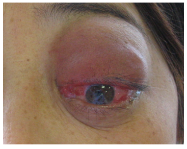
图1 患者外眼像显示左眼上睑高度红肿、结膜充血和眼球突出
Figure 1 Clinical photograph of the patient presenting with a swollen red upper eyelid, conjunctival congestion and proptosis
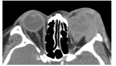
图2 横轴位CT图像显示左眼眶内不规则团块影,侵及邻近软组织和外侧骨壁
Figure 2 Axial CT demonstrating irregular mass involving soft tissue and bone in the left orbit
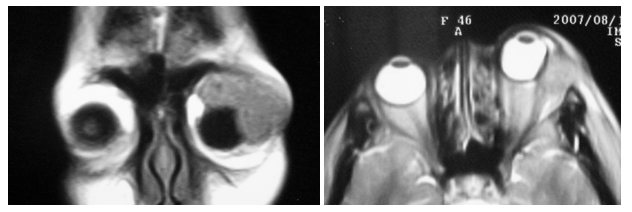
图3 MRI图像显示左眼眶外上方肿物,T1中低信号,T2中高信号
Figure 3 MRI showing mass with iso/hypo-intensity on T1WI, and iso/hyper-intensity on T2WI in the superotemporal aspect of the orbit
1.2 病理检查
送检肿物呈碎块状,堆积面积呈4 cm × 4 cm ×1.7 cm,灰黄色。镜下肿瘤细胞排列成岛状、片块状或条索状,瘤细胞间有大量分化成熟的淋巴细胞和浆细胞浸润。肿瘤细胞体积较大、多边形或梭形、细胞界限清楚或不清楚,细胞质嗜酸,胞核呈椭圆形,核染色质较淡,有明显的核仁。瘤细胞有明显的异型性,可见较多的病理性核分裂像(图4)。肿瘤细胞侵及眼外肌纤维。部分区域可见残留的泪腺组织,腺泡上皮基本正常,腺泡间有较多的分化成熟的淋巴细胞和浆细胞浸润,未见到良性上皮细胞岛状增生。免疫组织化学染色结果显示:肿瘤细胞细胞角蛋白AE1/AE3和上皮膜抗原(epithelial membrane antigen,EMA)呈阳性表达,增殖细胞核抗原(proliferating cell nuclear antigen,PCNA)阳性细胞>50%,细胞增殖抗原Ki67阳性细胞>30% (图5);肿瘤间质中的淋巴细胞分别对白细胞分化抗原3(cluster of differentiation 3,CD3) 或白细胞分化抗原20(cluster of differentiation 20,CD20)呈阳性表达。病理诊断:左眼眶部泪腺淋巴上皮癌。2014年6月对患者进行电话随访,患者术后在当地医院接受了进一步放射治疗(以下简称放疗)和化学治疗(以下简称化疗)(放疗方案不详;化疗药物为长春新碱、环磷酰胺和卡铂,具体剂量不详),目前除左侧颌下淋巴结肿大外,无眼眶内肿物复发或其他全身病变。
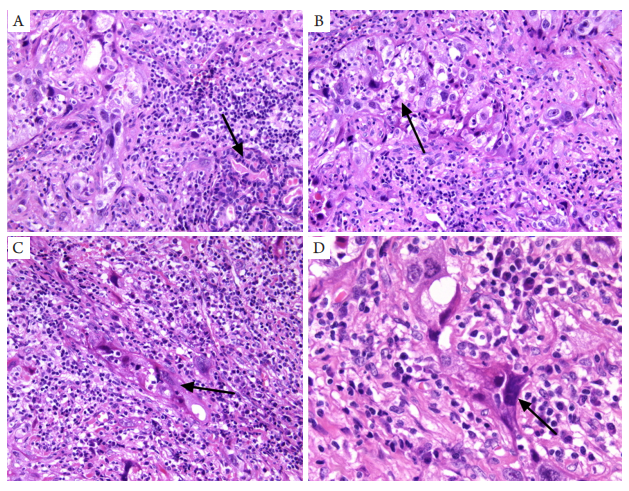
图4 泪腺淋巴上皮癌的组织病理学特征(HE染色)
Figure 4 Histopathological characteristics of lymphoepithelial carcinoma of orbital lacrimal gland (HE staining)
(A) The remaining lacrimal gland tissue could be found in the tumor (arrow, ×200); (B-D) Tumor cells with obvious atypia were arranged in lumps and strips, which were surrounded by a lot of lymphocytes and plasma cells (arrows indicate tumor cells; B-C, ×200; D, ×400).
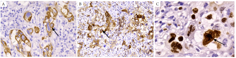
图5 泪腺淋巴上皮癌的免疫组织化学特征(免疫组织化学染色)
Figure 5 Immunohistochemical characteristics of lymphoepithelial carcinoma of orbital lacrimal gland (immunohistochemistry staining)
(A) AE1/AE3 was positive in tumor cells (arrow; ×400); (B) EMA was positive in tumor cells (arrow; ×200); (C) Ki67 was positive in a part of tumor cells (arrow; ×400).
1. Leon Barnes, John W. Eveson, Peter Reichart, 等. 头颈部肿瘤病理
学和遗传学[M]. 刘红刚, 高岩, 主译. 北京:人民卫生出版社,
2006: 292-294.
Leon Barnes, John W. Eveson, Peter Reichart, et al. Pathology
and genetics of head and neck tumours[M]. Translated by LIU
HG, GAO Y. Beijing: People’s Health Publishing House,
2006: 292-294.
2. Bloching M, Hinze R, Berghaus A. Lymphepithelioma-like carcinoma
of the lacrimal gland[ J]. Eur Arch Otorhinolaryngol, 2000, 257(7):
399-401.
3. Rao NA, Kaiser E, Quiros PA, et al. Lymphoepithelial carcinoma of the
lacrimal gland[ J]. Arch Ophthalmol, 2002, 120(12): 1745-1748.
4. Liu YT, Lin CI, Kao SC, et al. Lymphoepithelial carcinoma of the
lacrimal sac[ J]. Eye (Lond), 2009, 23(7): 1612-1615.
5. Khouchani M, Elmorabit B, Elomrani A, et al. Lymphoepithelial
carcinoma of the conjunctiva: an unusual location[ J]. Cancer
Radiother, 2012, 16(3): 219-221.
6. Whaley RD, Carlos R, Bishop JA, et al. Lymphoepithelial carcinoma
of salivary gland EBV-association in endemic versus non-endemic
patients: A report of 16 cases[ J]. Head Neck Pathol, 2020, 14(4):
1001-1012.
7. Leiser Y, Peled M, Wolff A, et al. Lymphoepithelial carcinoma--Review
of the treatment modalities and report of a rare case in the sublingual
gland[ J]. J Oral Maxillofac Surg, 2014, 72(4): 823-828.
8. Zhang G, Tang J, Pan Y, et al. CT features and pathologic characteristics
of lymphoepithelial carcinoma of salivary glands[ J]. Int J Clin Exp
Pathol, 2014, 7(3): 1004-1011.
9. 吴静, 林素暇, 杨秋霞, 等. 涎腺淋巴上皮样癌的影像表现[ J]. 中
华放射学杂志, 2012, 46(8): 747-749.
WU J, LIN SX, YANG QX, et al. Imaging features of
lymphoepithelioid carcinoma of salivary gland[ J]. Chinese Journal of
Radiology, 2012, 46(8): 747-749.
10. 梅银娥, 吴湣, 周春泉, 等. 下颌下腺淋巴上皮癌1例报告及文献
复习[ J]. 口腔颌面外科杂志, 2014, 24(2): 158-161.
MEI Y’e, WU M, ZHOU CQ, et al. Lymphoepithelial
carcinoma of submandibular gland: A case report and literature
review[ J]. Journal of Oral and Maxillofacial Surgery, 2014, 24(2):
158-161.
'%20fill='white'%20fill-opacity='0.01'/%3e%3cmask%20id='mask0_3477_29692'%20style='mask-type:luminance'%20maskUnits='userSpaceOnUse'%20x='0'%20y='0'%20width='16'%20height='16'%3e%3crect%20id='&%23232;&%23146;&%23153;&%23231;&%23137;&%23136;_2'%20x='16'%20width='16'%20height='16'%20transform='rotate(90%2016%200)'%20fill='white'/%3e%3c/mask%3e%3cg%20mask='url(%23mask0_3477_29692)'%3e%3cpath%20id='&%23232;&%23183;&%23175;&%23229;&%23190;&%23132;'%20d='M14%205L8%2011L2%205'%20stroke='%23333333'%20stroke-width='1.5'%20stroke-linecap='round'%20stroke-linejoin='round'/%3e%3c/g%3e%3c/g%3e%3c/svg%3e)