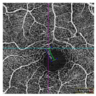
Figure 1 The navigation line is located in the center of the image as the fixation point, measuring the distance between the fixation point and the center of the macular fovea
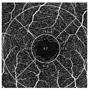
图2 Angio-PlexTM软件自动分析黄斑中心凹血流数据
Figure 2 Automatic blood flow analysis of the superficial retinal vascular plexus macular fovea data using Angio-plex software
1. 爱尔眼科医院集团科研基金(AFQ1708D2)。This work was supported by Science Research Foundation of Aier Eye Hospital Group, China (AFQ1708D2).
2. 爱 尔 眼 科 医 院 集 团 科 研 基 金 (AFQ1708D2)。This work was supported by Science Research Foundation of Aier Eye Hospital Group, China (AFQ1708D2)
参考文献
1. Koylu MT, Ozge G, Kucukevcilioglu M, et al. [J]. Semin Ophthalmol, 2017, 32(5): 553-558.
2. Godts DJM, Mathysen DGP. Amblyopia with eccentric fixation: is inverse occlusion still an option?[J]. J Binocul Vis Ocul Motil, 2019, 69(4): 131-135.
3. 赵堪兴. 斜视弱视学[M]. 北京: 人民卫生出版社, 2011.
4. Strabismus and amblyopia[M]. Beijing: People's Medical Publishing House, 2011.
5. Lei J, Durbin MK, Shi Y, et al. Repeatability and reproducibility of superficial macular retinal vessel density measurements using optical coherence tomography angiography en face images[J]. JAMA Ophthalmol, 2017, 135(10): 1092-1098.
6. Pilotto E, Frizziero L, Crepaldi A, et al. Repeatability and reproducibility of foveal avascular zone area measurement on normal eyes by different optical coherence tomography angiography instruments[J]. Ophthalmic Res, 2018, 59(4): 206-211.
7. Oh IK, Oh J, Kim SW, et al. Fixation and photoreceptor integrity in optical coherence tomography[J]. Optom Vis Sci, 2012, 89(7): E1000-E1008.
8. Nakamoto Y, Takada R, Tanaka M, et al. Quantification of eccentric fixation using spectral-domain optical coherence tomography[J]. Ophthalmic Res, 2018, 60(4): 231-237.
9. García-García Má, Belda JI, Schargel K, et al. Optical coherence tomography in children with microtropia[J]. J Pediatr Ophthalmol Strabismus, 2018, 55(3): 171-177.
10. Yilmaz I, Ocak OB, Yilmaz BS, et al. Comparison of quantitative measurement of foveal avascular zone and macular vessel density in eyes of children with amblyopia and healthy controls: an optical coherence tomography angiography study[J]. J AAPOS, 2017, 21(3): 224-228.
11. Sobral I, Rodrigues TM, Soares M, et al. OCT angiography findings in children with amblyopia[J]. J AAPOS, 2018, 22(4): 286-289.
12. Araki S, Miki A, Goto K, et al. Foveal avascular zone and macular vessel density after correction for magnification error in unilateral amblyopia using optical coherence tomography angiography[J]. BMC Ophthalmol, 2019, 19(1): 171.
13. Demirayak B, Vural A, Onur IU, et al. Analysis of macular vessel density and foveal avascular zone using spectral-domain optical coherence tomography angiography in children with amblyopia[J]. J Pediatr Ophthalmol Strabismus, 2019, 56(1): 55-59.
14. Zhang T, Xie S, Liu Y, et al. Effect of amblyopia treatment on macular microvasculature in children with anisometropic amblyopia using optical coherence tomographic angiography[J]. Sci Rep, 2021, 11(1): 39.
16. Advances in OCTA measurement of retinal vascular density and the foveal avascular zone in myopia[J]. Chinese Journal of Optometry Ophthalmology and Visual Science, 2021, 23(2): 150-155.
17. Do?uizi S, Y?lmazo?lu M, K?z?ltoprak H, et al. Quantitative analysis of retinal microcirculation in children with hyperopic anisometropic amblyopia: an optical coherence tomography angiography study[J]. J AAPOS, 2019, 23(4): 201.
18. Pujari A, Chawla R, Mukhija R, et al. Assessment of macular vascular plexus density using optical coherence tomography angiography in cases of strabismic amblyopia[J]. Indian J Ophthalmol, 2019, 67(4): 520-521.
19. Nishikawa N, Chua J, Kawaguchi Y, et al. Macular microvasculature and associated retinal layer thickness in pediatric amblyopia: magnification-corrected analyses[J]. Invest Ophthalmol Vis Sci, 2021, 62(3): 39.