白内障是常见的晶状体疾病,目前在发展中国家,白内障占到了伤残调整寿命年的90%以上[4],在世界范围内,白内障是首位致盲眼病。眼科AI在白内障的研究较为成熟深入,主要应用的是“裂隙灯+AI”的模式。Lin等[5]基于裂隙灯照相资料,使用深度学习算法创建了一个AI诊疗平台CC-Cruiser,主要用于先天性白内障的分诊。该算法包含了用于筛查先天性白内障的识别网络、对先天性白内障严重程度评估的分级网络,以及协助眼科医生进行治疗的辅助决策网络,对先天性白内障的诊断准确率达到了98.25%,治疗建议准确率达到了92.86%,目前已在多家协作医院完成临床试点应用。在进一步的多中心临床验证中,虽然发现CC-Cruiser的诊断和治疗判断准确率(分别为87.4%和70.8%)显著低于专家教授(分别是99.1%和96.7%),但CC-Cruiser诊断平均用时(2.79 min)显著短于专家教授(8.53 min),同时患者也对CC-Cruiser提供的整体医疗服务质量表示满意。对于诊断准确率较低,目前主要认为归因于儿童患者配合度低、眼睑睫毛遮挡镜头、裂隙灯强光使晶状体显像浑浊等,亟需进一步的改良优化。鉴于聚焦特定亚型白内障的人工智能系统可能在真实临床应用中存在局限性,Wu等[6]在进一步的实验室研究中建立并验证了一个通用的AI平台以分诊不同类型的白内障。该平台在初步试验中表现出了较好的性能,接收者操作特征曲线下面积(area under curve,AUC)均达到了99%以上。与传统模式相比,眼科医生在AI的辅助下服务的患者人数提高了10.2倍。“裂隙灯+AI”的模式,在白内障相关的AI研究中表现出了一定的应用潜力。
特征性的眼底改变是青光眼的标志之一,“眼底彩照+AI”的模式亦是研究的热点。Li等[13]和Liu等[14]报道了利用卷积神经网络训练的基于眼底彩照的深度学习模型,目前该模型尚处于实验室研究阶段。在实验室眼底照片验证中,诊断灵敏度均在95%以上,特异性均在90%以上,具有一定的临床应用潜力。同处实验室研究的还有Cheung等[15]报道的利用眼底彩照预测OCT参数的模型,该研究方向不仅希望能通过“眼底彩照+AI”的模式筛查诊断青光眼,还希望能通过眼底彩照获得更客观定量的病情评估。研究人员利用眼底彩照及其对应的OCT测量参数作为参考标准来训练深度学习模型,验证时,要求模型自主提取眼底图像特征,如视网膜神经纤维层(retinal nerve fibre layer,RNFL)厚度,Bruch?s膜开口距离与最小盘沿宽度(Bruch?s membrane opening-minimum rim width,BMO-MRW)等,依据眼底彩照直接预测OCT参数。Medeiros等[16]研究显示:该预测模型得出的RNFL厚度和真实测量值之间呈现强相关性,预测的平均绝对误差约为7 μm,使用这些预测值区分青光眼和正常眼的AUC(0.944)与使用实际RNFL厚度值的AUC(0.940)几乎相同。Thompson等[17]研究表明预测模型得出的BMO-MRW值和观测值之间亦呈强相关性,平均绝对误差为27.8 μm,使用预测值和真实值区分青光眼和正常眼的AUC相近,分别为0.945和0.933。可以设想,对于缺乏OCT等医疗设备的偏远地区以及基层医院,该预测模型可为临床医生提供可参考的OCT预测数据,以帮助临床医生获得更客观定量的病情评估,从而进一步提高眼底图像检测青光眼的应用价值。
2.3 青光眼的“视网膜扫描+AI”模式
王亚星等[18]认为,目前诊断青光眼时主要依据视野、眼底照片、OCT等,这种诊断模式于病变本身而言具有相对滞后性,当结构及相应功能学改变或进展出现时,病变实已进展到严重阶段,鉴于筛查诊断的重要价值是争取在临床前期或疾病早期发现患者,这种滞后性可能会削减眼科AI在青光眼的临床应用价值。基于该现状,Cordeiro等[19]设计出一种利用AI的快速且高敏的测试方法——凋亡视网膜细胞检测(detection of apoptosing retinal cells,DARC),可识别快速进展甚至存在失明风险的青光眼患者,该方法通过静脉注射荧光染色剂使其附着在视网膜细胞上,使凋亡视网膜细胞显影,再通过“视网膜扫描+AI”的模式得到客观的DARC计数,受损细胞越多则DARC计数越高。在二期临床试验中研究者使用AI对参与者进行了随访分析,发现所有DARC计数较高者均为进展性青光眼,相较金标准OCT检测,该技术能提前18个月预测青光眼的进行性损害,可大大提前干预的时间节点。目前,DARC已被英国药品和保健品管理局、美国食品药物管理局批准,纳入抗青光眼药物临床试验的终点指标。在合理的范围内将筛查发现、做出预警的时间提前,将有利于增加眼病的干预时间窗,增强眼科AI实用性。
1. 本科教学质量工程项目[教务(2021)93号]。This work was supported by the Undergraduate Teaching Quality Engineering Project, China [(2021) No. 93].
2. 本科教学质量工程项目 [ 教务 (2021)93 号 ]。This work was supported by the Undergraduate Teaching Quality Engineering Project, China [(2021) No. 93].
参考文献
1. Chiquita S, Rodrigues-Neves AC, Baptista FI, et al. The Retina as a Window or Mirror of the Brain Changes Detected in Alzheimer's Disease: Critical Aspects to Unravel[J]. Mol Neurobiol, 2019, 56(8): 5416-5435.
3. Discipline advantage of medical artificial intelligence in ophthalmology research[J]. Chinese Journal of New Clinical Medicine, 2020, 13(2): 127-129.
5. Applying artificial intelligence in ophthalmic real-world practice: opportunities and challenges[J]. Journal of Shandong University. Health Sciences, 2020, 58(11): 1-10.
6. Shanmugam PM, Barigali A, Kadaskar J, et al. Effect of lanosterol on human cataract nucleus[J]. Indian J Ophthalmol, 2015, 63(12): 888-890.
7. Lin H, Li R, Liu Z, et al. Diagnostic efficacy and therapeutic decision-making capacity of an artificial intelligence platform for childhood cataracts in eye clinics: a multicentre randomized controlled trial[J]. EClinicalMedicine, 2019, 9: 52-59.
8. Wu X, Huang Y, Liu Z, et al. Universal artificial intelligence platform for collaborative management of cataracts[J]. Br J Ophthalmol, 2019, 103(11): 1553-1560.
9. Resnikoff S, Pascolini D, Etya'ale D, et al. Global data on visual impairment in the year 2002[J]. Bull World Health Organ, 2004, 82(11): 844-851.
11. Standardized design and application guideline for artificial intelligence-aided screening system for glaucoma based on fundus image in China (2020)[J]. Chinese Journal of Ophthalmology, 2020, 56(6): 423-432.
12. Sikorski BL, Laudencka A. Comparison of advanced threshold and SITA fast perimetric strategies[J]. J Ophthalmol, 2020, 2020: 7139649.
13. Bengtsson B, Olsson J, Heijl A, et al. A new generation of algorithms for computerized threshold perimetry, SITA[J]. Acta Ophthalmol Scand, 1997, 75(4): 368-375.
14. Heijl A, Patella VM, Chong LX, et al. A new SITA perimetric threshold testing algorithm: construction and a multicenter clinical study[J]. Am J Ophthalmol, 2019, 198: 154-165.
15. Li F, Song D, Chen H, et al. Development and clinical deployment of a smartphone-based visual field deep learning system for glaucoma detection[J]. NPJ Digit Med, 2020, 3: 123.
16. Li Z, He Y, Keel S, et al. Efficacy of a deep learning system for detecting glaucomatous optic neuropathy based on color fundus photographs[J]. Ophthalmology, 2018, 125(8): 1199-1206.
17. Liu H, Li L, Wormstone IM, et al. Development and validation of a deep learning system to detect glaucomatous optic neuropathy using fundus photographs[J]. JAMA Ophthalmol, 2019, 137(12): 1353-1360.
18. Carol Y. Cheung, 冉安然. 青光眼影像人工智能深度学习研究现状与展望[J]. 山东大学学报(医学版), 2020, 58(11): 24-32.
19. Cheung, RAN Anran. Artificial intelligence deep learning in glaucoma imaging: current progress and future prospect[J]. Journal of Shandong University. Health Sciences, 2020, 58(11): 24-32.
20. Medeiros FA, Jammal AA, Thompson AC. From machine to machine: An OCT-trained deep learning algorithm for objective quantification of glaucomatous damage in fundus photographs[J]. Ophthalmology, 2019, 126(4): 513-521.
21. Thompson AC, Jammal AA, Medeiros FA. A deep learning algorithm to quantify neuroretinal rim loss from optic disc photographs[J]. Am J Ophthalmol, 2019, 201: 9-18.
23. Main problems and countermeasures of ophthalmic artificial intelligence research[J]. Ophthalmology in China, 2021, 30(2): 81-84.
24. Cordeiro MF. DARC (Detection of Apoptosing Retinal Cells) as a means for early detection of retinal neuronal damage, and how that can lead to a change in diagnosis and treatment[J]. Invest Ophthalmol Vis Sci, 2018, 59(9): 2581.
28. Research advances in telemedicine program for diabetic retinopathy screening[J]. International Eye Science, 2021, 21(2): 257-261.
29. Early photocoagulation for diabetic retinopathy. ETDRS report number 9. Early Treatment Diabetic Retinopathy Study Research Group[J]. Ophthalmology, 1991, 98(5 Suppl): 766-785.
31. Research status and prospect of deep learning algorithm-based artificial intelligence in assisted diagnosis of diabetic retinopathy[J]. Chinese Journal of Experimental Ophthalmology, 2019, 37(8): 684-688.
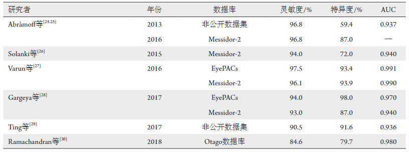
32. Abràmoff MD, Folk JC, Han DP, et al. Automated analysis of retinal images for detection of referable diabetic retinopathy[J]. JAMA Ophthalmol, 2013, 131(3): 351-357.
33. Abràmoff MD, Lou Y, Erginay A, et al. Improved automated detection of diabetic retinopathy on a publicly available dataset through integration of deep learning[J]. Invest Ophthalmol Vis Sci, 2016, 57(13): 5200-5206.
34. Solanki K, Ramachandra C, Bhat S, et al. EyeArt: automated, high-throughput, image analysis for diabetic retinopathy screening[J]. Invest Ophthalmol Vis Sci, 2015, 56(7): 1429-1429.
35. Gulshan V, Peng L, Coram M, et al. Development and validation of a deep learning algorithm for detection of diabetic retinopathy in retinal fundus photographs[J]. JAMA, 2016, 316(22): 2402-2410.
36. Gargeya R, Leng T. Automated identification of diabetic retinopathy using deep learning[J]. Ophthalmology, 2017, 124(7): 962-969.
37. Ting DSW, Cheung CY, Lim G, et al. Development and validation of a deep learning system for diabetic retinopathy and related eye diseases using retinal images from multiethnic populations with diabetes[J]. JAMA, 2017, 318(22): 2211-2223.
38. Ramachandran N, Hong SC, Sime MJ, et al. Diabetic retinopathy screening using deep neural network[J]. Clin Exp Ophthalmol, 2018, 46(4): 412-416.
39. Yang WH, Zheng B, Wu MN, et al. An evaluation system of fundus photograph-based intelligent diagnostic technology for diabetic retinopathy and applicability for research[J]. Diabetes Ther, 2019, 10(5): 1811-1822.
40. Natarajan S, Jain A, Krishnan R, et al. Diagnostic accuracy of community-based diabetic retinopathy screening with an offline artificial intelligence system on a smartphone[J]. JAMA Ophthalmol, 2019, 137(10): 1182-1188.
41. Sosale B, Sosale AR, Murthy H, et al. Medios—an offline, smartphone-based artificial intelligence algorithm for the diagnosis of diabetic retinopathy[J]. Indian J Ophthalmol, 2020, 68(2): 391-395.
'%20fill='white'%20fill-opacity='0.01'/%3e%3cmask%20id='mask0_3477_29692'%20style='mask-type:luminance'%20maskUnits='userSpaceOnUse'%20x='0'%20y='0'%20width='16'%20height='16'%3e%3crect%20id='&%23232;&%23146;&%23153;&%23231;&%23137;&%23136;_2'%20x='16'%20width='16'%20height='16'%20transform='rotate(90%2016%200)'%20fill='white'/%3e%3c/mask%3e%3cg%20mask='url(%23mask0_3477_29692)'%3e%3cpath%20id='&%23232;&%23183;&%23175;&%23229;&%23190;&%23132;'%20d='M14%205L8%2011L2%205'%20stroke='%23333333'%20stroke-width='1.5'%20stroke-linecap='round'%20stroke-linejoin='round'/%3e%3c/g%3e%3c/g%3e%3c/svg%3e)