Role of Retinal Nerve Fiber Layer Thickness and Optic Disk Measurement by OCT on Early Diagnosis of Glaucoma
'%20fill='white'%20fill-opacity='0.01'/%3e%3cmask%20id='mask0_3477_29692'%20style='mask-type:luminance'%20maskUnits='userSpaceOnUse'%20x='0'%20y='0'%20width='16'%20height='16'%3e%3crect%20id='&%23232;&%23146;&%23153;&%23231;&%23137;&%23136;_2'%20x='16'%20width='16'%20height='16'%20transform='rotate(90%2016%200)'%20fill='white'/%3e%3c/mask%3e%3cg%20mask='url(%23mask0_3477_29692)'%3e%3cpath%20id='&%23232;&%23183;&%23175;&%23229;&%23190;&%23132;'%20d='M14%205L8%2011L2%205'%20stroke='%23333333'%20stroke-width='1.5'%20stroke-linecap='round'%20stroke-linejoin='round'/%3e%3c/g%3e%3c/g%3e%3c/svg%3e)
关键词
摘要
Purpose: Glaucoma is an eye disease that can lead to irreversible optic nerve damage and cause blindness.Optical co- herence tomography(OCT)allows an early diagnosis of glau- coma by the measurements ofthe retinal nerve fiber and optic disc parameters.A retrospective study was designed to analyze the effects of the measurement of the retinal nerve fiber layer (RNFL) thickness and the optic disc tomography by spectral- domain OCT on the early diagnosis of suspected glaucoma and primary open angle glaucoma(POAG).
Methods: This was a clinical case-control study.The RNFL thickness around the optic disc and optic disk tomographic pa- rameters of the control(n=51,98 eyes),suspected glaucoma (n=81,146 eyes),and POAG groups(n=55,106 eyes)were measured by OCT.The parameters included superior,inferi- or,nasal and temporal mean RNFL thickness,disc area (DA),cup area(CA),rim area(RA),disc volume(DV), cup volume(CV),rim volume(RV),cup/disc area ratio (CA/DA),rim/disc area ratio(RA/DA),cup/disc volume ratio(CV/DV)and rim/disc volume ratio(RV/DV).
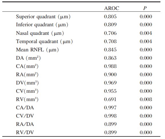
Results: Superior,nasal,and mean RNFL parameters,DA, CA,RA,DV,CV,CA/DA,RA/DA,CV/DV and RV/DV significantly differed among three groups by single-factorial ANOVA.Inferior and temporal RNFL thickness significantly differed between the control and POAG groups.No significant difference was observed in RV among three groups.In the POAG group,the maximum area under the ROC curve (AROC)of mean RNFL thickness was 0.845.The maximum AROC of optic disk parameters was RA/DA(0.998),followed by CA/DA(0.997).The AROC of CA,RA,CV,and DV were all>0.900.
Conclusion: OCT may serve as a useful diagnostic modality in distinguishing suspected glaucoma from POAG.
全文
Introduction
Glaucoma is an ocular disease that may result in irreversible optic nerve damage and even blindness, manifested as retinal ganglion cell(RGC)apoptosis. Loss of RGCs and axons are structurally character- ized as topical and/or diffusive thinning of retinal nerve fiber layer(RNFL)and progressive thinning of optic disc rim,and functionally manifest as visual acuity declines and visual field defects[1-3]. Optic disc injury is generally considered to precede visual acu- ity decline and visual field defects[4,5]. Therefore, RNFL thickness and optic disc parameters play a pivotal role in the early diagnosis of glaucoma.
Optical coherence tomography(OCT) is a nonin- vasive and reproducible auxiliary diagnostic method of glaucoma and it can quantitatively evaluate the RNFL thickness and optic disc parameters.The optic disc rim is defined as the area above the upward line perpendicular to RPE layer,while below the perpen- dicular line is the optic cup as seen with OCT. In this study,RNFL thickness and optic disc parameters were assessed by OCT and statistically compared among normal,glaucoma-suspected,and early POAG patients.
Materials and methods
Clinical data
Patients undergoing OCT examination of the optic disc at the Department of Ophthalmology,the Second Affiliated Hospital of Guangzhou Medical University between May and September, 2014 were selected for this clinical trial.All participants had refractive power<±5.0D, best corrected visual acuity ≥0.5, transparent refractive media and suffered from no retinal diseases or alternative optic neuropathy induced by glaucoma.Inclusion criteria:
Normal group: (1)naked or corrected visual acu- ity ≥1.0; (2)intraocular pressure ≤21 mmHg; (3) having no family history of glaucoma or other optic nerve diseases.
POAG-suspected group: those who met at least one of the three criteria were enrolled in this group: (1) those with a peak value of 24-h intraocular pres- sure>21 mmHg,with open chamber angle,without glaucomous fundus changes or visual field defects; (2) those with RNFL defects or optic disc morpho- logical changes,a peak value of 24-h intraocular pressure<21 mmHg,without glaucomous visual field defects and the chamber angle is open;and(3) those with open chamber angle,with high-risk factors of glaucoma,with a family history of glaucoma and those presenting with visual field defects with un- known causes.
POAG group: those who met one of the two cri- teria were enrolled in this group[7]: (1) those with a peak value of 24-h intraocular pressure>21 mmHg, with glaucomous fundus injury and/or glaucomous visual field defects,with open chamber angle,and other factors causing high intraocular pressure were excluded; and (2) those with a peak value of 24h intraocular pressure ≤21 mmHg,with glaucomous fun- dus injury and/or glaucomous visual field defects, with open chamber angle,and other diseases leading to fundus and visual field defects were excluded.
Examination methods
Conventional examination:visual acuity, intraoc- ular pressure,fundus,automatic computer optome- try,and anterior segment slit-lamp tests.
OCT examination:the pupil diameter of each eye was dilated to approximately 5mm using 1% tropicamide eye drops.The patients’affected eyes gazed at the screen with their lower mandible on the bracket.The OCT examination was performed at optic disc scanning mode.The experienced operator carefully observed the scanning site on the monitor screen with optic disc as the scanning center using Topcon 3D-OCT1000 equipment.The boundary between optic disc and optic cup was 120 μm above the RPE layer.
The RNFL thickness of four quadrants including the superior,inferior,nasal,and temporal sites was measured to calculate the mean RNFL thickness. Optic disc parameters consisted of DA,CA,RA CA/DA ratio,RA/DA ratio,DV,CV,RV,CV/DV ratio,and RV/DV ratio.
Statistical analysis
SPSS 18.0 statistical software was used for data analysis. RNFL thickness of each quadrant,mean RNFL thickness,and optic disc parameters were statistically compared among three groups using singlefactorial ANOVA.The optimal diagnostic index was determined by the area under ROC.P<0.05 was con- sidered as statistical significance.
Results
In the normal group,51 individuals(98 eyes) were enrolled,including 27 males(53 eyes)and 24 females(45 eyes),aged 43.21±13.53 years,on av- erage.In the glaucoma-suspected group,8l patients (146 eyes)were included:43 males(79 eyes)and 38 females(67 eyes),aged 45.38±12.46 years,on average.In the POAG group,55 patients(106 eyes) were enrolled,including 29 males(56 eyes)and 26 females(50 eyes),aged 46.15±11.76 years on average.
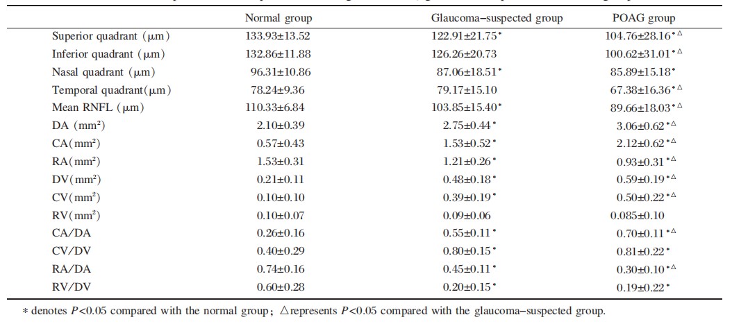
Comparison of different parameters among the three groups revealed that the RNFL thickness of each quadrant and mean RNFL thickness were sig- nificantly thinner in the POAG group than in the normal groups(all P<0.05).The DA,CA,DV, CV,CA/DA,and CV/DV ratio in the POAG group were significantly increased,whereas the RA,RA/ DA and RV/DV ratios were significantly decreased compared to the normal group(all P<0.05).The RN- FL thickness of the superior and nasal quadrant and mean RNFL thickness were significantly attenuated compared to the normal group and,the DA,CA, DV,CV,CA/DA,and CV/DV ratio were dramati- cally increased and the RA,RA/DA,and RV/DV ratios were significantly reduced in the glaucoma- suspected group(all P<0.05). Compared with the glaucoma-suspected group, the RNFL thickness of the superior,inferior and temporal quadrant and mean RNFL thickness were significantly thinner,the DA,CA,DV and CV were significantly increased, and the RA,RA/DA and RV/DV ratio were dramatically reduced in the POAG group(all P<0.05), as illustrated in Table 1.
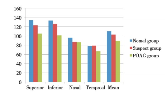
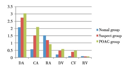
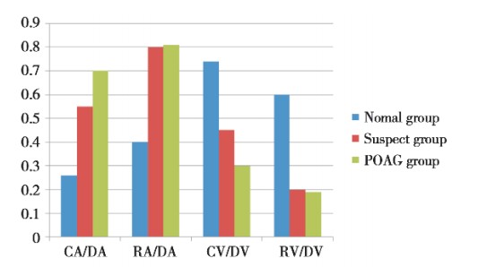
The RNFL thickness of each quadrant and mean RNFL thickness decreased progressively in all groups(Figure 1).The DA,CA,DV,and CV in three groups were elevated,whereas the RA was de- clined progressively.No significant pattern was ob- served in the RV among three groups(Figure 2). In all groups,the CA/DA and CV/DV ratio increased progressively, whereas the RA/DA and RV/DV ra- tio was reduced progressively(Figure 3)
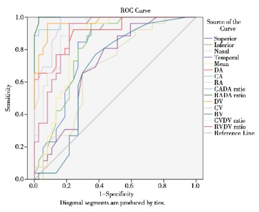
The optimal diagnostic index of POAG during OCT examination was determined as below:the area under the ROC curve(AROC)was calculated. A larger AROC was associated with a higher diagnostic value. AROC=0.5 denoted no diagnostic value.In the POAG group,the AROC of each parameter was of diagnostic value(P<0.05).Among the RNFL thick- nesses,the AROC of the mean RNFL thickness was the largest,at up to 0.845.The AROC of the RA/ DA ratio was the highest(0.998) among optic disc parameters, followed by CA/DA ratio(0.997), and the AROCs of CA,DV,DA,RA were all>0.900, as illustrated in Table 2 and Figure 4.
Discussion
Glaucomatous optic nerve damage substantially results from retinal ganglion cells and axon pathological changes. Making an early diagnosis of POAG is difficult due to the slow progression of the disease. Therefore,delivery of early diagnosis is essential for the treatment of POAG.The morphological changes of the glaucomous optic disc are generally viewed as preceding the visual field defects,which are mainly characterized as decreased peripapillary RNFL thickness, increased CA/DA ratio,and loss of rim. An OCT examination could objectively and quantita- tively measure the RNFL thickness and optic disc parameters with high accuracy and good repro- ducibility.This examination is of clinical significance in diagnosing early POAG-1.
In this study,the RNFL thickness of each quad- rant presented with"twin peaks,"shaped as thicker at the superior and inferior sites and thinner at nasal and temporal sides.The RNFL thickness of each quadrant and mean RNFL thickness were thinner in the glaucoma-suspected and POAG groups than in the normal counterparts,which is consistent with previous findings[12-14].Moreover,compared with those in normal counterparts,the DA,DV,CA,CV,CA/ DA,and CV/DV ratio were increased in glaucoma- suspected and POAG patients,whereas the RA, RV,RA/DA,and RV/DV ratio were decreased, which is almost consistent with the findings reported by Hu et al[15] .
The mean RNFL thickness played the most crucial role in diagnosing glaucoma,which is basically in accordance with previous findings[12,16]. The parameters including CV/DV,RA/DA,and RV/DV ratio, which were not investigated in previous research, were measured and statistically compared among different populations in this study.These parameters are of clinical significance in the diagnosis of glau- coma.Among all optic disc parameters,the RA/DA and CA/DA ratios,and CA,DV,DA and RA were of relatively significant diagnostic value.The AROC of the RA/DA ratio was the largest,suggesting that it probably served as an optimal parameter for early diagnosis of glaucoma.The loss of cup rim was also one of the vital characteristics of early glaucoma. The RA/DA ratios in the three groups were 0.74± 0.16,0.45±0.11,and 0.30±0.10,indicating the pos- sibility of suspected glaucoma when the RA/DA ra- tio was<0.45.Visual field and dynamic intraocular pressure examination and regular follow-up should be conducted.
The possibility of early glaucoma should be con- sidered for an RA/DA ratio <0.3.Considering the function and purpose of the RA/DA ratio,it was not regarded as a diagnostic index in this study,whereas it could provide evidence for the early diagnosis of glaucoma.The parameters including CV/DV,RA/DA,and RV/DV ratio,which were not investigated in previous research,were measured and statistically compared among different populations in this study. These parameters are of clinical significance in the diagnosis of glaucoma.
Due to the slow progression of POAG,auxiliary examinations with relatively high sensitivity should be performed to make an early diagnosis and subse- quent follow-up.At present,perimetry is still the gold standard for the diagnosis of glaucoma. However, the visual field progression of POAG is significantly slower than those of optic disc and optic nerve changes. Hence, OCT examination plays a pivotal role in the early diagnosis of glaucoma. Wollstein et al[17]. conducted a 4.7-year follow-up of the stability of visual field and OCT examinations in glaucoma-suspected and POAG patients,and sug- gested that 66%of patients had stable examination outcomes, 22%with progression detected by OCT, 9%with progression by perimetry and 3% with progression by both perimetry and OCT examination. Mitra Sehi[18]and Serbecic[19]have demonstrated that OCT examination is superior to perimetry in terms of evaluating the progression of POAG.
To sum up, the results from this study confirm that OCT examination showed high reproducibility and sensitivity,was convenient to operate,and ap- peared to reflect the changes in the optic disc pa- rameters. OCT examination might therefore be applied to conduct screening and diagnosis of glaucoma by evaluating the changes in the RNFL thickness and optic disc parameters in glaucoma-suspected and early glaucoma patients,thereby contributing to early diagnosis and treatment of POAG.
基金
参考文献
1. Alasil T,Wang K,Yu F,et al.Correlation of retinal nerve fiber layer thickness and visual fields in glaucoma:a bro- ken stick model.Am J Ophthalmol,2014,157(5):953- 959.
2. Li L,Zhao JL,Liu XL.Correlation between neuroretinal rim area/retinal nerve fiber layer thickness and differen- tial light sensitivity in visual field in primary open angle glaucoma.Zhongguo Yi Xue Ke Xue Yuan Xue Bao, 2009,31(5):607-611.
3. Anderson RS.The psychophysics of glaucoma:improving the structure/function relationship.Prog Retin Eye Res, 2006,25(1):79-97. 4 Quigley HA, Katz J, Deri.
4. Quigley HA,Katz J,Derick RJ,et al.An evaluation of optic disc and nerve fiber layer examinations in monitor- ing progression of early glaucoma damage.Ophthalmolo- gy,1992,99(1):19-28.
5. Pederson JE,Anderson DR.The mode of progressive disc cupping in ocular hypertension and glaucoma.Arch Oph- thalmol,1980,98(3):490-495.
6. Leung CK,Chong KK,Chan WM,et al.Comparative study of retinal nerve fiber layer measurement by Stratus OCT and GDx VCC,Ⅱ:structure/function regression analysis in glaucoma.Invest Ophthalmol Vis Sci,2005,46 (10):3702-3711.
7. Ophthalmology of Chinese medical association branch of the glaucoma group.The diagnosis and treatment of pri- mary glaucoma specialist consensus(2014).Chinese Journal of Ophthalmology,2014(5):382-383.
8. Li S,Wang X,Li S,et al.Evaluation of optic nerve head and retinal nerve fiber layer in early and advance glauco- ma using frequency-domain optical coherence tomogra- phy.Graefes.Arch Clin Exp Ophthalmol,2010,248(3): 429-434.
9. Li ZM,Huang XW,Huang H.Application of OCT in the early diagnosis of glaucoma.Modern Journal of Integrated Traditional Chinese and Western Medicine,2014.23(6): 651-652.
10. Liu X,Ling Y,Luo R,et al.Optical coherence tomogra- phy in measuring retinal nerve fiber layer thickness in normal subjects and patients with open-angle glaucoma. Chin Med J(Engl),2001,114(5):524-529.
11. Chen H,et al.Correlation between stratus OCT and GDx VCC in early glaucoma,ocular hypertension and glauco- ma suspect eyes.Joumal of Optometry,2012,5(1):24- 30.
12. Ji BL,You YA,Fang AW,et al.The significance of the retinal nerve fiber layer and macular thickness measure- ments in the early diagnosis of glaucoma using optical co- herence tomography.Chinese Journal of Optometry & Ophthalmology,2008,10(1):54-58.
13. Mwanza JC,Warren JL,Budenz DL.Combining spectral domain optical coherence tomography structural parame- ters for the diagnosis of glaucoma with early visual field loss.Invest Ophthalmol Vis Sci,2013,54(13):8393-8400.
14. Wong E,Yoshioka N,Kalloniatis M,et al.Cirus HD- OCT short-term repeatability of clinical retinal nerve fiber layer measurements.Optom Vis Sci,2014. 2015,92(1): 83-88.
15. Hu,SJ,Qiu L.Comparing glaucomatous optic neuropathy in primary open angle by optical coherence tomography. Journal of Clinical Ophthalmology,2007.15(1):26-28.
16. Wang,XZ,Li SN,Wu GW,et al.Effects of optic disc topography and retinal nerve fiber layer thickness measurement by spectral-domain OCT on diagnosis of glaucoma. Chinese Joumal of Experimental Ophthalmology,2011,29 (9):820-824.
17. Wollstein G,Ishikawa H,Wang J,et al.Comparison of three optical coherence tomography scanning areas for detection of glaucomatous damage.Am J Ophthalmol, 2005,139(1):39-43.
18. Sehi M,Zhang X,Greenfield DS,et al.Retinal nerve fiber layer atrophy is associated with visual field loss over time in glaucoma suspect and glaucomatous eyes.Am J Oph- thalmol,2013,155(1):73-82 el.
19. Serbecic N,Beutelspacher SC,Aboul-Enein FC,et al.Re- producibility of high-resolution optical coherence tomog- raphy measurements of the nerve fibre layer with the new Heidelberg Spectralis optical coherence tomography.Br J Ophthalmol,2011,95(6):804-810.