
多发性硬化相关视神经炎亚临床进展的监测与疾病修饰治疗策略优化:从病例启示到文献综述
'%20fill='white'%20fill-opacity='0.01'/%3e%3cmask%20id='mask0_3477_29692'%20style='mask-type:luminance'%20maskUnits='userSpaceOnUse'%20x='0'%20y='0'%20width='16'%20height='16'%3e%3crect%20id='&%23232;&%23146;&%23153;&%23231;&%23137;&%23136;_2'%20x='16'%20width='16'%20height='16'%20transform='rotate(90%2016%200)'%20fill='white'/%3e%3c/mask%3e%3cg%20mask='url(%23mask0_3477_29692)'%3e%3cpath%20id='&%23232;&%23183;&%23175;&%23229;&%23190;&%23132;'%20d='M14%205L8%2011L2%205'%20stroke='%23333333'%20stroke-width='1.5'%20stroke-linecap='round'%20stroke-linejoin='round'/%3e%3c/g%3e%3c/g%3e%3c/svg%3e)
关键词
摘要
目的:探讨亚临床病变监测在多发性硬化相关视神经炎(multiple sclerosis-related optic neuritis, MS-ON)治疗决策中的重要性及不同疾病修饰治疗(disease-modifying therapy, DMT)药物的疗效差异。
方法:报告1例30岁女性MS-ON患者,以左眼急性视神经炎首发,最终确诊为多发性硬化的临床病程、眼科检查及治疗经过。
结果:该例MS-ON患者在急性期经糖皮质激素冲击治疗后视力部分恢复,但遗留视野缺损。启用富马酸二甲酯(dimethyl fumarate, DMF)治疗后9个月内,虽无主观视力下降主诉,但光相干断层扫描(optical coherence tomography, OCT)显示左眼视网膜神经纤维层(retinal nerve fiber layer, RNFL)持续变薄,视野平均偏差(mean deviation, MD)值波动后恶化,最终出现临床复发。停用DMF,转换为奥法妥木单抗(ofatumumab, OFA)治疗,后随访1年,患者左眼最佳矫正视力(best corrected visual acuity, BCVA)恢复至1.0,视野显著改善,且OCT显示RNFL和黄斑区神经节细胞层(ganglion cell layer, GCL)厚度维持稳定,无复发及不良事件发生。
结论:本病例表明,在MS-ON患者中,即使临床稳定期也可能存在进行性的亚临床视神经损伤,如RNFL持续变薄,即“阴燃病灶”。常规依赖主观症状和BCVA评估可能遗漏此类进展。系统性监测,尤其是OCT RNFL/GCL和视野检查对于早期识别治疗不足至关重要。当DMF等中效DMT无法控制亚临床进展并导致临床复发时,及时转换为高效DMT可有效控制疾病活动,保护视神经结构和功能,实现长期稳定,这突出了基于亚临床指标优化DMT策略在MS-ON管理中的关键作用。
全文
文章亮点
1. 关键发现
• 多发性硬化相关视神经炎在中效疾病修饰治疗期间出现亚临床神经损伤需及时换用高效药物。
2. 已知与发现
• 视神经炎是多发性硬化的常见临床表现,疾病修饰治疗可延缓疾病进展。
• 部分多发性硬化相关视神经炎患者临床稳定期仍存在亚临床视网膜神经纤维层持续变薄。
3. 意义与改变
• 多发性硬化相关视神经炎的管理除关注主观症状与常规视力评估外,应整合光相干断层扫描和视野等指标以早期识别疾病亚临床进展;发现持续性亚临床损伤后及时调整治疗策略,是保护视神经功能并实现长期稳定的关键。
多发性硬化( multiple sclerosis, MS)是一种常见的中枢神经系统自身免疫性脱髓鞘疾病。多发性硬化相关视神经炎(multiple sclerosis-related optic neuritis, MS-ON),是MS常见的临床表现,表现为视力下降、视野缺损及眼球运动疼痛。目前,针对MS的治疗策略主要分为高效和低效疾病修饰治疗(disease-modifying therapy, DMT),早期使用高效DMT可以减少残疾进展和脑部病变。本文报告了1例MS-ON患者亚临床病情控制情况,强调了监测亚临床病变的必要性及对治疗策略做出相应调整的重要性。
1. 临床资料
患者女,30岁,因“左眼中心视野小片状缺损,伴眼球转动痛及头晕1个月”,于2023年2月到当地医院眼科就诊。患者于2022年11月曾感染新型冠状病毒,并行人工流产手术,否认其他全身系统性疾病史。眼科检查:右眼最佳矫正视力(best corrected visual acuity, BCVA)为1.2,左眼BCVA 为0.6。双眼眼前节检查未见明显异常。瞳孔检查:左眼存在相对性传入性瞳孔障碍(relative afferent pupillary defect, RAPD)。视觉电生理检查:图形视觉诱发电位(pattern visual evoked potential, PVEP)显示,右眼各视角方格形觉反应正常;左眼60′方格下记录不到明确波形,30′方格P100波潜伏期重度延迟,且其P100波振幅在30′及15′方格均较右眼严重降低(图1)。视野(visual field, VF)检查,右眼视野平均偏差(mean deviation, MD)值为1.36 dB(RE),左眼视野出现中心暗点,其MD值为27.30 dB(LE)(图2A)。荧光素眼底血管造影(fundus fluorescein angiography, FFA)示造影早期左眼视盘荧光充盈晚于动脉充盈,左眼视盘荧光强于右眼;视网膜及血管未见异常荧光,拱环结构完整。右眼未见异常(图2B)。诊断:左眼球后视神经炎。治疗,予甲泼尼龙冲击(第1—3天予 500 mg/d,第4—6天予 125 mg/d),然后予泼尼松龙口服(初始 60 mg/d),1个月内逐渐减量至8 mg/d。治疗后,患者自觉眼球运动疼痛消退,左眼视力改善。出院左眼BCVA为0.8,眼底检查,左眼视盘颞侧苍白(图3A),左眼视野弥漫性光敏感度下降,旁中心及鼻侧视野缺损,其MD值为10.06 dB (LE)(图3B)。光相干断层扫描(optical coherence tomography, OCT),右眼视网膜神经纤维层(retinal nerve fiber layer, RNFL)平均厚度为 94 μm (RE),左眼RNFL为112 μm (LE);右眼黄斑区神经节细胞层(ganglion cell layer, GCL)平均厚度为78 μm (RE),左眼GCL为66 μm(LE)。
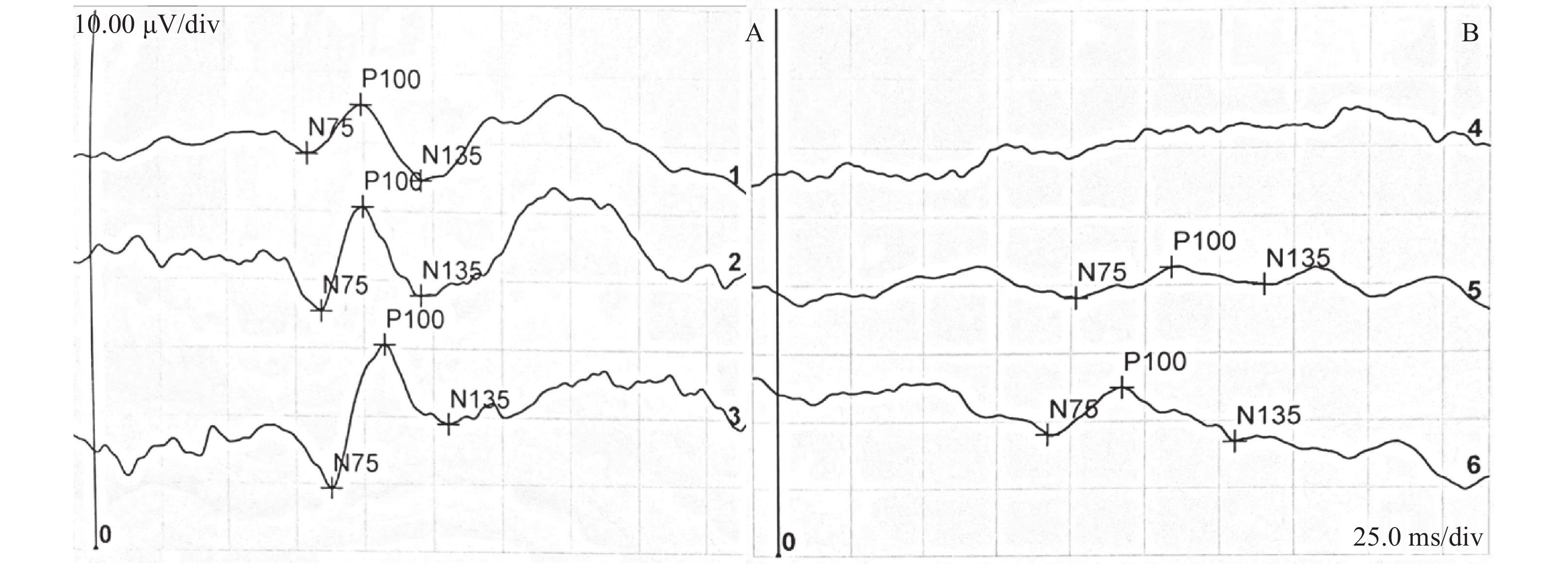
(A)右眼各视角方格形觉反应正常,波形重复性好,P100波潜伏期及振幅均在正常范围。(B)左眼异常波形,60′视角方格下未记录到可重复波形,30′视角方格P100波潜伏期重度延迟,且其P100波振幅在30′及15′视角方格均较右眼严重降低。
(A) The right eye shows normal pattern visual responses at each check size, with good waveform reproducibility, and both P100 latency and amplitude within normal ranges. (B) The left eye shows abnormal waveforms: no reproducible waveform is recorded at 60′ check size; at 30′ check size, the P100 latency is severely delayed, and the P100 amplitude is markedly reduced compared with the right eye at both 30′ and 15′ check sizes.
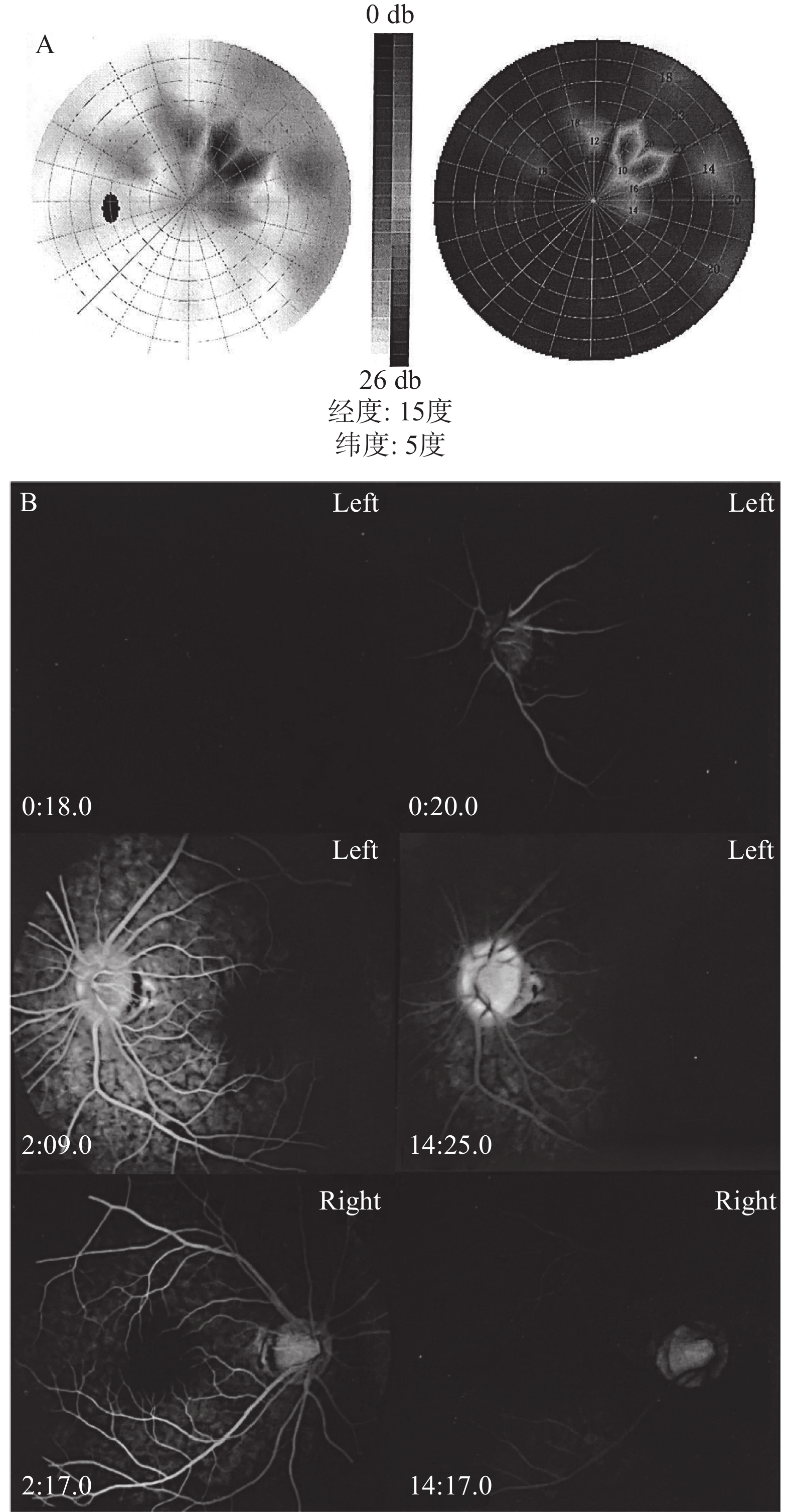
(A)左眼视中心视野有小片状缺损,鼻上、颞上、鼻下方弥漫性光敏感度降低。(B)双眼荧光素眼底血管造影像,造影早期左眼视盘荧光充盈晚于动脉充盈,左眼视盘荧光强于右眼;视网膜及血管未见异常荧光,拱环结构完整;右眼未见异常。
(A) The visual field of the left eye shows a small paracentral scotoma with diffuse reduction of light sensitivity in the superonasal, superotemporal, and inferonasal areas. (B) Fundus fluorescein angiography images of both eyes. In the early phase of angiography, the left optic disc shows later fluorescence filling than arterial filling, and the left optic disc exhibits stronger fluorescence than the right eye. No abnormal fluorescence is observed in the retina or blood vessels, and the foveal avascular zone (FAZ) architecture is intact. The right eye shows no abnormalities.
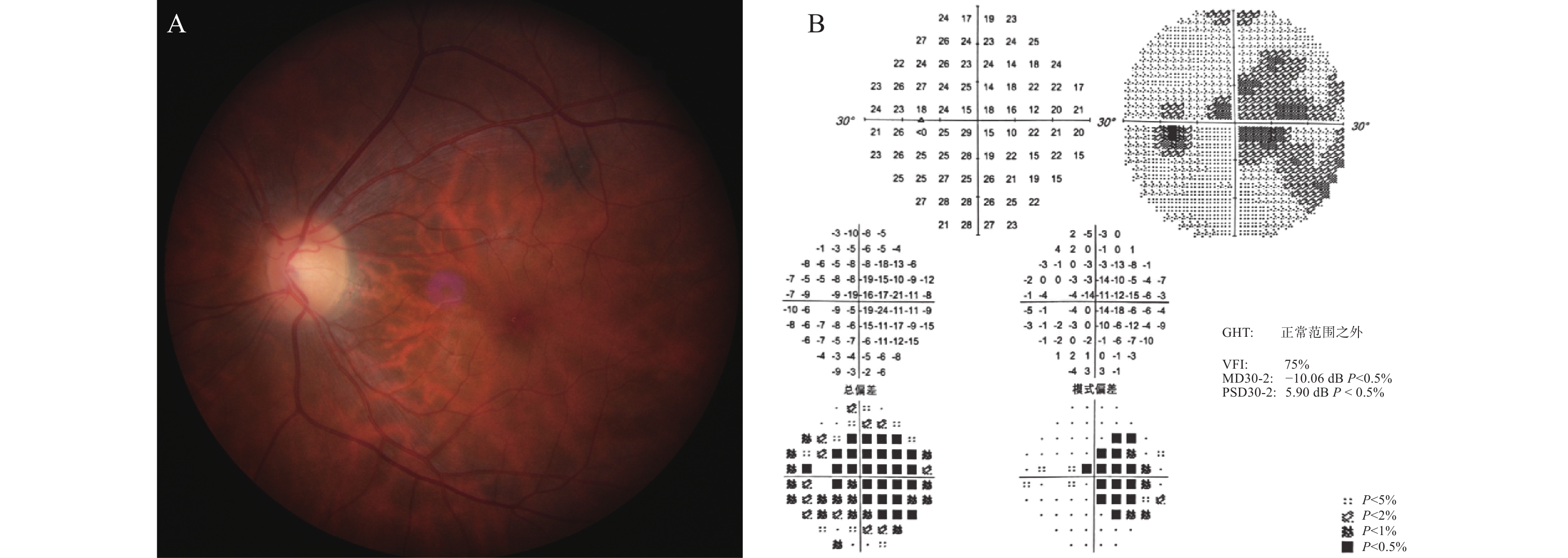
(A)彩色眼底像,见左眼视盘颞侧苍白。(B)激素冲击及口服治疗1个月后视野影像,见左眼视野弥漫性光敏感度下降,旁中心及鼻侧视野缺损。
(A) Color fundus photograph shows temporal pallor of the left optic disc. (B) Visual field image obtained one month after steroid pulse and oral therapy shows diffuse reduction of light sensitivity, paracentral and nasal visual field defects in the left eye.
2023年4月行血液检查,抗可溶性抗原(extractable nuclear antigen, ENA)抗体均(-),排除系统性红斑狼疮(systemic lupus erythematosus, SLE)、混合性结缔组织病、干燥综合征(Sjögren's syndrome, SS)、进行性系统性硬化症、多发性肌炎(polymyositis, PM)等自身免疫性疾病。血清及脑脊液检查示水通道蛋白4抗体(aquaporin-4 immunoglobulin G, AQP4 IgG)阴性、髓鞘少突胶质细胞糖蛋白抗体(myelin oligodendrocyte glycoprotein IgG, MOG IgG)阴性。行腰穿脑脊液检查,显示寡克隆带(oligoclonal bands, OCB)阳性。磁共振成像(magnetic resonance imaging, MRI)显示(起病3个月),头颅双侧颞叶颞角旁白质、胼胝体压部见线状、小斑片状T2加权像(T2-weighted image, T2WI)及液体衰减反转恢复序列(fluid-attenuated inversion recovery, FLAIR)高信号,皮质轻度萎缩,考虑脱髓鞘改变可能(图4A、B);眼眶MRI示左侧视神经在FLAIR压脂序列信号较右侧稍高,考虑视神经炎病变(图4C)。诊断:MS。予富马酸二甲酯(dimethyl fumarate, DMF)口服,初始剂量120 mg每天2次,14 d后以240 mg每天2次维持;泼尼松龙口服(初始剂量60 mg/d),序贯减量1个月后,停用激素。
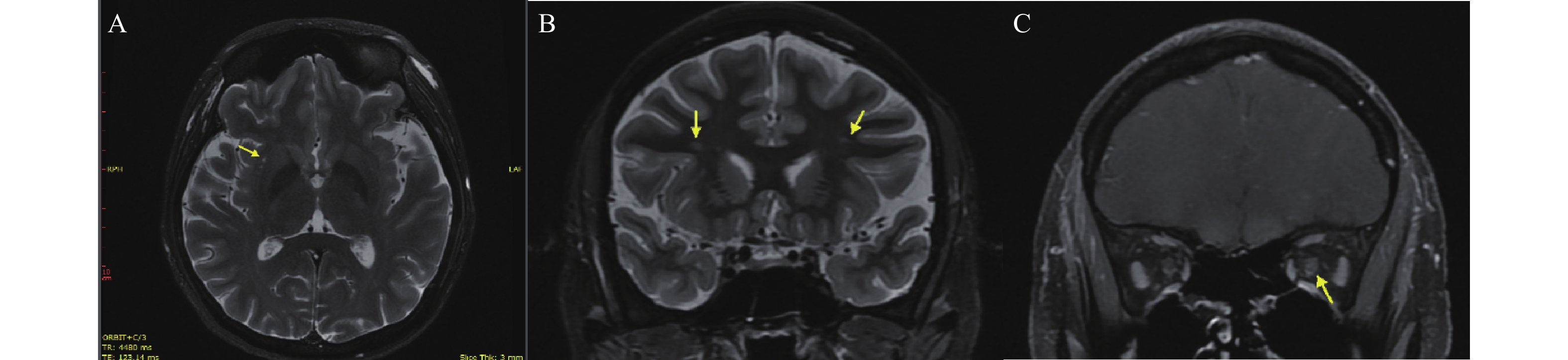
(A、B)双侧颞叶颞角旁白质、胼胝体压部见线状、小斑片状T2W1及FLAIR高信号,皮质轻度萎缩,考虑脱髓鞘改变可能。(C) 示左侧视神经FLAIR抑脂信号可疑升高,考虑视神经炎病变。
(A, B) Linear and small patchy hyperintensities on T2-weighted imaging and FLAIR are observed in the periventricular white matter of the bilateral temporal lobes and the splenium of the corpus callosum, with mild cortical atrophy, suggesting possible demyelinating changes. (C) FLAIR fat-suppressed signal of the left optic nerve is suspiciously elevated, indicating optic neuritis involvement.
2023年7月(经DMF治疗3个月,停用激素2个月后),患者自觉左眼视力稍下降。眼科检查:左眼BCVA为0.63,RAPD呈阳性;视野MD为4.30 dB (LE)(图5A)。微视野检查显示右眼正常,左眼视网膜光敏感度稍下降,其平均阈值为26.8 dB (LE)(图5B)。OCT检查:左眼RNFL平均厚度持续变薄,降至79 μm (LE),左眼GCL厚度为66 μm(LE)。头颅及眼眶MRI复查(对比2023年4月MRI):显示侧脑室旁出现新发脱髓鞘灶,并见后角旁“中央静脉征”(图6);同时,双侧视神经于压脂序列呈现高信号。上述影像学病灶,结合患者新发的视力症状、RAPD体征、视野旁中心暗点及OCT证实的进行性RNFL变薄,考虑DMF对视神经炎症控制不佳。诊断: MS-ON复发。DMF基础上加用激素60mg/d,并序贯减量。
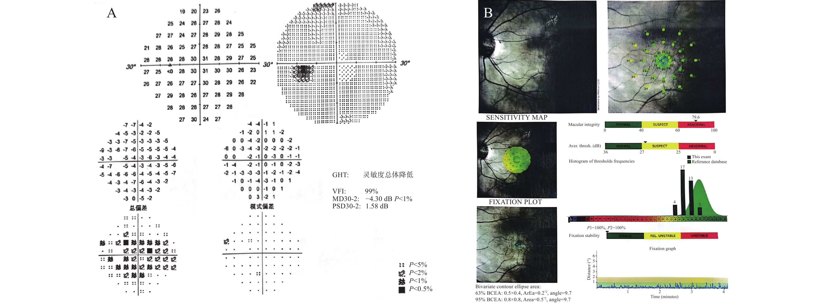
(A)左眼视野像,左眼旁中心暗点伴视敏度下降。(B)左眼微视野像,左眼视网膜光敏感度稍下降。
(A) Visual field image of the left eye shows a paracentral scotoma with decreased visual sensitivity. (B) Microperimetry image of the left eye shows a slight decrease in retinal light sensitivity.
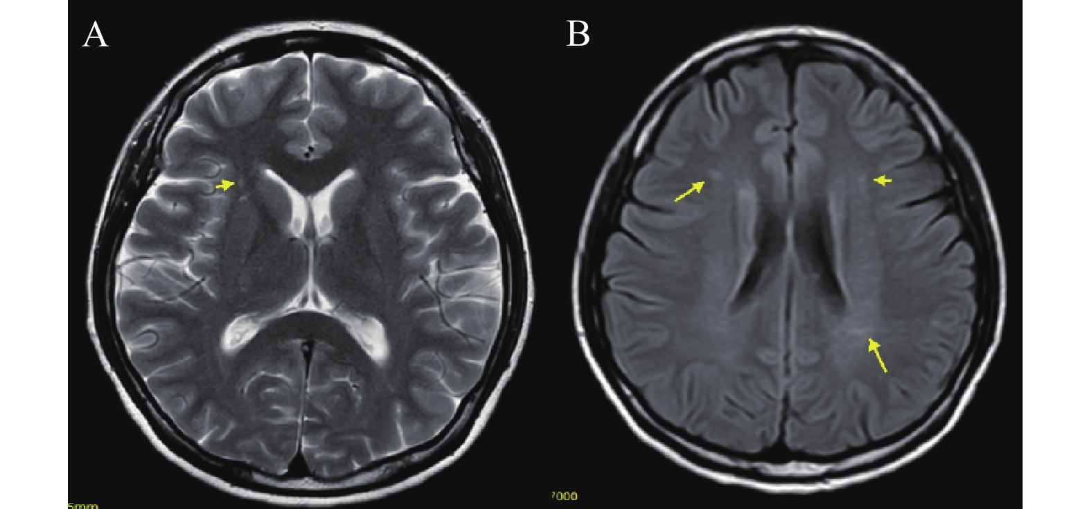
对比2023年4月MRI,可见侧脑室旁白质区出现新发斑片状高信号脱髓鞘病灶(A),部分病灶可见“中央静脉征”(B),符合多发性硬化活动期影像学特征。
Compared with the MRI from April 2023, new patchy hyperintense demyelinating lesions are observed in the periventricular white matter (A), and some lesions show the "central vein sign" (B), which is consistent with the imaging features of active multiple sclerosis.
2024年1月(经DMF继续治疗5个月后),患者视力维持稳定,左眼BCVA 为1.0。治疗期间患者左眼视野在短暂改善后又出现恶化,视野MD值由4.30 dB (LE)(2023年7月)提升到1.40 dB (LE)(2023年9月),之后再度下降到3.56 dB (LE)(2024-1)。左眼RNFL持续变薄,厚度由98 μm (RE)下降至71 μm (RE)。处理:停用DMF,改奥法妥木单抗(ofatumumab, OFA)皮下注射治疗,初始给药为20 mg每周1次,经过3次治疗后,CD19+B 细胞比例从22.56%(2023年3月)耗尽到0.24%(2024年3月)。此后延长给药间隔,并监测外周血淋巴细胞水平。左眼最佳矫正视力维持为1.0,VF 持续改善,恢复至正常视野,MD值为1.35 dB (LE)(图7A,图8);左眼RNFL与黄斑GCL平均厚度维持稳定(图7B、C,图8),未见明显不良事件。
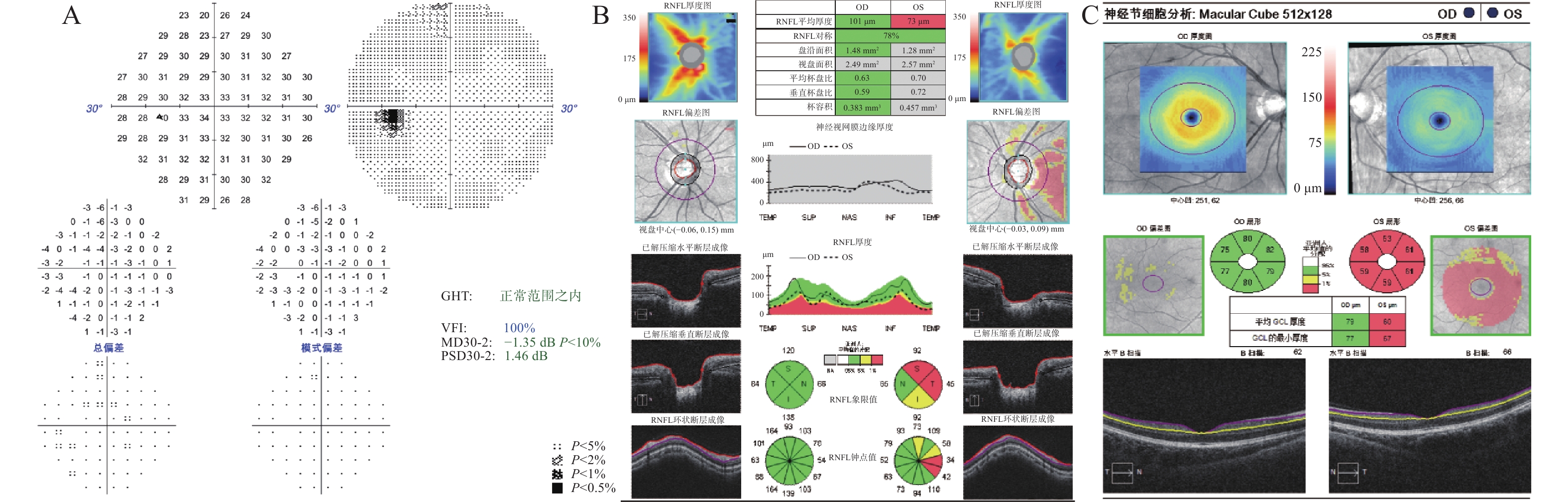
(A)左眼恢复至正常视野。B、C示双眼光相干断层扫描像,(B)显示左眼RNFL平均厚度及各象限厚度,可见颞侧较其他象限下降更为严重。(C)为GCL平均厚度值,右眼正常,左眼严重降低。
(A) The left eye shows recovery to a normal visual field. (B, C) Optical coherence tomography images of both eyes. (B) Shows the average RNFL thickness and quadrant thicknesses of the left eye, revealing that the temporal quadrant is more severely reduced than the other quadrants. (C) Shows the average GCL thickness, which is normal in the right eye but severely reduced in the left eye.
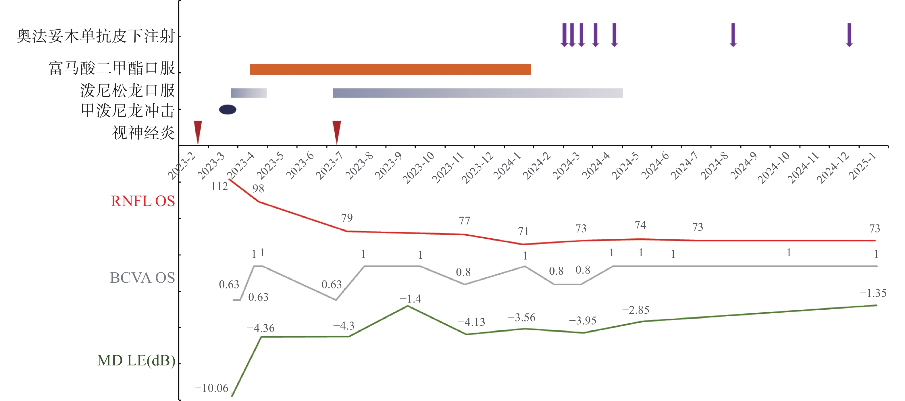
较高的视野MD和BCVA值表示更好的视觉功能,较高的RNFL值表示视网膜结构损伤更轻。患者两次视神经炎发作期间,接受激素冲击治疗后BCVA和MD值有所恢复,RNFL层持续变薄,阴燃病灶持续存在。自2024年3月起接受OFA皮下注射治疗,此后BCVA和MD维持在正常范围,RNFL厚度维持稳定。
Higher visual field MD and BCVA values indicate better visual function, and higher RNFL value indicates less severe retinal structural damage. During the two episodes of optic neuritis, the patient's BCVA and MD values partially recovered after steroid pulse therapy, while RNFL thickness continued to thin and smoldering lesions persisted. Since receiving subcutaneous ofatumumab (OFA) therapy from March 2024, BCVA and MD have remained within normal ranges, and RNFL thickness has remained stable.
2. 讨论
MS-ON是多发性硬化患者中最为常见的症状之一,高达34%~75%的特发性视神经炎(idiopathic optic neuritis, IDON)在10~15年内进展为MS[1-2]。在不伴视神经炎的多发性硬化患者(multiple sclerosis without optic neuritis, MS-NON)中,也发现有VEP异常,RNFL变薄,或视野缺损的亚临床损伤[3-4],提示潜在的神经退行性过程仍在持续。如何发现亚临床损伤,及时控制病情,避免神经系统损伤及功能残疾累积,是临床需要面对的重要问题[5-6]。
本病例经历2次MS-ON发作后,眼部症状在DMF治疗期间基本消失,在其后6个月的随访期内,虽未曾出现主观不适症状,但OCT监测结果显示RNFL持续变薄,视野MD值波动后恶化,最终出现临床复发,提示DMF未能有效控制其亚临床进展及潜在的疾病活动[7- 8]。DMF作为中效DMT[9-10],主要通过激活Nrf2通路发挥抗氧化和抗炎作用,但在部分活动性较高的MS患者中,其免疫抑制强度可能不足以完全遏制B细胞介导的炎症及后续的轴索损伤[11-13]。
考虑到患者存在持续亚临床进展并出现复发,将其DMT转换为B细胞靶向的高效药物OFA。OFA作为全人源CD20单抗[14-16],能有效清除外周CD20+B细胞,减少抗原呈递、炎性因子释放及可能的中枢异位淋巴滤泡形成,从而保护前视路轴突[17-21]。ASCLEPIOS I/II 试验显示,OFA在减少MS复发和延缓功能障碍方面显著优于特立氟胺[15, 22]。DMT药物的选择,和药物之间的转换需要基于患者的具体情况和治疗反应[23- 24]。本例患者在初始使用DMF进行维持治疗期间,患者虽未有明确的自觉视力下降症状,但RNFL厚度持续变薄,说明视神经损伤未被控制,并在最终出现临床复发。考虑其复发原因,对治疗方案重新评估。转换为OFA治疗后,患者RNFL与GCL厚度趋于稳定,视野明显改善。随访一年无复发,进一步印证了针对B细胞的高效治疗在控制MS相关前视路亚临床进展方面的潜在优势。
MS-ON的病情随访中存在一些问题。首先是亚临床病变的早期识别的挑战。特别是临床表现,如视力视野稳定时,可能存在神经损伤仍在继续,即所谓的阴燃病灶。其次,现有的疗效评估一般只检测BCVA,而忽视了对视野和RNFL、GCL的检查,有可能遗漏进行性发展的神经纤维损伤。最后,应根据视力、视野、OCT对现行治疗方案有效性进行及时及定期的评估[25-26],必要时转换治疗药物。例如本病例患者的定期随访及时发现阴燃病灶,使病情得到有效控制,显示了定期随访和及时干预的重要性。
综上所述,在MS-ON患者的治疗随访过程中,除了急性期临床症状的改善外,还应重视亚临床病变的监测,并及时给予治疗策略的调整。
声明
在论文撰写中无使用生成式人工智能,所有作者对内容的真实性、完整性和科学性负责。所有科学贡献和智力劳动均由所有作者共同完成。
利益冲突
所有作者均声明不存在利益冲突。
开放获取声明
本文适用于知识共享许可协议(Creative Commons),允许第三方用户按照署名(BY)-非商业性使用(NC)-禁止演绎(ND)(CC BY-NC-ND)的方式共享,即允许第三方对本刊发表的文章进行复制、发行、展览、表演、放映、广播或通过信息网络向公众传播,但在这些过程中必须保留作者署名、仅限于非商业性目的、不得进行演绎创作。
基金
1. 国家自然科学基金(81870656)。This work was supported by the National Natural Science Foundation, China (81870656).