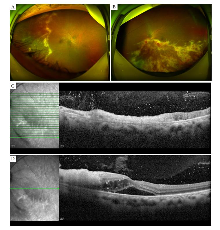
Ultra-wide-angle photographic shows (A) a yellowish-white granular lesion in the temporal retina of the right eye, with cheese-like exudation at the border of the lesion; (B) large yellowish-white cheese-like exudation in the entire retinal layer along the blood vessels below the optic disc of the left eye. OCT shows (C) retina atrophy and thinning in the left eye, with unclear gradation and strong punctured reflection in the vitreous cavity; (D) the retinal neuroepithelial fluid dark area in the macular area of the left eye, with cystic edema in the neurosensory layer and strong punctured reflection.
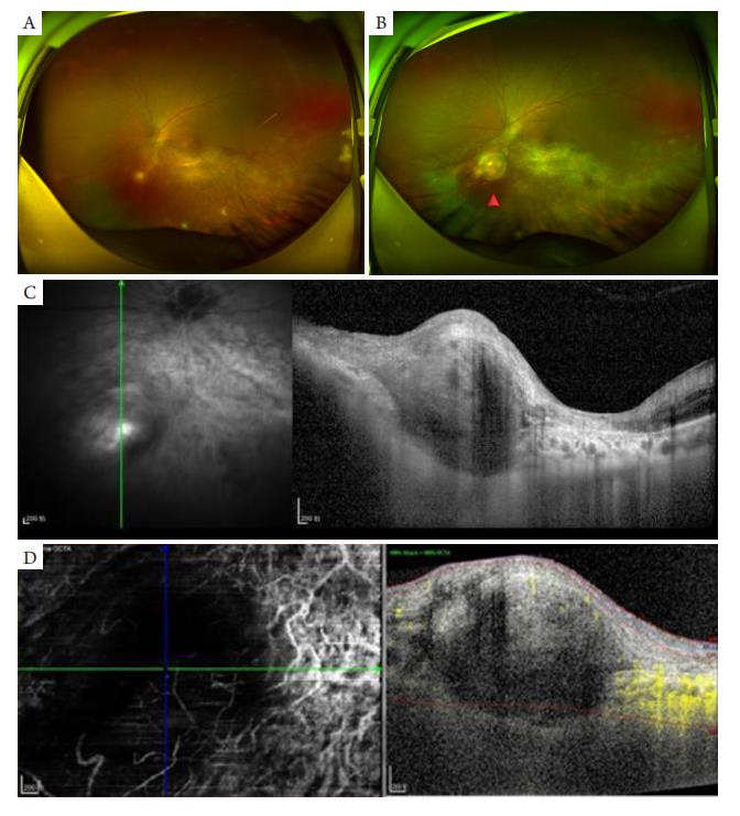
Ultra-wide-angle photographic shows (A) yellowish-white exudation and hemorrhage absorption in the left eye, and yellowish-white lesions with a size of 2/3PD appeared in the lower retina; (B) a yellow-white elevated lesion of about 5PD below the optic disc nose (red triangle), with tortuous retinal vessels above. OCT shows (C) the structure of the retinal layers and choroid capillary layer in the lesion area was poorly visualized, the stromal choroid was thickened with low reflection, the dome-shaped retina was elevated, the subretinal heterogeneous medium to low reflection signal was visible; (D) the choroid capillary plexus was absent in the lesion and there’s no blood perfusion.
表1 患者全身CD4值及HIV病毒载量变化情况
Table 1 Changes in CD4 value and HIV viral load of patients
1. 云南省眼部临床医学中心开放课题 (YJZX-05);云南省高层次人才培养计划万人计划青年拔尖人才计划(YNWR-QNBJ-2020-267)。This work was supported by the Open project of Yunnan Clinical Eye Medical Center (YJZX-05), and Yunnan High-level Talent Training Program, Ten Thousand Talents Program, Youth Top-notch Talent Program (YNWR-QNBJ-2020-267), China
参考文献
1. 李鹏宇, 陈莉华, 贾皇超, 等. 中医药治疗艾滋病免疫功能重建不
良的现状与思考[ J]. 河南预防医学杂志, 2022, 33(1): 12-16.
LI Pengyu, CHEN Lihua, JIA Huangchao, et al. The current situation
and thinking of incomplete immune reconstitution in HIV infection
treated with traditional Chinese medicine[ J]. Henan Journal of
Preventive Medicine, 2022, 33(1): 12-16.
2. Brice?o O, Chávez-Torres M, Peralta-Prado A, et al. Associations
between recent thymic emigrants and CD4+ T-cell recovery after short-
term antiretroviral therapy initiation[ J]. AIDS, 2020, 34(4): 501-511.
3. 沈鸿洁, 张嘉桢, 陈雪峰, 等. 以眼后节表现为主的梅毒性眼病临
床分析[ J]. 中国眼耳鼻喉科杂志, 2021, 21(3): 173-177.
SHENG Hongjie, ZHANG Jiazhen, CHEN Xuefeng, et al. Clinical
features of ocular posterior segment syphilis[ J]. Chinese Journal of
Ophthalmology and Otorhinolaryngology, 2021, 21(3): 173-177.
4. Goldberg DE, Smithen LM, Angelilli A, et al. HIV-associated
retinopathy in the HAART era[ J]. Retina, 2005, 25(5): 633-649.
5. Khalili Pour E, Riazi-Esfahani H, Ebrahimiadib N, et al. Acquired
immunodef icienc y sy ndrome presented as at y pical ocular
toxoplasmosis[ J]. Case Rep Ophthalmol Med, 2021, 2021: 5512408.
6. Flores Herrera MF, Dauby N, Maillart E, et al. Multimodal imaging in
AIDS-related ocular cryptococcosis[ J]. Case Rep Ophthalmol Med,
2021, 2021: 8894075.
7. 毛菲菲, 孙挥宇, 李丹. 获得性免疫缺陷综合征合并隐球菌性脑膜
炎的眼部病变特征分析[J]. 中华眼科杂志, 2015, 51(5): 364-368.
MAO Feifei, SUN Huiyu, LI Dan. Ophthalmic manifestations in acquired
immune deficiency syndrome patients with cryptococcal meningitis[ J].
Chinese Journal of Ophthalmology, 2015, 51(5): 364-368.
8. Salman A, Parmar P, Rajamohan M, et al. Subretinal fluid analysis in the
diagnosis of choroidal tuberculosis[ J]. Retina, 2003, 23(6): 796-799.
9. 曾苗, 陈晓, 宋艳萍, 等. 脉络膜结核瘤1例[ J]. 中华实验眼科杂
志, 2017, 35(9): 791-792.
ZENG Miao, CHEN Xiao, SONG Yanping. Choroid tuberculoma: a
case report[ J]. Chinese Journal of Experimental Ophthalmology, 2017,
35(9): 791-792.
10. 毛羽, 彭晓燕. 脉络膜结核瘤的临床特征及疗效观察[ J]. 眼科,
2019, 28(5): 336-340.
MAO Yu, PENG Xiaoyan. Clinical features and therapeutic outcomes
of choroidal tuberculoma[ J]. Ophthalmology in China, 2019, 28(5):
336-340.
11. Yashiro S, Nishijima T, Yamamoto Y, et al. Spectral domain optical
coherence tomography and fundus autofluorescence findings
in cytomegalovirus retinitis in HIV-infected patients[ J]. Jpn J
Ophthalmol, 2018, 62(3): 373-389.
12. Shi Y, Lu H, He T, et al. Prevalence and clinical management of
cytomegalovirus retinitis in AIDS patients in Shanghai, China[ J]. BMC
Infect Dis, 2011, 11: 326.
13. 杜葵芳, 黄晓婕, 陈超, 等. 获得性免疫缺陷综合征合并巨细胞病
毒性视网膜炎超广角眼底影像特征分析[ J]. 中华眼底病杂志,
2020, 36(9): 669-674.
DU Kuifang, HUANG Xiaojie, CHEN Chao, et al. Analysis of ultra-
wide-field fundus manifestations of cytomegalovirus retinitis in
acquired immunodeficiency syndrome patients[ J]. Chinese Journal of
Ocular Fundus Diseases, 2020, 36(9): 669-674.
14. Port AD, Orlin A, Kiss S, et al. Cytomegalovirus retinitis: a review[ J]. J
Ocul Pharmacol Ther, 2017, 33(4): 224-234.
15. Sudharshan S, Babu RB, Nair N, et al. Combined infection of ocular
tuberculoma and cytomegalovirus retinitis in the same eye of a patient
with human immunodeficiency virus[ J]. Indian J Ophthalmol, 2020,
68(9): 1965-1967.
16. Arej N, Fadlallah A, Chelala E. Choroidal tuberculoma as a presenting
sign of tuberculosis[ J]. Int Med Case Rep J, 2016, 9: 365-368.
17. 毕晓达, 赵娟, 司艳芳, 等. 脉络膜结核瘤的临床观察[ J]. 临床眼
科杂志, 2020, 28(6): 509-512.
BI Xiaoda, ZHAO Juan, SI Yanfang, et al. The clinical observation of
choroidal tuberculoma[ J]. Journal of Clinical Ophthalmology, 2020,
28(6): 509-512.
18. Butler NJ, Thorne JE. Current status of HIV infection and ocular
disease[ J]. Curr Opin Ophthalmol, 2012, 23(6): 517-522.
19. Sudharshan S, Kaleemunnisha S, Banu AA, et al. Ocular lesions in
1,000 consecutive HIV-positive patients in India: a long-term study[ J].
Ophthalmic Inflamm Infect, 2013, 3(1): 2.
'%20fill='white'%20fill-opacity='0.01'/%3e%3cmask%20id='mask0_3477_29692'%20style='mask-type:luminance'%20maskUnits='userSpaceOnUse'%20x='0'%20y='0'%20width='16'%20height='16'%3e%3crect%20id='&%23232;&%23146;&%23153;&%23231;&%23137;&%23136;_2'%20x='16'%20width='16'%20height='16'%20transform='rotate(90%2016%200)'%20fill='white'/%3e%3c/mask%3e%3cg%20mask='url(%23mask0_3477_29692)'%3e%3cpath%20id='&%23232;&%23183;&%23175;&%23229;&%23190;&%23132;'%20d='M14%205L8%2011L2%205'%20stroke='%23333333'%20stroke-width='1.5'%20stroke-linecap='round'%20stroke-linejoin='round'/%3e%3c/g%3e%3c/g%3e%3c/svg%3e)