





HIGHLIGHTS
• This Proteome-wide mendelian randomization (PW-MR) study identified 24 plasma proteins linked to diabetic retinopathy (DR), including 18 previously unrecognized ones, offering new avenues for drug targeting. Four proteins highly supported by evidence from co-location analyses were identified, along with an overview of their status in drug development.
• The study focused on cis-pQTLs that directly influence protein functionality and meticulously filtered instrumental variables to reduce weak instrumental biases. By combining bidirectional MR, Bayesian co-localization, and phenotype scanning, confounding factors and reverse causation have been effectively minimized.
• Highlighting the synergy between proteomics and genomics, the study underscored the potential for novel drug targets. The overlap of causal proteins in different diseases suggested broader implications for drug target identification.
INTRODUCTION
Glaucoma is the leading cause of irreversible blindness worldwide, characterized by the degeneration of the retinal ganglion cell and optic nerve that is commonly associated with elevated intraocular pressure (IOP).[1] Primary open-angle glaucoma (POAG), the most prevalent form, is projected to affect 80 million individuals by 2040.[2] Despite the potential for conventional treatment methods (medication, laser, or surgery) to lower IOP, it does not offer universal protection against disease progression.[3] This underscores the critical need for early detection and innovative therapies to combat the growing prevalence of glaucoma among an aging demographic worldwide.[4]
Proteins play a crucial role in both health and disease and serve as primary targets for numerous drugs. The plasma proteome, consisting of a wide range of circulating molecules, serves as a reflection of an individual’s systemic health, shaped by genetics, lifestyle, and environmental factors.[5] Advances in proteomics have deepened our understanding of the role of plasma proteome in diseases such as dementia, heart failure, and cancer, thus solidifying its importance as a vital resource for drug discovery.[6-8] Remarkably, in 2017, three-quarters of newly approved drugs targeted human proteins, underscoring the critical role of proteomics in driving innovation for future therapies.[9]
Mendelian randomization (MR), which simulates the conditions of randomized controlled trials, is instrumental in identifying causal relationships between variables such as plasma proteins and POAG, while minimizing confounding factors.[10] Although MR has been used to explore the causality of specific biomarkers in POAG,[11-13] there is a notable scarcity of proteome-wide studies for identifying new POAG-related molecules. Recent genome-wide association studies (GWAS) have profiled approximately 4,800 plasma proteins,[5, 14-19] thereby providing a basis for proteome-wide MR (PW-MR) studies aimed at investigating disease etiology. Therefore, this study aims to identify potential therapeutic targets for POAG, explore novel therapeutic mechanisms, and systematically rank these proteins for the advancement of POAG therapies.
METHODS
Study Design
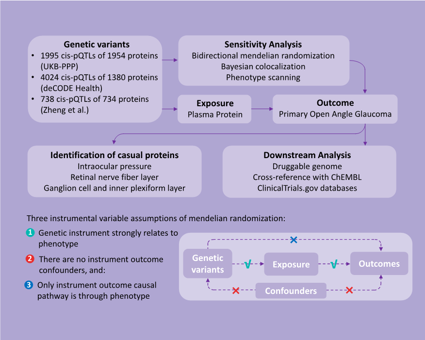
This study employed a two-sample PW-MR approach using protein quantitative trait loci (pQTL) data from seven extensive proteomics investigations to explore their correlation with POAG (Figure 1). Rigorous sensitivity analyses were conducted to reinforce the causal link between plasma proteins and POAG. To validate the association of proteins with POAG, their causal connections with various POAG-related traits were established. The study culminated in a comprehensive evaluation of the druggability of these proteins, with a specific focus on their potential as therapeutic agents. Ethical clearances from the original studies, which relied on publicly available summary data, obviated the need for additional ethical approval or patient consent for this study.
Data Sources
Instrumental variables (IVs) for plasma proteins, specifically cis-pQTLs, were primarily derived from two large-scale genome-wide association study (GWAS) datasets: the UK Biobank Pharma Proteomics Project (UKB-PPP) and the deCODE Health study.[15- 20] A total of 734 plasma proteins associated with cis-pQTLs were identified from Zheng et al.'s research.[21] The selection criteria included genome-wide significance (P < 5×10⁻⁸), independence (linkage disequilibrium [LD] clump r 2 < 0.001), and proximity, with cis-pQTLs located within 1 Mb of their protein-coding genes. Following these guidelines, we cataloged 1,995 cis-pQTLs for 1,954 plasma proteins from the UK Biobank Pharma Proteomics Project (UKB-PPP) (Olink platform, n = 54,306) and 4,024 cis-pQTLs for 1,380 circulating proteins from the deCODE Health study (Somascan platform, n = 35,559). In addition, 738 cis-pQTLs for 734 plasma proteins based on the Olink and Somascan platform reported by Zheng et al., included studies by Sun et al., Emilsson et al., Suhre et al., Yao et al., and Folkersen et al. (eTable 1 in the supplement).
Further, data on single nucleotide polymorphisms (SNPs) linked to plasma proteins and their associations with POAG were obtained from the FinnGen R10 study. This study comprised 8,530 cases of European descent and 391,275 controls.[22] We also integrated the largest GWAS dataset for three established POAG-related phenotypes: intraocular pressure (IOP),[23] retinal nerve fiber layer (RNFL) thickness, and ganglion cell-inner plexiform layer (GCIPL) thickness,[24] thereby ensuring racial homogeneity in the exposure and outcome datasets.
STATISTICAL ANALYSIS
Two-sample MR Analysis
Leveraging cis-pQTLs for plasma proteins sourced from three comprehensive databases, we undertook a meticulous evaluation of genetic instrument reliability. This involved calculating the variance proportion (R ²) explained by the genetic IVs for each protein as well as evaluating the association strength between these IVs and risk factors utilizing F-statistics. Proteins without available IVs were excluded to ensure data consistency. By conducting thorough screening, we found 5,722 cis-pQTLs for 2,412 proteins with an R² greater than 0.1% to be valid genetic instruments for our analysis.
In our primary analysis, we used plasma proteins as the exposure and POAG as the outcome, utilizing the “TwoSampleMR” software package for computation. We derived association estimates between plasma proteins and POAG utilizing the Wald ratio method (for proteins associated with a single cis-pQTL) and the inverse variance-weighted (IVW) MR method (for proteins linked to multiple cis-pQTLs). Confidence intervals (CIs) were determined through the delta method, with results expressed as MR odds ratios (ORs) and corresponding 95% CIs for an increase of one standard deviation (SD) in gene-predicted plasma protein levels.[25] To mitigate the risk of false positives due to multiple testing, we applied the Benjamini–Hochberg correction, targeting a false discovery rate (FDR) of 0.05 and a more stringent Bonferroni correction, setting significance at P < 2.07 × 10⁻⁵ (0.05 / 2,412).
Reverse Causality Detection
To address the possibility of reverse causation, we utilized bidirectional MR to assess the relationship between POAG risk and the levels of causal plasma proteins. Effect estimates were derived using the MR-inverse variance weighted (MR-IVW) method, and were further validated using MR-Egger, weighted median, simple mode, and weighted mode techniques. Statistical significance was established at P < 0.05, based on comprehensive GWAS data from three previous plasma protein studies.
Bayesian Colocalization Analysis
We conducted colocalization analysis to discern the impact of horizontal pleiotropy from genetic IVs on the identified causal links.[26] For this analysis, we utilized a Bayesian framework to evaluate posterior probabilities for five scenarios regarding the genetic overlap between plasma proteins and POAG. The scenarios ranged from no shared causal variants (H0) to specific causal variants for proteins (H1) or POAG (H2), independent causal variants affecting both (H3), and a common causal variant for both (H4). A posterior probability of (PH4) ≥ 0.8 indicates strong colocalization, whereas 0.8 > PH4 ≥ 0.5 suggests moderate support. This was performed using the “coloc” package in R (version 4.4.1).
Phenotype Scanning
Utilizing the “phenoscanner” R package, we explored large-scale genetic association studies for links between our identified pQTLs and POAG-related traits.[27] SNPs that showed pleiotropy met stringent criteria: genome-wide significance (P < 5×10⁻⁸), association studies in European populations, and links to established POAG risk factors, including metabolic traits, proteins, or clinical features.
Identification in POAG-related Traits
To validate identified plasma proteins and investigate their potential as therapeutic targets, we conducted two-sample MR analyses to assess the relationship between plasma proteins and POAG-related phenotypes. Utilizing SNPs from an extensive database of plasma protein cis-pQTLs as IVs, we focused on three major GWAS datasets for POAG traits: intraocular pressure (IOP), retinal nerve fiber layer (RNFL) thickness, and ganglion cell-inner plexiform layer (GCIPL) thickness. Through comprehensive MR analyses, we estimated the associations between these proteins and POAG-related phenotypes using both the Wald ratio and inverse variance-weighted MR methods. The results are reported as MR ORs with 95% CIs for an increase in plasma protein levels by one standard deviation, with significance set at P < 0.05.
Downstream Analysis of Drug Target Proteins
We explored plasma proteins as potential drug targets, and examined the druggability of MR-prioritized proteins against an updated list of drug-gene candidates.[28] The ChEMBL database (version 33) was consulted for drug molecule classifications, approved indications, and clinical trial outcomes,[29] alongside a detailed review through the https://www.ClinicalTrials.gov website for additional relevant information. This druggability assessment categorized proteins into four groups: approved (for proteins with at least one approved drug), in development (for proteins under clinical investigation), druggable (for proteins identified as potential targets but not yet listed in the drug database), and not currently listed as druggable (for proteins not listed as potential targets in the drug database).
Data Availability
Summary-level statistics for cis-pQTLs are available from the original publications. POAG GWAS summary statistics can be accessed via the FinnGen consortium (https://www.finngen.fi). Summary statistics for IOP, RNFL, and GCIPL can be retrieved from the GWAS Catalog (https://www.ebi.ac.uk/gwas).
RESULTS
Mendelian Randomization Between Plasma Proteins and POAG
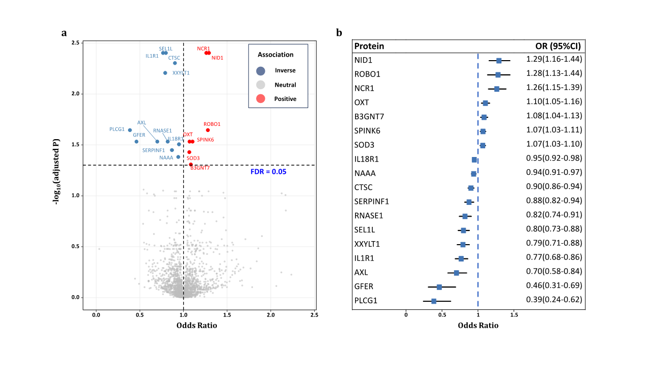
In the MR analysis, utilizing these 2,412 plasma proteins as study exposures and implementing FDR correction, we identified 18 plasma proteins significantly associated with POAG risk (Table 1). For each protein, an increment of one SD in genetically predicted levels altered the odds of POAG, with ORs ranging from 0.39 (95% CI: 0.24–0.62; P = 7.70×10⁻⁵) for phospholipase C gamma 1 (PLCG1) to 1.29 (95% CI: 1.16–1.44; P = 6.72 ×10⁻⁶) for nidogen-1 (NID1). Among these proteins, 7 exhibited positive associations with POAG risk, while 11 displayed negative associations (Table 1, Figure 2). After applying a Bonferroni correction for multiple testing (P < 2.07×10⁻⁵), six proteins remained significantly linked to POAG. Notably, all analyzed proteins demonstrated F-statistic values above 10, thereby effectively eliminating the possibility of weak instrumental variable bias.
|
Protein |
Chr |
Position |
rs number cis-pQTL |
EAF |
EA |
MR analysis |
Source
|
||||||
|
β (SE) |
OR |
95%CI |
P value |
R2 |
F statistics |
|
|||||||
|
AXL |
19 |
41233275 |
rs66841352 |
0.40 |
C |
-0.36(0.09) |
0.70 |
0.58-0.84 |
1.62×10⁻⁴ |
0.014 |
473.03 |
UKB-PPP |
|
|
B3GNT7 |
2 |
231398416 |
rs2290130 |
0.23 |
A |
0.08(0.02) |
1.08 |
1.04-1.13 |
3.77×10⁻⁴ |
0.169 |
6888.59 |
UKB-PPP |
|
|
GFER |
16 |
1973321 |
rs61516948 |
0.13 |
T |
-0.77(0.20) |
0.46 |
0.31-0.69 |
1.50×10⁻⁴ |
0.003 |
89.38 |
UKB-PPP |
|
|
PLCG1 |
20 |
41168825 |
rs753381 |
0.47 |
C |
-0.95(0.24) |
0.39 |
0.24-0.62 |
7.70×10⁻⁵ |
0.002 |
70.03 |
deCODE |
|
|
SEL1Lb |
14 |
81506097 |
rs11499034 |
0.01 |
C |
-0.22(0.05) |
0.80 |
0.73-0.88 |
5.05×10⁻⁶ |
0.044 |
1538.02 |
UKB-PPP |
|
|
SERPINF1 |
17 |
1666253 |
rs62088172 |
0.35 |
T |
-0.13(0.04) |
0.88 |
0.82-0.94 |
2.54×10⁻⁴ |
0.088 |
316.65 |
Sun |
|
|
CTSCa, b |
11 |
88337746 |
rs11600158 |
0.10 |
G |
-0.10(0.02) |
0.90 |
0.86-0.94 |
1.06×10⁻⁵ |
0.146 |
5787.36 |
UKB-PPP |
|
|
88066714 |
rs55897509 |
0.92 |
A |
0.085 |
509.91 |
Emilsson |
|||||||
|
IL18R1a |
2 |
102377596 |
rs10190555 |
0.77 |
G |
-0.05(0.01) |
0.95 |
0.92-0.98 |
1.86×10⁻⁴ |
0.228 |
9955.02 |
UKB-PPP |
|
|
101625575 |
rs115232861 |
0.03 |
C |
0.006 |
226.86 |
deCODE |
|||||||
|
103035044 |
rs1420106 |
0.78 |
G |
0.270 |
1221.57 |
Sun |
|||||||
|
102877724 |
rs183611009 |
0.02 |
G |
0.006 |
227.53 |
deCODE |
|||||||
|
102030778 |
rs187572594 |
0.02 |
G |
0.002 |
63.20 |
deCODE |
|||||||
|
101829431 |
rs55715763 |
0.06 |
G |
0.004 |
133.11 |
deCODE |
|||||||
|
102264346 |
rs55871806 |
0.15 |
C |
0.088 |
3399.96 |
deCODE |
|||||||
|
102549866 |
rs72995641 |
0.21 |
A |
0.031 |
1126.2 |
deCODE |
|||||||
|
101525580 |
rs75094400 |
0.02 |
C |
0.003 |
101.34 |
deCODE |
|||||||
|
102971459 |
rs80339564 |
0.03 |
C |
0.003 |
116.82 |
deCODE |
|||||||
|
IL1R1a, b |
2 |
102156623 |
rs3917238 |
0.30 |
T |
-0.27(0.06) |
0.77 |
0.68-0.86 |
4.22×10⁻⁶ |
0.015 |
528.97 |
UKB-PPP |
|
|
102138349 |
rs6706048 |
0.28 |
C |
0.012 |
440.56 |
deCODE |
|||||||
|
102746276 |
rs7588201 |
0.72 |
A |
0.007 |
39.29 |
Emilsson |
|||||||
|
NAAAa |
4 |
75932965 |
rs112197434 |
0.23 |
T |
-0.06(0.02) |
0.94 |
0.91-0.97 |
3.00×10⁻⁴ |
0.141 |
5525.12 |
UKB-PPP |
|
|
76102346 |
rs12331871 |
0.17 |
G |
0.016 |
568.16 |
deCODE |
|||||||
|
75906793 |
rs1394919 |
0.29 |
A |
0.148 |
6148.47 |
deCODE |
|||||||
|
76209167 |
rs76229059 |
0.17 |
G |
0.005 |
190.32 |
deCODE |
|||||||
|
76848231 |
rs9996608 |
0.30 |
T |
0.119 |
445.59 |
Sun |
|||||||
|
NCR1a, b |
19 |
55419632 |
rs143981324 |
0.90 |
T |
0.23(0.05) |
1.26 |
1.15-1.39 |
1.99×10⁻⁶ |
0.013 |
74.26 |
Emilsson |
|
|
54904812 |
rs7255591 |
0.09 |
C |
0.031 |
1073.76 |
UKB-PPP |
|||||||
|
NID1a, b |
1 |
236099073 |
rs17554536 |
0.18 |
A |
0.26(0.06) |
1.29 |
1.16-1.44 |
6.72×10⁻⁶ |
0.002 |
67.92 |
deCODE |
|
|
236104574 |
rs58074293 |
0.02 |
A |
0.011 |
379.94 |
deCODE |
|||||||
|
236067017 |
rs76183323 |
0.01 |
A |
0.012 |
402.08 |
UKB-PPP |
|||||||
|
OXTa |
20 |
3067658 |
rs6115776 |
0.40 |
C |
0.10(0.03) |
1.10 |
1.05-1.16 |
1.61×10⁻⁴ |
0.059 |
2208.44 |
deCODE |
|
|
3100953 |
rs857244 |
0.46 |
C |
0.001 |
41.77 |
deCODE |
|||||||
|
3049890 |
rs877172 |
0.66 |
T |
0.125 |
776.73 |
Emilsson |
|||||||
|
RNASE1a |
14 |
20789957 |
rs11624082 |
0.26 |
T |
-0.20(0.05) |
0.82 |
0.74-0.91 |
1.16×10⁻⁴ |
0.001 |
50.25 |
deCODE |
|
|
20814883 |
rs12897030 |
0.30 |
T |
0.016 |
592.52 |
deCODE |
|||||||
|
21280678 |
rs17254387 |
0.69 |
A |
0.022 |
72.52 |
Sun |
|||||||
|
20828671 |
rs188152481 |
0.02 |
C |
0.002 |
63.19 |
deCODE |
|||||||
|
20722147 |
rs2002078 |
0.41 |
T |
0.004 |
158.96 |
deCODE |
|||||||
|
20627658 |
rs75124405 |
0.01 |
A |
0.002 |
66.29 |
deCODE |
|||||||
|
ROBO1a |
3 |
78946828 |
rs331172 |
0.49 |
G |
0.25(0.06) |
1.28 |
1.13-1.44 |
6.78×10⁻⁵ |
0.003 |
88.58 |
deCODE |
|
|
78725134 |
rs3773233 |
0.21 |
T |
0.014 |
485.44 |
deCODE |
|||||||
|
78735620 |
rs3773244 |
0.20 |
A |
0.020 |
687.56 |
UKB-PPP |
|||||||
|
79787885 |
rs62256944 |
0.10 |
A |
0.002 |
58.34 |
deCODE |
|||||||
|
77848463 |
rs73103884 |
0.02 |
G |
0.001 |
45.09 |
deCODE |
|||||||
|
78741405 |
rs73111654 |
0.01 |
A |
0.002 |
81.48 |
deCODE |
|||||||
|
79369917 |
rs7639769 |
0.46 |
T |
0.003 |
100.95 |
deCODE |
|||||||
|
SOD3a |
4 |
24800212 |
rs1799895 |
0.01 |
G |
0.06(0.02) |
1.07 |
1.03-1.10 |
2.42×10⁻⁴ |
0.133 |
5170.20 |
UKB-PPP |
|
|
24802616 |
rs2695234 |
0.08 |
G |
0.031 |
1145.21 |
deCODE |
|||||||
|
25235769 |
rs313548 |
0.30 |
A |
0.003 |
109.25 |
deCODE |
|||||||
|
25664005 |
rs76930256 |
0.02 |
T |
0.005 |
190.96 |
deCODE |
|||||||
|
24444429 |
rs77514718 |
0.01 |
C |
0.014 |
505.01 |
deCODE |
|||||||
|
24639073 |
rs80234081 |
0.03 |
C |
0.021 |
751.96 |
deCODE |
|||||||
|
SPINK6a |
5 |
148263754 |
rs116664727 |
0.13 |
A |
0.07(0.02) |
1.07 |
1.03-1.11 |
1.41×10⁻⁴ |
0.006 |
223.76 |
deCODE |
|
|
148186561 |
rs12717962 |
0.55 |
G |
0.072 |
2630.07 |
UKB-PPP |
|||||||
|
147294723 |
rs137864680 |
0.02 |
A |
0.001 |
50.58 |
deCODE |
|||||||
|
148310721 |
rs139120114 |
0.01 |
C |
0.014 |
511.80 |
deCODE |
|||||||
|
147603178 |
rs1432688 |
0.92 |
G |
0.155 |
605.02 |
Sun |
|||||||
|
149047717 |
rs148402978 |
0.02 |
T |
0.003 |
113.83 |
deCODE |
|||||||
|
147734214 |
rs148632877 |
0.01 |
T |
0.002 |
70.11 |
deCODE |
|||||||
|
148183699 |
rs74765295 |
0.01 |
T |
0.007 |
248.34 |
deCODE |
|||||||
|
147882756 |
rs75666474 |
0.05 |
A |
0.003 |
93.34 |
deCODE |
|||||||
|
148453118 |
rs75666559 |
0.04 |
T |
0.007 |
259.13 |
deCODE |
|||||||
|
148057440 |
rs7719473 |
0.03 |
A |
0.057 |
2144.61 |
deCODE |
|||||||
|
XXYLT1a, b |
3 |
195287123 |
rs1538767 |
0.46 |
C |
-0.23(0.05) |
0.79 |
0.71-0.88 |
1.58×10⁻⁵ |
0.016 |
564.92 |
deCODE |
|
|
195619664 |
rs534994266 |
0.09 |
A |
0.002 |
80.40 |
deCODE |
|||||||
|
194783033 |
rs55947051 |
0.84 |
T |
0.010 |
56.46 |
Emilsson |
|||||||
|
195072715 |
rs7635512 |
0.16 |
C |
0.007 |
240.77 |
deCODE |
|||||||
Source indicates the protein GWAS providing the estimate of the effect of the cis-pQTL on protein level. Results express changes in primary open angle glaucoma risk per 1-SD increase in protein level. EA=effect allele; EAF=effect allele frequency; β=effect on diabetic retinopathy; OR=odds ratio.
a Mendelian randomization using the inverse variance weighted methods.
b Plasma protein passed Bonferroni correction (P < 2.12×10⁻⁵).
Multiple Sensitivity Analyses
Table 2. Summary of reverse causality detection, Bayesian co-localization analysis and phenotype scanning on 18 potential causal proteins.
|
Protein |
Bidrectional MR |
Co-localization |
Previously reported associations |
||
|
OR(95%CI) |
P value |
PPH3 |
PPH4 |
||
|
SEL1L |
1.00(0.98-1.02) |
0.80 |
0.002 |
0.995 |
lipid metabolism |
|
ROBO1 |
1.00(0.98-1.03) |
0.92 |
0.065 |
0.865 |
NA |
|
AXL |
1.00(0.99-1.00) |
0.12 |
0.052 |
0.858 |
NA |
|
NID1 |
1.00(0.97-1.02) |
0.73 |
0.039 |
0.806 |
NA |
|
GFER |
1.01(0.99-1.03) |
0.34 |
0.125 |
0.804 |
NA |
|
NCR1 |
1.00(0.97-1.02) |
0.81 |
0.335 |
0.505 |
NA |
|
SERPINF1 |
0.96(0.91-1.02) |
0.20 |
0.366 |
0.476 |
Body measurement |
|
CTSC |
0.99(0.97-1.01) |
0.36 |
0.323 |
0.431 |
NA |
|
SPINK6 |
1.00(0.98-1.02) |
0.94 |
0.399 |
0.260 |
NA |
|
B3GNT7 |
1.01(0.99-1.04) |
0.25 |
0.322 |
0.210 |
Body measurement |
|
RNASE1 |
0.97(0.93-1.02) |
0.29 |
0.117 |
0.193 |
Cervix uteri disease |
|
NAAA |
0.99(0.97-1.01) |
0.36 |
0.181 |
0.116 |
NA |
|
IL1R1 |
1.00(0.97-1.02) |
0.75 |
0.354 |
0.076 |
Blood cell, asthma, hayfever, rhinitis, eczema, bronchitis, emphysema |
|
IL18R1 |
1.02(1.00-1.03) |
0.07 |
0.340 |
0.026 |
Blood cell, inflammatory bowel disease |
|
PLCG1 |
1.01(0.96-1.06) |
0.68 |
0.092 |
0.025 |
Blood cell, lipid metabolism |
|
XXYLT1 |
0.96(0.91-1.02) |
0.20 |
0.520 |
0.022 |
NA |
|
SOD3 |
0.99(0.97-1.01) |
0.42 |
0.506 |
0.009 |
Vascular dementia |
|
OXT |
1.02(0.96-1.08) |
0.54 |
0.018 |
0.004 |
NA |
OR=odds ratio; NA=not applicable.
Colocalization analysis was employed to address potential confounding from linkage disequilibrium (LD), thereby determining whether the levels of causal plasma proteins shared genetic origins with POAG. This analysis confirmed strong co-localization evidence (PH4 ≥ 0.8) for SEL1L, ROBO1, AXL, NID1, and GFER, with NCR1 showing moderately strong evidence (0.8 > PH4 ≥ 0.5), thereby suggesting shared genetic determinants between these proteins and POAG (eFigure in the supplement).
Furthermore, phenotypic scanning of cis-pQTLs for POAG-associated proteins revealed no direct links between the IVs and POAG or its risk factors. However, we identified associations of cis-pQTLs for certain proteins with other health conditions: IL1R1 (rs3917238, rs6706048, and rs7588201) and IL18R1 (rs10190555 and rs1420106) with blood components; SEL1L (rs11499034) and PLCG1 (rs753381) with specific diseases; RNASE1 (rs75124405) with cervical diseases; SOD3 (rs80234081) with vascular dementia; IL18R1 with inflammatory bowel disease; and IL1R1 with immune disorders such as asthma, hay fever, allergic rhinitis, and eczema. Modifying the significance threshold to P < 1 × 10⁻⁵ did not identify any cis-pQTLs for plasma proteins related to POAG or its risk factors.
Identification of Plasma Proteins in IOP, RNFL, and GCIPL
The MR validation for 18 plasma proteins in relation to POAG traits was processed (eTable2 in the supplement). Among these, nine proteins demonstrated genetic associations with at least one POAG-related phenotype. Notably, pigment epithelium-derived factor (SERPINF1) was the only protein found to be significantly associated with IOP outcomes. Four proteins namely dipeptidyl peptidase 1 (CTSC), serine protease inhibitor Kazal-type 6 (SPINK6), interleukin-1 receptor type 1 (IL1R1), and ribonuclease pancreatic (RNASE1) were found to show significant causal relationships with RNFL thickness. Moreover, five proteins including interleukin-18 receptor 1 (IL18R1), namely N-acylethanolamine-hydrolyzing acid amidase (NAAA), IL1R1, phospholipase C gamma 1 (PLCG1), and sel-1 homolog 1 (SEL1L) were significantly associated with GCIPL thickness.
Directional consistency was observed for IL18R1, IL1R1, PLCG1, RNASE1, and SPINK6 across their associations with POAG-related traits and POAG itself. Remarkably, an increase of one standard deviation in genetically predicted levels of IL1R1 revealed a significant positive causal correlation with both RNFL (OR = 1.42; 95% CI, 1.11–1.83; P = 0.01) and GCIPL (OR = 1.42; 95% CI, 1.02–1.84; P = 0.04), thereby indicating IL1R1’s protective effect on these eye structures and its beneficial role in POAG, as highlighted in the primary MR analysis.
The Druggability and Development Status of POAG-causal Protein
To assess the druggability and advancement of plasma protein targets associated with POAG, we undertook a comprehensive review of druggable genes, the ChEMBL drug discovery database, and ClinicalTrials.gov. We identified 18 drug targets and sorted them into four categories based on their development stage: approved (targets with drugs already , authorized for use), in development (targets undergoing clinical trials), druggable (potential targets), and not currently listed as druggable (not potential targets) (Table 3). This analysis revealed five plasma proteins as prime candidates for future drug development efforts: tyrosine-protein kinase receptor UFO (AXL), dipeptidyl peptidase 1 (CTSC), interleukin-18 receptor 1 (IL18R1), oxytocin (OXT), and interleukin-1 receptor type 1 (IL1R1).
Drugs targeting AXL, such as gilteritinib, have received approval for specific neoplasms, including acute myeloid leukemia. Similarly, IL1R1 has played a key role in treating immune disorders, such as cryopyrin- associated periodic syndromes and rheumatoid arthritis, with anakinra being a notable example. Apart from these, the other three highlighted proteins are associated with drugs in Phase I–III clinical trials; however, there is no direct evidence of their use in developing treatment for POAG. The drug development status for the remaining 13 plasma proteins remains unclear. Our findings suggest AXL, GFER, SERPINF1, CTSC, IL18R1, IL1R1, NAAA, NCR1, NID1, OXT, RNASE1, SOD3, and SPINK6 as significant druggable targets that warrant further investigation for drug development.
Table 3. Summary of druggability and clinical development activity for primary open angle glaucoma associated with causal associations on MR analysis.
|
Target |
Compound name |
Clinical development activities |
|
Approved |
||
|
AXL |
GILTERITINIB (CHEMBL3301622)b |
Approved for neoplasms, acute myeloid leukemia, myeloid leukemia; Phase II: myelodysplastic syndromes, blast crisis; Phase I: non-smal-cell lung carcinoma, kidney diseases, liver diseases |
|
IL1R1 |
ANAKINRA (CHEMBL1201570)b |
Approved: immune system diseases; cryopyrin-associated periodic syndromes, rheumatoid arthritis; Phase III: metabolic syndrome, giant cell arteritis, pneumonia, severe acute respiratory syndrome, myocardial infarction, mucocutaneous lymph node syndrome, familial mediterranean fever, influenza, dermatitis, allergic contact; Phase II: brain injuries, lymphoma, follicular, multiple myeloma, pericarditis, diabetes mellitus, Still's disease, uveitis, hvidradenitis suppurativa, myositis, alcoholic hepatitis, macrophage activation syndrome, gout, dermatomyositis, sarcoidosis, amyotrophic lateral sclerosis, infections, hypoglycemia, type 1 diabetes mellitus, osteoarthritis, juvenile arthritis, heart failure, cerebral hemorrhage, polycystic ovary syndrome, myocarditis, labyrinth diseases, chronic fatigue syndrome, chronic renal insufficiency; Phase I: castration-resistant prostatic neoplasms, pancreatic neoplasms, knee injuries, blepharitis, HIV infections, multiple sclerosis, breast neoplasms, pulmonary hypertension, pain, smoldering multiple, myeloma, asthma, rectal neoplasms, chronic B-cell leukemia lymphocytic, hearing loss, neoplasms |
|
In development |
||
|
AXL |
BEMCENTINIB (CHEMBL3809489)b |
Phase II: adenocacinoma of lung; mesothelioma; severe acute respiratory; inflammatory breast neoplasms; Phase I: myelodysplastic syndromes; acute leukemia myeloid; pancreatic neoplasms; non-small-cell lung carcinoma; melanoma |
|
AXL |
ENAPOTAMAB VEDOTIN (CHEMBL4297987)b |
Phase I: non-small-cell lung carcinoma |
|
AXL |
BPI-9016 (CHEMBL3545236)b |
Phase I: non-small-cell lung carcinoma; neoplasms |
|
CTSC |
BRENSOCATIB (CHEMBL3900409)b |
Phase III: cystic fibrosis; COVID-19; Phase II: bronchiectasis |
|
IL18R1 |
IBOCTADEKIN (CHEMBL2108034)b |
Phase II: melanoma; Phase I: neoplasms; lymphoma non-hodgin; lymphoma |
|
IL1R1 |
MEDI-8968 (CHEMBL2109607)b |
Phase II: hidradenitis suppurativa; chronic obstructive pulmonary disease |
|
IL1R1 |
AMG-108 (CHEMBL2109458)b |
Phase II: rheumatoid arthritis; diabetes mellitus; osteoarthritis |
|
OXT |
OXYTOCINc |
Phase III: Prader-Willi syndrome (NCT02804373), socially adaptive mirroring (NCT03640156); Phase II: central diabetes insipidus (NCT06036004), schizophrenia (NCT01699997) |
|
Druggablea |
||
|
AXL, GFER, SERPINF1, CTSC, IL18R1, IL1R1, NAAA, NCR1, NID1, OXT, RNASE1, SOD3, SPINK6 |
||
|
Not currently listed as druggablea |
||
|
B3GNT7, PLCG1, SEL1L, ROBO1, XXYLT1 |
||
a Data from druggable gene list.
b Data from ChEMBL release 33 (compound ID in brackets).
DISCUSSION
Summary of Findings
This PW-MR study reveals 18 plasma proteins as potential therapeutic targets for POAG, thereby effectively addressing concerns related to pleiotropy and reverse causality. Co-localization analysis reveals that elevated genetic expressions of SEL1L, AXL, and GFER decrease POAG risk, while higher levels of ROBO1, NID1, and NCR1 increase it. Validation confirms that nine proteins are causally associated with POAG related phenotypes, with IL18R1, IL1R1, PLCG1, RNASE1, and SPINK6 consistently linked to POAG. The assessment of durability and clinical development activities prioritized 18 causal plasma proteins. These findings provide novel insights into POAG drug development strategies and emphasize the potential of PW-MR in unraveling the etiological biology of complex diseases.
MR-derived novel biomarkers and potential targets for POAG
Our study identified 18 proteins as novel POAG targets, with specific emphasis on SEL1L, ROBO1, AXL, NID1, and GFER for their strong co-localization evidence. SEL1L, as the protein most significantly associated with POAG risk—is integral to the endoplasmic reticulum-associated degradation (ERAD) system, which mitigates ER stress by eliminating misfolded proteins.[30-32] This feature proposes the capacity of SEL1L to impede the advancement of POAG, thus emphasizing the cruciality of additional assessment via meticulous experimental and clinical investigations to substantiate these proteins as potential therapeutic objectives for POAG.
In addition, ROBO1, which is integral to neurogenesis and angiogenesis, may be associated with POAG risk through its involvement in the SLIT/ ROBO1 pathway and SRGAP2 expression.[33-36] Studies have demonstrated that an increase in ROBO1 levels is closely linked to POAG, which may lead to retinal neovascularization and the possible onset of secondary glaucoma.[37-38] These results underscore the imperative for additional investigations into the involvement of ROBO1 in POAG neuroprotection and angiogenesis.
Nidogen-1 (NID1), a key basement membrane glycoprotein, has been found to be as positively associated with POAG risk.[39-40] Increased nidogen presence in glaucoma patients suggests its involvement in extracellular matrix dynamics and aqueous efflux obstruction.[41] The specific mechanisms by which NID1 contributes to POAG remain to be clarified, thereby highlighting a crucial area for future research. Our study found that higher levels of GFER, which is a protein vital for mitochondrial integrity, is correlated with reduced POAG risk.[42-44] This suggests GFER’s protective role in preventing oxidative stress and mitochondrial dysfunction, both of which contributes in glaucoma progression.[44-45] The potential of GFER as a therapeutic target for POAG highlights the need for additional research into its mechanisms of action.
AXL, known for its regulatory functions in cancer and inflammation,[46] emerges as a neuroprotective factor in POAG through its interaction with the APOE gene and microglial regulation.[47-48] This study proposed the involvement of AXL in reducing neurodegeneration in glaucoma, thus advocating for further investigation to validate AXL as a promising therapeutic approach in the management of POAG.
Multiple Sensitivity Analysis for MR Assumptions
To ensure the scientific validity and robustness of our results, we conducted a rigorous sensitivity analysis to confirm the assumptions of the MR. Initially, our focus was on cis-pQTLs within 1 Mb of protein-coding genes, specifically selected for their direct impact on protein expression. We adhered to stringent selection criteria including linkage disequilibrium (LD) clustering (r 2< 0.001), a significance threshold (P ≤ 5.0 × 10⁻⁸), and F-statistics over 10 to ensure the relevance of genetic IVs. Further, we guaranteed MR independence by examining associations between specific proteins (e.g., IL1R1, SERPINF1, and SOD3) and various traits, but we found no confounding influences on our MR analysis. To address the exclusion restriction assumption, we utilized the Bayesian co-localization analysis, identifying shared variants of five proteins (SEL1L, ROBO1, AXL, NID1, and GFER) with POAG, while acknowledging the method’s high false-negative rate (~60%), which did not detract from our main conclusions. Moreover, a bidirectional MR analysis was conducted to eliminate the possibility of reverse causation, ultimately confirming the absence of it among the studied proteins. This comprehensive approach emphasizes our commitment to maintaining the accuracy of MR assumptions, even though we acknowledge the ongoing potential impact of reverse causation.
Validation of Causal Proteins
The primary aim of this study was to establish a causative correlation between particular proteins and POAG-associated characteristics, with the intention of confirming potential pharmaceutical targets and elucidating the fundamental mechanisms of operation. By analyzing data from the most extensive GWAS database for IOP, RNFL, and GCIPL, our MR study revealed significant causal links between various plasma proteins and POAG-related traits, further validating them as potential targets for medication. This holds particular significance as the field pivots its attention towards not only decreasing IOP, but also delving into neuroprotective treatments that combat the apoptosis of retinal ganglion cells (RGCs), encompassing factors such as inflammation, neuroglial activation, neurotrophic deficiencies, and oxidative stress. Notably, IL1R1—part of the IL-1receptor family known for its role in inflammation and microglia activation—was associated with an increase in RNFL and GCIPL thickness, thereby suggesting a protective effect against POAG.[49-51] This positions IL1R1 as a promising candidate for neuroprotective therapeutic strategies in POAG, although further investigation is required to fully understand the roles of these proteins in POAG pathogenesis.
Scientific and Clinical Implications
This study provides critical insights that have profound implications for both medical research and clinical practice, emphasizing the value of a comprehensive approach in biomarker testing for identifying novel drug targets. Our analysis spanned 2,412 plasma proteins related to POAG, which is merely a fraction of the total proteome and, thus, emphasizes the untapped potential in the vast protein landscape. The findings from the TwoSampleMR (PW-MR) analysis not only shed light on potential drug mechanisms associated with these proteins but also validate the causal influence of certain proteins on POAG and its related traits. This correlation lays a genetic groundwork for developing neuroprotective therapies tailored to the molecular mechanisms of POAG.
Furthermore, our results indicate that proteins causally associated with POAG might also influence susceptibility to other diseases, thereby indicating the possibility of shared pathological pathways across different conditions. Interestingly, certain identified proteins are already recognized in the treatment of other diseases or are in clinical trials, thereby hinting at the potential for repurposing existing drugs for POAG treatment. Moreover, PW-MR analysis emerges as a powerful preclinical tool for drug target prioritization and the anticipation of possible adverse effects, thereby facilitating the discovery of novel therapeutic agents with superior efficacy and safety profiles. This study emphasizes the necessity of exploring the proteome more comprehensively to unveil novel pathways and therapeutic targets for POAG and beyond.
Strengths and Limitations
This study has several key strengths. First, it leverages the latest comprehensive GWAS data from European populations, thereby effectively reducing population stratification bias and enhancing the identification of causal associations. Rigorous validation through a suite of sensitivity analyses, including two-way MR, co-localization analyses, and phenotypic scanning, was instrumental. In particular, co-localization analyses were crucial in identifying shared genetic variations between exposures and outcomes. And MR validation analysis reinforced the causal relationships between plasma proteins and POAG-related phenotypes as well as affirming the viability of potential drug targets.
However, this study is not without its limitations. While single IV MR analysis may prompt concerns over incidental findings, our approach mitigated this by selecting strong, highly correlated cis-pQTLs, backed by robust SNPs, thereby ensuring reliability. We also employed multiple protein quantification techniques, such as Olink and Somascan, to address technique-specific biases.[52] Although our plasma protein data predominantly originated from the UK, the diverse origins of most cis-pQTLs bolster the generalizability of our findings, despite the potential for sample overlap. Further, this study's focus on individuals of European descent (due to data availability) highlights the necessity for future GWAS on biomarkers in non-European populations to enable more inclusive cross-ethnic MR analyses.
CONCLUSION
In conclusion, our systematic MR analysis of the plasma proteome has identified 18 promising drug targets for POAG, with robust co-localization support for five (SEL1L, ROBO1, AXL, NID1, GFER). These findings provide an unprecedented genetic insight into the etiology of POAG, thereby highlighting potential therapeutic targets and avenues. Our findings also emphasize the vast potential for future research in population genomics, proteomics, and their interplay with disease to discover new drug candidates. The next steps involve experimental and clinical studies to validate the functions and therapeutic efficacy of these proteins and, thus, mark a significant leap toward innovative POAG treatments.