




Introduction
Pterygium is a growth of conjunctiva in a triangular shape as a result of collagen degeneration and fibrovascular proliferation. It gradually extends from either the nasal or the temporal side of the sclera to the cornea, which can lead to the obstruction of vision in serious casesl1, 2. There are two types of pterygium:primary and recurrent. Primary pterygium occurs due to chronic degeneration of the conjunctival epithelium and subconjuctival connective tissue. Recurrent pterygium is a more aggressive fibrovascular growth that can result in lesions in the corneal and conjunctival tissue; recurrent pterygium can manifest from months to years after the first removal of the primary pterygium. Pterygium can be caused by chronic exposure to ultraviolet light, vitamin A deficiency, tear film disturbances, and micro trauma to the eyes2, 4. If left untreated, pterygium can cause damage to the visual axis, reduce vision, and cause irregular astigmatism. Thus, the best treatment pterygium is to excise the tissue via surgery1.
Many techniques have been used in pterygium surgery; however, only a few effectively reduce it from recurrence. Recently, amniotic membrane transplant (AMT) is the latest innovation that is being used in treating pterygium. Human amniotic membrane is derived from the fetal inner amniotic membranes, which are composed of:an intact epithelial structure, an intact dense basement membrane, and a loose collagen layer with fibroblasts12, 13. The amniotic membrane is used to cover the conjunctional defect after the removal of pterygium tissue. It is fitted to cover the defective conjunctiva by trimming off the excess portion. After the surgery, once covered by the epithelium, the amniotic membrane becomes visibly indistinguishable from the conjunctival tissue. It is secure to the corneal edge of the defect by either fibrin glue or Vicryl 8-0 sutures.
Suturing using needle and thread is the traditional way to close a wound. Many suture alternatives, including Fibrin glue, have been proposed in order to find a method ideal for surgery. TISSEEL is a type of Fibrin glue that consist of a sealer protein (human fibrinogen from pooled plasma), a fibrinolysis inhibitor solution (bovine), thrombin (human), and calcium chloride14. TISSEEL mimics the body's own response to injury by creating a coagulum (or clot) that makes the tissues adhere to each other. TISSEEL becomes coagulum within minutes of delivery;therefore, the amniotic membrane must be placed into position quickly3, 6, 7.
Many studies have proved that amniotic membrane transplantation is a safe and effective technique for pterygium surgery. The present research is intended to study how different methods of attaching amniotic membrane to the sclera using either TISSEEL or sutures can lead to different outcomes in terms of complications and recurrence.
Methods
TISSEEL preparation14
Pre-warming TISSEEL Kit with FIBRINOTHERM
1. Plugged the Fibrinotherm Heating and Stirring Device into an electrical socket and activated the warmer.
2. Placed all 4 vials from the TISSEEL Kit into the pre-warmed wells of the Fibrinotherm and allowed the vials to warm for 5 minutes.
Preparation for Sealer Protein Solution with FIBRINOTHERM
1. Removed the caps from the Sealer Protein Concentrate and the Fibrinolysis Inhibitor Solution vials; transferred the Fibrinolysis Inhibitor Solution into the Sealer Protein Concentrate vial.
2. Placed the vial into the largest opening of the Fibrinotherm device, switched on the stirrer and waited until all Sealer Protein Concentration was dissolved.
Preparation of Thrombin Solution with FIBRINOTHERM
1. Removed the caps from the Thrombin and the
Calcium Chloride Solution vials;then transferred the
contents of Calcium Chloride solution into the
Thrombin vial.
2. Placed the vial into the Fibrinotherm, then waited until all the Thrombin concentration was dissolved.
3. Kept the Thrombin Solution at 37℃ until application.
Excision of pterygium
Regardless of which technique was being used after the pterygium excision, all pterygium surgery patients underwent pterygium tissue removal. Firstly, the eye that underwent surgery was anesthetized topically and subconjuctivally. For the topical application, one drop of 0.2% Tetracaine solution was applied to the eye. After 15 minutes, the eye was sterilized and draped. An eyelid speculum was inserted, and then the pterygium tissue was outlined using surgical marking. A 0.3 ml dose of 2%Lidocaine with epinephrine was injected into the sub-conjunctiva to balloon the pterygium tissue, which led to the partition of the conjunctiva from the sclera so the conjunctiva could be excised while maintaining the sclera. Wescott scissors and No. 12 Miltex scalpel were used to remove pterygium tissue;cautery was used to stop any bleeding2, 4, 5.With recurrent cases, extra care was needed during surgery. There were scars on both the cornea and on the sclera underlying the pterygium;the excision on the sclera side was more difficult, resulting in more bleeding and in a greater danger of the surgeon unintentionally cutting the underlying tissue. During the excision of the pterygium tissue, the surgeon had to be careful not to cut the underlying rectus muscle, which was often adhered to a current pterygium. Amniotic membrane transplant using TISSEEL
After the removal of the pterygium tissue, a matching size of amniotic membrane with conjunctival defect was cut. Then, the amniotic membrane was placed onto the bare sclera with the epithelial structure down. The amniotic membrane was adhered with the defective conjunctiva with TISSEEL4, 10.
Amniotic membrane transplant using sutures
After removal of the pterygium tissue, a matching size of amniotic membrane with defective conjuncti va was cut. Then, the amniotic membrane was placed onto the bare sclera with the epithelial structure down. The amniotic membrane was sutured onto the superficial sclera with 8-0 Vicryl Suture1.Figure 1 Race distribution of pterygium patients who underwent surgery with amniotic membrane transplant using either TISSEEL or Sutures
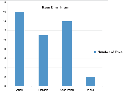
Figure 2 Age distribution of pterygium patients who underwent surgery with amniotic membrane transplant using either TISSEEL or sutures.
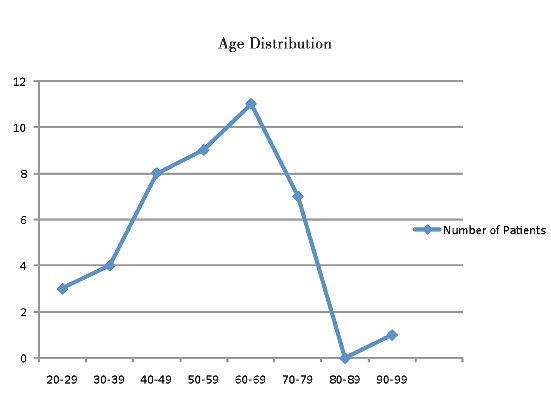
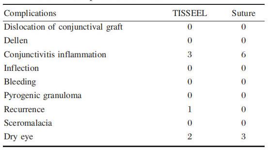
Table 1 Complications of TISSEEL versus suture
Results
Forty-three eyes of 43 patients, ranging from twenty-to ninety-years-old, with pterygium, received operation. Forty eyes had primary pterygium and three had recurrent. After the operation, patients were followed up from one week up to one year. At two weeks post-operation, 9 out of 43 patients (20.9%)in both groups had showed signs of conjunctivitis inflammation;of those, three eyes were in the TISSEEL group and six eyes were in the sutures group. After several followed ups and weeks of using anti-inflammatory and antibiotic drops, all eyes with conjunctivitis inflammation healed completely. P value of chi-square test was <0.05, which showed a significant differences in the two groups.The recurrence rate was 4.2% (1 in 24 eyes)in the TISSEEL group. The patient with the recurrence underwent another operation. Later follow-up showed no other sign of complication or recurrence. There were two dry eyes in the TISSEEL group and three dry eyes in the sutures group. P value >0.05 showed that no significant differences existed between the two groups.
The outcome of amniotic membrane transplant in patients of both groups was positive:the eyes healed without any problems. There was no dislocation of conjunctival graft, dellen, inflammation, infection, bleeding, pyogenic granuloma, or scleromalacia in either group.
Discussion
Pterygium can cause redness, swelling, itching, irritation, and blurring of vision. Most significantly, pterygium can cause loss of eyesight when it grows over the central area of the cornea. According to previous study, pterygium occurs twice as often in males as in females. It rarely takes place in persons under 20 s. However, persons aged between twenty to forty years have the highest incidence of pterygium, and persons older than forty have the highest prevalence of cases. The distribution of pterygium cases in the population varies with geographical location:it decreases with the upper latitudes and increases with the lower latitudes. Most importantly, pterygium is more likely to occur in people who are exposed to more ultraviolet light2 .As in any other operation, pterygium surgery has risks and complications. Though complications are rare, they may include, but are not limited to, bleeding, infection, double vision, droopy eyelids, dislocation of conjunctival grafts, Dellen, pain, inflammation, recurrence, scleromalacia, and pyogenic granuloma. The goals of pterygium surgery are to remove the pterygium tissue, restore the conjunctival tissue, and prevent recurrence. Thus, amniotic membrane transplant is an effective new technique, which is being used in pterygium surgery to deliver the best results1, 4, 10, 12.
Amniotic membrane is derived from the innermost layer of the human amniotic fetal membranes. The advantage of using amniotic membrane is that it has unique properties, including anti-adhesive effects, bacteriostatic properties, wound protection, and pain reduction. Additionally, the amniotic membrane has a lack of immunogenicity, which will not cause an immune reaction to the transplant. The uniqueness of the amniotic membrane is that it is regenerative rather reparative1, 4, 12.
AMT has been used to treat ocular surface abnormality since over five decade ago. Recently, the role of AMT in ocular disorders has been re-evaluated. Amniotic membrane has been successfully used in patients with persistent epithelial defects, pterygium, or symblepharon, and for ocular surface reconstruction. The amniotic membrane has been found to facilitate epithelization, to maintain a normal epithelial phenotype, to reduce inflammation, scarring, and vascularization12.
As a natural basement membrane, amniotic membrane contains various matrix proteins, which facilitate the adhesion, migration, differentiation, and prevention of the apoptosis of the epithelial cell. The amniotic membrane is capable of binding growth factors;this may help to promote wound healing. Subsequently, the amniotic membrane matrix inhibits extracellular matrix production and scar formation by the fibroblasts. Therefore, in the promotion of conjunctival epithelial wound healing, the suppression and extracellular matrix production by pterygium fibroblast are thought to be the major mechanisms by which an amniotic membrane graft inhibits pterygium recurrence. Other possible mechanisms include inhibition of inflammation by inhibiting chemokines expression by fibroblasts, and interleukin-1 expression by epithelial cells, inhibition of neovascularization by inhibiting vascular endothelial cell growth, presence of anti-angiogeni/anti-inflamatory proteins, and protease inhibitors. Possibly, the inhibition of postoperative inflammation and of vascular cells activation and invasion by amniotic membrane may also contribute to reduction of pterygium recurrence8, 12.
In this study, all patients underwent AMT using either TISSEEL or sutures. The recurrence rate for AMT with TISSEEL was 4.2%; by contrast, there were no recurrences for AMT with sutures. Amniotic membrane had been successfully used in patients for restoration of the ocular surface. As its mechanism of action becomes more fully understood, its application will become more refined, with more appropriate usage of this valuable technique.
Advantages and disadvantages of fibrin glue and suture
Suturing is an old, basic method to close wounds after surgery;it has some disadvantages in ophthalmic use, such as irritation, infection, inflammation, and allergy. Suturing a corneal laceration inflicts trauma to the cornea just from the multiple needle passes. Sutured wounds typically result in astigmatism and uneven healing. Additionally, sutures are a potential cause for infection, and they can incite inflammation and neovascularization in the cornea, which can ultimately result in corneal scarring. Suturing also requires additional operating time and technical skill. Finally, the prompt removal of suture is necessary, particularly those made of monofilament nylon material, to avoid the risks of suture abscess and neovascularization3, 5, 9.TISSEEL is a new suture alternative. The advantages of TISSEEL over sutures are less inflammation, faster surface rehabilitation, shorter surgical time, and less discomfort for the patients. The disadvantage of TISSEEL is the hypothetical risk of viral or prion disease transmission. Prion diseases transmission is hypothetically possible because of bovine component in TISSEEL. However, there has never been any report of the transmission. The transmission of Hepatitis B, Hepatitis C, and HIV is also possible;currently, there has been no report of these3, 7, 9, 11.
While Fibrin glue proved to be efficient in adjuncting tissue after surgery, it also has adverse effect, such as14:
Hypersensitivity or Allergic/Anaphylactoid reactions: in specific cases, these reactions can lead to severe anaphylaxis.
● Potential transmission of viral diseases from the donor plasma
TISSEEL is applied as a thin layer. Excessive clot thickness may negatively interfere with the product's efficacy and the wound healing process.
There have been rare reports14 of:
● Immune system disorders:hypersensitivity, anaphylactic responses
● Cardiac disorders:bradycardia, tachycardia, hypertension, thromboembolic complications
● Respiratory, thoracic, and mediastinal disorders:dyspnea
● Gastrointestinal disorders:nausea
● Skin and subcutaneous tissue disorders:urticaria, pruritus
● General disorders and administration site condition:flushing
Post-operative complications of amniotic membrane transplant
Complications are rare, but if they occur, the amnion membrane can become loosened or dislocated;hemorrhaging can occur under the membrane and cause early disintegration. The data in Figure l of this study show that there were a greater number of pterygium cases in Asian, Asian Indian, and Hispanic patients than in Caucasian patients. Since most of the participants in the study were non-Caucasians, this could contribute to certain groups having a higher occurrence rate than the others. Additionally, before immigrating to the United States, many patients lived in regions with more exposure to ultraviolet light. Besides the contribution of geographical and racial factors, age also played an important role in pterygium occurrence. Figure 2 shows that more patients in the range of forty to seventy-nine years old have pterygium.
Conclusion
Pterygium surgery reduced astigmatism and increased the spherical power of cornea. Amniotic membrane facilitated reJ-epithelization, and reduced scarring and vascularization;as a result, eyes appearedwhiter than when using conjunctival autographs. Amniotic membrane was 50% to 100% larger in size than conjunctival autografts.
Using amniotic membrane in pterygium surgery offered aesthetic improvement and decreased pain, dryness, and discomfort;it also resulted in a whiter eye after surgery. Using amniotic membrane decreased rehabilitation time, and did not cause the patient to miss work or important daily activities.
There were few complications during and post operation for either TISSEEL or sutures. Table l shows there were more cases of dry eye and inflammations in suture patients than in TISSEEL patients. However, though there was one recurrence case in TISSEEL group, the difference was insignificant.