





Previous researches1,2 reported that selective implantation of an aspherical intraocular lens(IOL) based on the corneal spherical aberration measured before surgery is able to minimize the patient's total spherical aberration to a value close to zero following the procedure, thereby yielding better visual function3,4. The equipment used to measure corneal spherical aberration includes corneal topography systems, the iTrace wavefront aberration analyzer, and the Pentacam HR anterior segment analyzer. In this study, we utilized the Pentacam analyzer to measure patients'corneal spherical aberration preoperatively and conducted aspherical IOP implantation according to each individual situation. The consistency between preoperative estimates and actual total spherical aberration measured postoperatively was statistically compared.
General data
Among the 82 eyes, 45 were implanted with the Akreos Advanced Optic IOL(AO) and the other 37 with the TecnisZA9003 IOL. Regardless of the type of IOL implant,all patients had a significant difference in corneal spherical aberration(6 mm diameter) before and after surgery(P<0.05), as shown in Table 1.


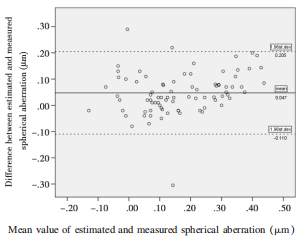
(d) between preoperative estimates and postoperative measurement. The dashed line represents the standard deviation of difference (SD=0.080) within 95%CI(d±1.96×SD).
Materials and methods
Study subjects
Forty-seven patients with age-related cataract ad- mitted to the Department of Ophthalmology at our hospital were enrolled in this study,including 20 males and 27 females, aged 51 to 84 years, (average 67±9 years). Inclusion criteria: patients with agerelated cataract aged from 50 to 85 years; preoperative axial length: 22~26 mm; normal cognitive ability; full exposure of palpebral fissure; normal tear film; and those who can be followed up over time. Exclusion criteria: those with a history of ocular trauma, fundus diseases, glaucoma, and corneal defects; those undergoing corneal refractive surgery; those with keratoconus or dry eyes;those with exclusion systemic connective tissue diseases and severe autoimmune diseases,such as systemic lupus erythematosus and rheumatoid arthritis,etc.Postoperative complications, such as IOP elevation and endophthalmitis, etc. were observed.Clinical observations
1.Slit-lamp microscopy was conducted preoperatively and at 1 d, 1 week, and 3 months postoperatively to evaluate the severity of corneal edema, wound healing, and anterior chamber reaction. 2. The Pentacam HR anterior segment analyzer(Oculus, Germany)was used to measure corneal spherical aberration before and 3 months after surgery. No surface anesthesia was required for non-contact examinations. The examinees sat and gazed at the monochromatic slit blue light source at the center of rotating axis. The examiners utilized the controller to aim at and focus according to the instructions on the screen.Following the manufacturer's instructions, only measurements with an imaging quality of “OK” displayed on the screen were included. The diameter of corneal spherical aberration measurement was set at 6 mm. All procedures were completed by one single examiner. Three test results with good reproducibility and intact tear film(within 10s) were obtained and averaged. 3.At postoperative 3 months, the degree of IOL tilt and decentration was measured with the Pentacam HR anterior segment analyzer. The subjects with a mean decentration ≤0.3 mm and an angle of tilt ≤5 were enrolled in this study. 4.An Allegretto Wave Analyzer was used to measure ocular wavefront aberrations and the corneal spherical aberration measured under 6mm pupil diameter was analyzed with the installed software. Three best images were selected for each patient for parameter analysis and the average value was calculated.IOL selection and objective of postoperative total spherical aberration
Aspherical IOL implantation was aimed to allow postoperative total spherical aberration to range from 0 to 0.3 μm and maintain the function of accommodation⁵⁶.Methods:patients with corneal spherical aberration≤0.3 μm were implanted with Akreos AO lens with zero spherical aberration; those with corneal spherical aberration>0.3 μm were implanted with Tecnis Z9003 lens with -0.27 μm spherical aberration; IOL refraction was calculated from the measurements by optical coherence interferometry. Eyes with 0 to -0.50 D refraction were chosen.Estimates of postoperative total spherical aberration
Spherical aberration(SA) individual cornea was defined as the corneal spherical aberration under 6 mm diameter measured at 1 d preoperatively. Akreos AOems SAl=0 μm and Tecnis Z9003 lens SAlm=-0.27 μm. The simplified formula⁵, SAe=SAimividnal coma+ SALes was used to calculate postoperative total spherical aberration.Surgical approach
All surgical procedures were conducted under ocular surface anesthesia. The pattern of transparent corneal incision was employed. The main incision was made at the 10 o'clock position by a 3.0 mm disposable keratome, and an assisted incision was created at the 2 o'clock position of corneal limbus using a corneal acupuncture needle at a 15 angle. After the injection of viscoelastic agents into the anterior chamber,continuous curvilinear capsulorhexis was performed with a diameter of approximately 5.5 to 6.5 mm, phacoemulsification was conducted after water detachment,the remnant cortex was removed, lens-in-the-bag implantation was performed, and the viscoelastic agents were cleaned. All surgeries were conducted successfully in all patients by one single experienced physician.Statistical analysis
All data were analyzed using SPSS 17.0 statistical software. The data were expressed as mean ±SD. Enumeration data were statistically analyzed by t- test. Correlation analysis and Blant-Altman plot were used to assess the consistency and degree of agreement between preoperative estimate and postoperative measurement of spherical aberration.Results
General data
Among the 82 eyes, 45 were implanted with the Akreos Advanced Optic IOL(AO) and the other 37 with the TecnisZA9003 IOL. Regardless of the type of IOL implant,all patients had a significant difference in corneal spherical aberration(6 mm diameter) before and after surgery(P<0.05), as shown in Table 1.Differences between preoperative estimates and postoperative measurements of total spherical aberration
According to SAeye=SAimdvdua coma+SAnem², the preoperative estimate of total spherical aberration is the sum of the preoperative corneal spherical aberration (6 mm in diameter) and the spherical aberration after the implantation with an aspherical IOL. The measurement value was the total spherical aberration measured by an aberrometer. The difference was the estimated value subtracted from the measurement value. The mean estimated total spherical aberrati was (0.189±0.151)μm and the average measured value was (0.141±0.131)μm. The difference between the two values was statistically significant(t=5.347, P< 0.01). For patients implanted with the Akreos AO lens, the preoperative estimate significantly differed from the postoperative measurement as determined by a t-test(P<0.01). A statistical significance was equally noted in patients implanted with the Tecnis ZA9003 aspherical lens(P<0.05), as illustrated in Table 2.Table 1 General data of cataract patients
Table 2 Comparison of total sphe rical aberration before and 3 m onths after IOL implantation(μm, x±s)
Correlation between preoperatively estimated and postoperatively measured spherical aberration
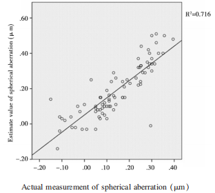
Preoperative estimates and postoperative measurements of spherical aberration were positively correlated for all patients(y=0.846, P<0.01), as shown in Figure 1.
Analysis of the degree of agreement between preoperatively estimated and postoperatively measured spherical aberration
A Bland-Altman plot indicated that 96.34%(79/ 82) of the points were distributed within 95%CI. A relatively high degree of agreement was found between preoperative and postoperative total spherical aberration in all patients.Figure 1 Correlation between estimated and measured spherical aberration
Discussion
Assessment of the degree of agreement
The paired t-test is used to assess the difference rather than the degree of agreement⁸. Consequently, the paired t-test has apparent limitations in evaluating the degree of agreement9. Bland-Altman plots are extensively used to evaluate the agreement among two different instruments or two measurements techniques by directly reflecting the outcomes9,10. In the present study, the Bland-Altman plot indicated a high degree of agreement between preoperative estimates and postoperative actual measurement of total spherical aberration, suggesting that the preoperative estimates of total spherical aberration can be used to evaluate patients'postoperative total spherical aberration.Figure 2 The solid line indicates the mean difference
(d) between preoperative estimates and postoperative measurement. The dashed line represents the standard deviation of difference (SD=0.080) within 95%CI(d±1.96×SD).