





Introduction
Blepharoptosis can either be a congenital or an acquired ptosis¹. An acquired ptosis is usually classified according to its pathogenesis as either neuro genic ,myogenic, mechanical, or aponeurogenic² . Sometimes the treatment of acquired ptosis is challenging. Finding the cause of acquired ptosis is important and should be established prior to surgery if possible.
Most cases of acquired blepharoptosis are secondary to aponeurotic causes, especially in elderly patients³. Trauma is another common cause and traumatic ptosis may be the result of damage to the levator palpebrae superioris(LPS) muscle or disruption of the neural pathway⁴. Contact lens-induced and traumatic ptosis were reported to be common causes of acquired blepharoptosis in young and middle-aged adults⁵. Uncommon causes of blepharoptosis include Horner syndrome⁶, myasthenia gravis⁷, fibrous dysplasia⁸, vernal conjunctivitis⁹, myocysticercosis¹, blephrospasm¹, conjunctival cyst"and post-surgical ptosis12,13.
Although some authors classified post-surgical ptosis together with post-traumatic ptosis, the latter is actually more complicated than post-surgical ptosis and the periorbital anatomical structures are often severely damaged. Some cases are difficult to repair. Post-surgical ptosis often receives more attention and requires correction as soon as possible. Analyzing the etiology and treatment of previous cases with acquired ptosis will help in the selection of the appropriate method for correction and avoid possible surgical complications.
The purpose of this study is to investigate the etiology and the treatment of acquired ptosis inpatients,especially secondary to surgery.
Physical examination of the eyes included evaluation of each palpebral fissure with regard to height, levator excursion,and levator function. Diagnosis, treatment, complications, and pathological results were also reviewed. All patients were examined by a neurologist. Photos(preoperative and post operation of less than one week) were copied with a digital camera from medical records.
Depending on the drooping of the upper lid covering the cornea, the ptosis was graded into mild(1-2 mm than the normal eye), moderate(3-4 mm than the normal eye), and severe(>4 mm than the normal eye). Usually, the upper lid margin covers 1~2 mm below the upper limbus in normal eyes. Levator function was graded into three levels:good(≥ 8mm), fair(5-7 mm),and poor(1-4 mm)¹ .
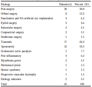
After obtaining detailed histories and physical examinations, the causes of acquired ptosis were classified according to etiology(Table 1). The leading causes were previous ocular surgery and post-surgical ptosis(20/65,30.8%),followed by trauma(17/ 65, 26.2%) and aponeurotic ptosis(12/65, 18.5%). Unknown etiology(2/65, 3.1%) was identified in two cases. Pathological results showed chronic inflammation of levator muscle in two patients and neurofibromatic infiltration of the levator muscle in one patient.
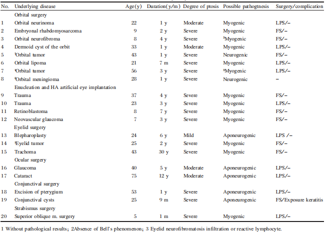
Post-surgical ptosis was easier to repair than was post-traumatic ptosis because the anatomical structure was slightly less damaged and LPS/FS surgery corrected ptosis satisfactorily.The levator palpebrae superioris and superior rectus muscles are known to have a common embryologic origin supplied by the third nerve. The LPS muscle initially shares a common muscle mass with the superior rectus⁴. This complex relationship means that the levator is easily damaged during ocular surgical procedures and is affected by periorbital tumors. The pathogenesis of ptosis may be myogenic, aponeurogenic, mechanical, or neurogenic. Although the mechanism of post- surgical ptosis is not entirely certain, some studies have pointed to the use of a speculum, bridle suture, and anesthesia during intraocular surgery secondary to levator aponeurosis dehiscence, eyelid edema or hematoma¹⁴. Excision of eyelid, conjunctival, or orbital tumors may result in scarring, symblepharon, or damage directly to the levator or the third nerve. Reactive lymphocyte proliferation and the infiltration or recurrence of the tumor may also be part of the mechanism, as shown in cases 3 and 7(Table 2). The enucleation technique, imbrications of the rectus muscles after enucleation over spherical implants, and implant migration can explain the ptosis after enucleation and HA artificial eye implantation¹⁵ .
Post-surgical ptosis was easier to repair than was post-traumatic ptosis because the anatomical structure was slightly less damaged and LPS/FS surgery corrected ptosis satisfactorily. The levator palpebrae superioris and superior rectus muscles are known to have a common embryologic origin supplied by the third nerve. The LPS muscle initially shares a common muscle mass with the superior rectus⁴. This complex relationship means that the levator is easily damaged during ocular surgical procedures and is affected by periorbital tumors. The pathogenesis of ptosis may be myogenic, aponeurogenic, mechanical, or neurogenic. Although the mechanism of post- surgical ptosis is not entirely certain, some studies have pointed to the use of a speculum, bridle suture, and anesthesia during intraocular surgery secondary to levator aponeurosis dehiscence, eyelid edema or hematoma¹⁴. Excision of eyelid, conjunctival, or orbital tumors may result in scarring, symblepharon, or damage directly to the levator or the third nerve. Reactive lymphocyte proliferation and the infiltration or recurrence of the tumor may also be part of the mechanism, as shown in cases 3 and 7(Table 2). The enucleation technique, imbrications of the rectus muscles after enucleation over spherical implants, and implant migration can explain the ptosis after enucleation and HA artificial eye implantation¹⁵.
Because the post-surgical ptosis can be transient, repair is often performed after six months when the ptosis was persistent.In the present study, severe ptosis occurred in a five-year-old girl after partial resection of obliquus superior and this was repaired in a month to avoid deprivation amblyopia. During surgery, we found the interior half of the levator to be cut off. Exploration of the levator is necessary before correction of ptosis. If a disinsertion or dehiscence of the levator aponeurosis is found, the aponeurosis is reattached to the tarsal plate.If the levator is attached but its function is deficient, a small levator resection is indicated.Repair is best accomplished under topical anesthesia with a satisfactory result of correction.If the levator is damaged severely and cannot be explored, FS operation is the best choice.Before a decision for correction, Bell's phenomenon must also be checked.Absence of a Bell's phenomenon possibly means a complication of exposure keratitis after the repair operation. In the present study, one patient without a Bell's phenomenon was not operated because of possible exposure keratitis. Two patients suffered from exposure keratitis, one after LPS surgery and another after FS operation.
Post-traumatic ptosis was the second identifiable cause in present study.The severity and complexity depended on the underlying etiology. Nearly half of these cases(41.2%) were vehicle accidents and resulted in severe ptosis. Other modes of trauma included knife(23.5%), umbrella(5.9%), iron wire (5.9%), falling injury(5.9%), pen(5.9%), fist (5.9%) and iron hook(5.9%). Most of the post- traumatic ptosis(82.4%) was severe. Only eight cases (47.1%)were corrected because the anatomical structures of the eyelid and the orbit in some cases were severely damaged. The mechanism of post-traumatic also was myogenic,aponeurogenic, neurogenic or mechanical. Traumatic ptosis following facial fractures involving the inferior or medial orbital wall may also result in enophthalmos. A sunken globe may affect the support of Whitnall's ligament, thereby altering eyelid mechanics.Enophthalmos also induces narrowing of the palpebral fissure and hence upper lid pseudoptosis¹⁶. Complex lacerations of the upper eyelid have a better chance of recovery of levator function when repaired primarily. If not, the correction is often performed six months after the trauma.In the current study, only three patients (17.6%)were corrected using LPS surgery and five patients were corrected(29.4%) using a FS operation.
Our study further supports existing data that aponeurogenic ptosis is the most common cause of acquired ptosis in elderly patients. Aponeurogenic ptosis is believed to have good levator function17. However, in the present study, four eyes had poor levator function, which may be associated with some other etiologies. The data showed that most ptosis was severe. This meant that the majority of patients in China with mild or moderate aponeurogenic ptosis had no desire for treatment if the floppy eyelid did not cover the pupil. Most patients(80%) had a good or fair levator function and were corrected by LPS surgery. The FS operation was performed in two patients with poor levators; one of them developed exposure keratitis after correction.
The present study was retrospective and long-term follow up results were not available. We focused on the complication of exposure keratitis, which often influences visual activity if it is not treated properly.
Patients and methods
The study was approved by the Ethics Committee of Sun Yat-sen University in China and complied with the tenets of the Declaration of Helsinki for biomedical research involving human subjects. The clinical records of 65 consecutive in-patients with acquired ptosis in the Zhongshan ophthalmic center and in the first affiliated hospital of Sun Yat-sen University from 2003 to 2011 were reviewed regarding the onset of ptosis and any history of possible causes for acquired ptosis(e.g., ocular trauma, previous surgery, and episodes of eyelid or orbital inflammation, systemic disorders, etc.). Congenital ptosis patients were excluded.Physical examination of the eyes included evaluation of each palpebral fissure with regard to height, levator excursion,and levator function. Diagnosis, treatment, complications, and pathological results were also reviewed. All patients were examined by a neurologist. Photos(preoperative and post operation of less than one week) were copied with a digital camera from medical records.
Depending on the drooping of the upper lid covering the cornea, the ptosis was graded into mild(1-2 mm than the normal eye), moderate(3-4 mm than the normal eye), and severe(>4 mm than the normal eye). Usually, the upper lid margin covers 1~2 mm below the upper limbus in normal eyes. Levator function was graded into three levels:good(≥ 8mm), fair(5-7 mm),and poor(1-4 mm)¹ .
Results
Etiology
A total years on average of 65 inpatients were identified who had acquired ptosis between the age of 2 and 82 years(36.1±21.6 years on average). The average duration of ptosis was 4.6 years, ranged from 2 days to 30 years.32(49.2%) patients were male and 33(50.8%) patients were female.After obtaining detailed histories and physical examinations, the causes of acquired ptosis were classified according to etiology(Table 1). The leading causes were previous ocular surgery and post-surgical ptosis(20/65,30.8%),followed by trauma(17/ 65, 26.2%) and aponeurotic ptosis(12/65, 18.5%). Unknown etiology(2/65, 3.1%) was identified in two cases. Pathological results showed chronic inflammation of levator muscle in two patients and neurofibromatic infiltration of the levator muscle in one patient.
Table 1 Etiology of acquired ptosis(n=65)
However, aponeurotic ptosis was the most common etiology identified in patients ≥50 years of age, occurring in 12(63.2%) of all 19 cases. In patients younger than 50 years(46 cases), post-surgical ptosis was noted in 17 patients (37.0%) and traumatic ptosis occurred inl6 patients(34.8%).
Post-surgical ptosis
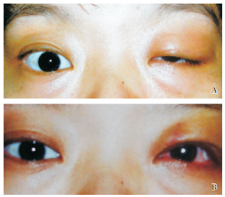
Out of the 20 cases of post-surgical ptosis, 11 (55.0%) were male and nine(45.0%)were female. The age of patients ranged between 5 and 75 years (29.3±18.6)years. The average age at initial presentation was 4.4 years, ranging from one month to 30 years. According to the fissure of palpebrae, one case(10.0%) was mild, four cases(20.0%) were moderate, and 15 cases(75.0%) were severe ptosis. Eight cases(40.0%) occurred after orbital surgery and four cases (20.0%) occurred after enucleation and HA artificial eye implantation. Three cases (15.0%) occurred after eyelid surgery and one case (5.0%) occurred after obliquus superior surgery. Ptosis after ocular and conjunctive surgery occurred in two patients(10.0%)(Table 2). In these cases, the LPS muscle of ten eyes(50.0%) was explored and corrected by reattaching it to the tarsal plate or shortening the LPS muscle. Nine eyes(45.0%) under went a frontalis suspension(FS) operation because the LPS muscle was missing in exploration. One (5.0%) patient without a Bell sign was not corrected to avoid exposure keratitis.One patient(5.0%) developed exposure keratitis after ptosis correction by FS operation(Figure 1).Traumatic ptosis
Eleven cases(64.7%) were male and six(35.3%) were female in 17 patients with traumatic ptosis. Patient age ranged from 2 to 63 years (26.7±17.2 on average). The average age at initial presentation was 2.4 years, ranging from two days to 14 years. According to the fissure of palpebrae, 3 cases(17.6%) were moderate and 14 cases(82.4%) were severe ptosis. In these cases, three patients(17.6%) were corrected by LPS surgery and five(29.4%) patients were corrected by FS operation; nine(52.9%) patients were not corrected for severe damage of eyelids or a short period of post-trauma(Table 3).Table 2 Post-surgical ptosis
Aponeurotic ptosis
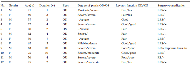
There were 12 cases(20 eyes) of aponeurotic pto- sis.In these patients, five(41.4%) were male and seven(58.3%) were female. Patient ages ranged between 50 and 82 years(65.7±9.8 on average). The average age at initial presentation was 5.4 years, ranging from one year to 20 years. Eight cases (66.7%) were bilateral and four cases were unilater- al(33.3%). According to the fissure of palpebrae, one eye(5.0%) was mild,4 eyes(20.0%) were moderate,and 15 eyes(75.0%) were severe ptosis. Among these 20 eyes, the levator function was good in seven eyes(35%), fair in nine eyes(45.0%) and poor in four eyes(20.0%). In these cases, eleven patients(91.7%) were corrected by LPS surgery and one patient(8.3%) underwent FS operation because of poor levator function(Table 4). One patient with severe ptosis and poor levator function experienced exposure keratitis after correction by LPS surgery. Because our study was retrospective and lacked long-term follow up data,the cases of under correction or over correction of ptosis could not be collect- ed in the operated patients.
Discussion
Our 65 consecutive cases of acquired ptosis revealed post-surgical ptosis as the first identifiable cause in twenty patients(30.8%) and post-traumatic ptosis as the second identifiable cause in seventeen patients(26.2%). Post-surgical ptosis and post-traumatic ptosis were the main causes in young and middle-aged patients(<50 years old), accounting for 67.4%. Aponeurogenic ptosis was the third identifiable cause in twelve patients,accounting for 18.5 percent. It was the first cause in elderly patients(≥ 50 years old), accounting for 63.2%. These three causes accounted for>3/4 of the total cases. LPS surgery was performed if levator function was fair or good;otherwise, FS operation was selected. Exposure keratitis complication occurred in 2 cases after surgical correction of ptosis.Post-surgical ptosis was easier to repair than was post-traumatic ptosis because the anatomical structure was slightly less damaged and LPS/FS surgery corrected ptosis satisfactorily.The levator palpebrae superioris and superior rectus muscles are known to have a common embryologic origin supplied by the third nerve. The LPS muscle initially shares a common muscle mass with the superior rectus⁴. This complex relationship means that the levator is easily damaged during ocular surgical procedures and is affected by periorbital tumors. The pathogenesis of ptosis may be myogenic, aponeurogenic, mechanical, or neurogenic. Although the mechanism of post- surgical ptosis is not entirely certain, some studies have pointed to the use of a speculum, bridle suture, and anesthesia during intraocular surgery secondary to levator aponeurosis dehiscence, eyelid edema or hematoma¹⁴. Excision of eyelid, conjunctival, or orbital tumors may result in scarring, symblepharon, or damage directly to the levator or the third nerve. Reactive lymphocyte proliferation and the infiltration or recurrence of the tumor may also be part of the mechanism, as shown in cases 3 and 7(Table 2). The enucleation technique, imbrications of the rectus muscles after enucleation over spherical implants, and implant migration can explain the ptosis after enucleation and HA artificial eye implantation¹⁵ .
Figure 1 One patient with exposure keratitis after ptosis correction. A: Ptosis occurred in the left eyelid after resection of conjunctival tumor;B:Conjunctival congestion due to exposure keratitis.
Table 4 Aponeurotic ptosis
Discussion
Our 65 consecutive cases of acquired ptosis revealed post-surgical ptosis as the first identifiable cause in twenty patients(30.8%) and post-traumatic ptosis as the second identifiable cause in seventeen patients(26.2%). Post-surgical ptosis and post-traumatic ptosis were the main causes in young and middle-aged patients(<50 years old), accounting for 67.4%. Aponeurogenic ptosis was the third identifiable cause in twelve patients,accounting for 18.5 percent. It was the first cause in elderly patients(≥ 50 years old), accounting for 63.2%. These three causes accounted for>3/4 of the total cases. LPS surgery was performed if levator function was fair or good;otherwise, FS operation was selected. Exposure keratitis complication occurred in 2 cases after surgical correction of ptosis.Post-surgical ptosis was easier to repair than was post-traumatic ptosis because the anatomical structure was slightly less damaged and LPS/FS surgery corrected ptosis satisfactorily. The levator palpebrae superioris and superior rectus muscles are known to have a common embryologic origin supplied by the third nerve. The LPS muscle initially shares a common muscle mass with the superior rectus⁴. This complex relationship means that the levator is easily damaged during ocular surgical procedures and is affected by periorbital tumors. The pathogenesis of ptosis may be myogenic, aponeurogenic, mechanical, or neurogenic. Although the mechanism of post- surgical ptosis is not entirely certain, some studies have pointed to the use of a speculum, bridle suture, and anesthesia during intraocular surgery secondary to levator aponeurosis dehiscence, eyelid edema or hematoma¹⁴. Excision of eyelid, conjunctival, or orbital tumors may result in scarring, symblepharon, or damage directly to the levator or the third nerve. Reactive lymphocyte proliferation and the infiltration or recurrence of the tumor may also be part of the mechanism, as shown in cases 3 and 7(Table 2). The enucleation technique, imbrications of the rectus muscles after enucleation over spherical implants, and implant migration can explain the ptosis after enucleation and HA artificial eye implantation¹⁵.
Because the post-surgical ptosis can be transient, repair is often performed after six months when the ptosis was persistent.In the present study, severe ptosis occurred in a five-year-old girl after partial resection of obliquus superior and this was repaired in a month to avoid deprivation amblyopia. During surgery, we found the interior half of the levator to be cut off. Exploration of the levator is necessary before correction of ptosis. If a disinsertion or dehiscence of the levator aponeurosis is found, the aponeurosis is reattached to the tarsal plate.If the levator is attached but its function is deficient, a small levator resection is indicated.Repair is best accomplished under topical anesthesia with a satisfactory result of correction.If the levator is damaged severely and cannot be explored, FS operation is the best choice.Before a decision for correction, Bell's phenomenon must also be checked.Absence of a Bell's phenomenon possibly means a complication of exposure keratitis after the repair operation. In the present study, one patient without a Bell's phenomenon was not operated because of possible exposure keratitis. Two patients suffered from exposure keratitis, one after LPS surgery and another after FS operation.
Post-traumatic ptosis was the second identifiable cause in present study.The severity and complexity depended on the underlying etiology. Nearly half of these cases(41.2%) were vehicle accidents and resulted in severe ptosis. Other modes of trauma included knife(23.5%), umbrella(5.9%), iron wire (5.9%), falling injury(5.9%), pen(5.9%), fist (5.9%) and iron hook(5.9%). Most of the post- traumatic ptosis(82.4%) was severe. Only eight cases (47.1%)were corrected because the anatomical structures of the eyelid and the orbit in some cases were severely damaged. The mechanism of post-traumatic also was myogenic,aponeurogenic, neurogenic or mechanical. Traumatic ptosis following facial fractures involving the inferior or medial orbital wall may also result in enophthalmos. A sunken globe may affect the support of Whitnall's ligament, thereby altering eyelid mechanics.Enophthalmos also induces narrowing of the palpebral fissure and hence upper lid pseudoptosis¹⁶. Complex lacerations of the upper eyelid have a better chance of recovery of levator function when repaired primarily. If not, the correction is often performed six months after the trauma.In the current study, only three patients (17.6%)were corrected using LPS surgery and five patients were corrected(29.4%) using a FS operation.
Our study further supports existing data that aponeurogenic ptosis is the most common cause of acquired ptosis in elderly patients. Aponeurogenic ptosis is believed to have good levator function17. However, in the present study, four eyes had poor levator function, which may be associated with some other etiologies. The data showed that most ptosis was severe. This meant that the majority of patients in China with mild or moderate aponeurogenic ptosis had no desire for treatment if the floppy eyelid did not cover the pupil. Most patients(80%) had a good or fair levator function and were corrected by LPS surgery. The FS operation was performed in two patients with poor levators; one of them developed exposure keratitis after correction.
The present study was retrospective and long-term follow up results were not available. We focused on the complication of exposure keratitis, which often influences visual activity if it is not treated properly.