
视网膜母细胞瘤钙化容积 CT 影像特征和临床应用价值

关键词
摘要
全文
文章亮点
1. 关键发现
• 网膜母细胞瘤瘤(retinoblastoma, RB)瘤内钙化沉积水平随着病程推进,在眼球内期、青光眼期至眼外期的不同阶段呈现逐级升高的态势。晶状体位置变化随病情进展、RB患者钙化程度逐渐增加。2. 已知与发现
• RB患者眼球内有肿块和钙化的占比高达95%,随着RB病情进展,肿瘤及瘤内钙化生长速度较快,受到免疫反应及血液供应不足等因素影响,极易造成组织细胞的坏死,从而产生钙化复合体或钙质沉着等使肿瘤内钙化灶体积不断增大;多层螺旋CT(multi-slice computed tomography, MSCT)对钙化灶高敏感性,且具有多方位快速成像的优势,能对肿瘤内钙化程度和肿瘤的空间位置,与邻近组织关系准确判断,在RB诊断和临床治疗中提供重要依据。3. 意义与改变
• 本文通过容积测量实现定量分析肿瘤容积和肿瘤内钙化容积在RB病情进展过程中不同分期的影像特征。旨在通过多参数、多角度分析RB肿瘤和肿瘤内钙化容积的CT影像特征获取更多的肿瘤的生长规律,以做到早期诊断,进一步提高患者生存率,保存眼球甚至视功能。1 资料与方法
1.1 临床资料
1.2 研究方法
1.3 影像检查设备及检查方法
|
扫描设备 |
工作站/软件 |
扫描范围 |
扫描条件 |
算法/矩阵 |
层厚/间距 |
窗宽/窗位 |
|
Philips Access CT |
IntelliSpaceIX/LX/Volume软件 |
眶上缘至硬腭平面 |
120kV/173mAs |
SB/512 |
1mm/0.5 mm |
300Hu/40 Hu |
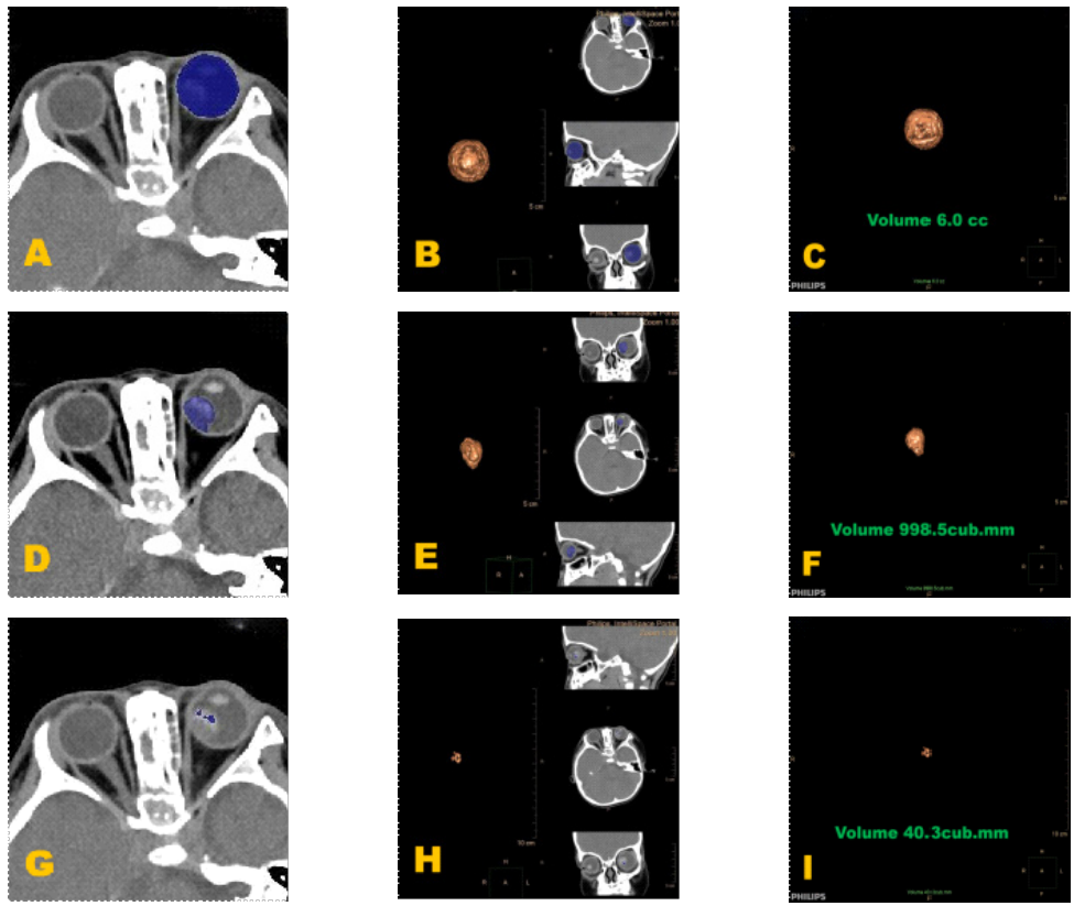
(A)眼球范围选取示图:在软组织窗轴位上选取眼球感兴趣区,蓝色区域为层面内眼球组织。(B)眼球范围选取及三维修正示图:在轴位软窗中选取眼球感兴趣区,蓝色区域为层面内眼球组织,并在冠状位和矢状位中修正范围。(C)眼球容积计算示意图:黄色图像为眼球三维图,图中绿色文字为眼球容积大小。(D)肿瘤范围选取示意图:在软组织窗轴位上选取肿瘤感兴趣区,蓝色区域为层面内肿瘤组织。(E)肿瘤范围三维修正示图:在冠状位和矢状位中修正范围。(F)肿瘤容积计算示意图:黄色图像为肿瘤三维图,图中绿色文字为肿瘤容积大小。(G)肿瘤内钙化范围选取示图:在软组织窗轴位上选取肿瘤内钙化感兴趣区,蓝色区域为层面内肿瘤内钙化。(H)肿瘤内钙化范围三维修正示图:在冠状位和矢状位中修正范围。(I)肿瘤内钙化容积计算示意图:黄色图像为肿瘤内钙化三维图,图中绿色文字为肿瘤内钙化容积大小。
(A) Schematic diagram of eye range selection: The eye region of interest is selected on the axial plane of soft tissue window. The blue area represents intra-layer eye tissue. (B) Schematic diagram of eyeball range selection and three-dimensional correction: select the eyeball region of interest (ROI) on the axial soft window. The blue area represents the eyeball tissue within the slice, and the range is corrected in the coronal and sagittal planes. (C) Schematic diagram of eyeball volume calculation: the yellow image is the 3D model of the eyeball, and the green text in the figure indicates the size of the eyeball volume. (D) Schematic diagram of tumor range selection: select the tumor ROI on the axial soft tissue window. The blue area represents the tumor tissue within the slice. (E) Schematic diagram of tumor range three-dimensional correction: correct the range in the coronal and sagittal planes. (F) Schematic diagram of tumor volume calculation: the yellow image is the 3D model of the tumor, and the green text in the figure indicates the size of the tumor volume. (G) Schematic diagram of calcification range selection in tumor: select the calcification ROI within the tumor on the axial soft tissue window. The blue area represents the calcified tissue within the tumor slice. (H) Schematic diagram of three-dimensional correction of calcification range in tumor: correct the range in the coronal and sagittal planes. (I) Schematic diagram of calcification volume calculation in tumor: the yellow image is the 3D model of tumor calcification, and the green text in the figure indicates the size of the calcification volume within the tumor.
1.4 统计学方法
2 结 果
2.1 眼别结果
|
参数 |
OD (n=60) |
OS (n=67) |
Statistic值 |
P值 |
|
钙化最高CT值 |
523.28 ± 285.26 |
565.13 ± 350.53 |
t (125)= -0.73 |
0.470 |
|
TV单位(mm³) |
2 338.78 ± 1 845.20 |
1 927.55 ± 1 168.92 |
t (125)= 1.52 |
0.130 |
|
CRV单位(mm³) |
215.40 (54.05-580.15) |
221.60 (78.90-702.70) |
Z = -0.93 |
0.35 |
|
EV单位(mm³) |
5 846.67 ± 1272.06 |
5 637.31 ± 892.36 |
t (125)= 1.08 |
0.280 |
|
CRV/EV |
0.041 (0.009-0.090) |
0.040 (0.014-0.125) |
Z = -0.88 |
0.38 |
|
TV/EV |
0.386 ± 0.207 |
0.343 ± 0.201 |
t (125)= 1.19 |
0.240 |
|
CRV/TV |
0.173 ± 0.169 |
0.234 ± 0.195 |
t (123)= -1.84 |
0.068 |
|
OD:右眼;OS:左眼;TV:肿瘤体积;CRV:钙化区域体积;EV:眼球总体积。 OD: right eye; OS: left eye; TV: tumor volume; CRV: calcified region volume within the tumor; EV: total eye volume. |
||||
2.2 性别结果
|
参数 |
女性 (n=56) |
男性 (n=71) |
Statistic值 |
P值 |
|
钙化最高CT值 |
533.13 ± 297.55 |
555.01 ± 339.78 |
t (125)= -0.38 |
0.700 |
|
TV/mm³ |
1 948.80 (1 022.10-2 527.15) |
2 341.00 (1 250.60-3 095.00) |
Z= -1.53 |
0.13 |
|
CRV/mm³ |
270.55 (96.15-641.85) |
175.00 (68.50-576.20) |
Z= 1.10 |
0.27 |
|
EV/mm³ |
5 439.29 ± 775.23 |
5 970.42 ± 1 239.40 |
t (125)= -2.80 |
0.006 |
|
CRV/EV |
0.047 (0.019-0.126) |
0.029 (0.011-0.087) |
Z= 1.44 |
0.15 |
|
TV/EV |
0.344 ± 0.193 |
0.379 ± 0.212 |
t (125)= -0.96 |
0.340 |
|
CRV/TV |
0.199 (0.087-0.311) |
0.099 (0.041-0.294) |
Z= 1.62 |
0.11 |
|
TV:肿瘤体积;CRV:钙化区域体积;EV:眼球总体积。 TV: tumor volume; CRV: calcified region volume within the tumor; EV: total eye volume. |
||||
2.3 年龄分组结果
|
参数 |
0~<1岁组(0岁至不满1岁) (n=77) |
1~<2岁组(1岁至不满2岁) (n=30) |
2~<3岁组(2岁至不满3岁) (n=12) |
≥3岁组(3岁及以上) (n=8) |
Statistic值 |
P值 |
|
钙化最高CT值 |
538.14±292.03 |
615.63±401.75 |
530.50±334.16 |
373.63±162.59 |
F (3,123)= 1.27 |
0.290 |
|
TV/mm³ |
2 179.20 (1 088.00-2 693.40) |
1 982.25 (1 100.20-3 022.90) |
969.05 (361.55-2 849.85) |
3 506.50 (2 284.85-4 104.65) |
χ2 (3)= 7.95 |
0.047 |
|
CRV/mm³ |
328.20 (102.40-642.50) |
278.25 (91.50-652.00) |
92.70 (23.90-180.45) |
84.80 (54.25-104.75) |
χ2 (3)= 11.24 |
0.010 |
|
EV/mm³ |
5 463.64±1 151.53 |
5 870.00±505.25 |
6 416.67±589.04 |
6 837.50±1 507.07 |
F (3,123)= 6.90 |
<0.001 |
|
CRV/EV |
0.055 (0.017-0.143) |
0.048 (0.014-0.111) |
0.015 (0.003-0.031) |
0.014 (0.008-0.018) |
χ2(3)= 13.35 |
0.004 |
|
TV/EV |
0.413 (0.215-0.523) |
0.353 (0.216-0.509) |
0.168 (0.054-0.464) |
0.481 (0.382-0.626) |
χ2 (3)= 5.85 |
0.12 |
|
CRV/TV |
0.198 (0.070-0.380) |
0.179 (0.043-0.298) |
0.117 (0.022-0.249) |
0.030 (0.012-0.054) |
χ2 (3)= 9.50 |
0.023 |
|
TV:肿瘤体积;CRV:钙化区域体积;EV:眼球总体积。 TV: tumor volume; CRV: calcified region volume within the tumor; EV: total eye volume. |
||||||
2.4 影像分期组结果
|
参数 |
眼内期组 (n=46) |
青光眼期组 (n=68) |
眼外期组 (n=13) |
Statistic值 |
P值 |
|
钙化最高CT值 |
452.09 ± 334.04 |
569.50 ± 303.92 |
749.15 ± 254.30 |
F (2,124)= 5.07 |
0.008 |
|
TV/mm³ |
915.65 (308.00-1 407.00) |
2 389.00 (1 967.20-3 020.00) |
3 095.00 (2 701.50-3 385.20) |
χ2 (2)= 47.47 |
<0.001 |
|
CRV/mm³ |
73.95 (17.50-221.60) |
359.00 (126.25-745.00) |
404.20 (314.40-849.50) |
χ2 (2)= 28.14 |
<0.001 |
|
EV/mm³ |
5 756.52 ± 825.34 |
5 552.94 ± 942.76 |
6 623.08 ± 1972.37 |
F (2,124)= 5.67 |
0.004 |
|
CRV/EV |
0.014 (0.003-0.043) |
0.062 (0.024-0.155) |
0.070 (0.050-0.102) |
χ2 (2)= 26.65 |
<0.001 |
|
TV/EV |
0.157 (0.057-0.255) |
0.462 (0.371-0.540) |
0.544 (0.403-0.584) |
χ2 (2)= 47.95 |
<0.001 |
|
CRV/TV |
0.137 (0.025-0.248) |
0.179 (0.055-0.336) |
0.119 (0.084-0.250) |
χ2 (2)= 1.41 |
0.49 |
|
TV:肿瘤体积;CRV:钙化区域体积;EV:眼球总体积。 TV: tumor volume; CRV: calcified region volume within the tumor; EV: total eye volume. |
|||||
2.5 晶状体位置变化结果
|
参数 |
眼内期组 (n=46) |
青光眼期组 (n=68) |
眼外期组 (n=13) |
Statistic值 |
P值 |
|
钙化最高CT值 |
452.09 ± 334.04 |
569.50 ± 303.92 |
749.15 ± 254.30 |
F(2,124)= 5.07 |
0.008 |
|
TV/mm³ |
915.65 (308.00-1 407.00) |
2 389.00 (1 967.20-3 020.00) |
3 095.00 (2 701.50-3 385.20) |
χ2 (2)= 47.47 |
<0.001 |
|
CRV/mm³ |
73.95 (17.50-221.60) |
359.00 (126.25-745.00) |
404.20 (314.40-849.50) |
χ2 (2)= 28.14 |
<0.001 |
|
EV/mm³ |
5 756.52 ± 825.34 |
5 552.94 ± 942.76 |
6 623.08 ± 1 972.37 |
F(2,124)= 5.67 |
0.004 |
|
CRV/EV |
0.014 (0.003-0.043) |
0.062 (0.024-0.155) |
0.070 (0.050-0.102) |
χ2 (2)= 26.65 |
<0.001 |
|
TV/EV |
0.157 (0.057-0.255) |
0.462 (0.371-0.540) |
0.544 (0.403-0.584) |
χ2 (2)= 47.95 |
<0.001 |
|
CRV/TV |
0.137 (0.025-0.248) |
0.179 (0.055-0.336) |
0.119 (0.084-0.250) |
χ2 (2)= 1.41 |
0.49 |
|
TV:肿瘤体积;CRV:钙化区域体积;EV:眼球总体积。 TV: tumor volume; CRV: calcified region volume within the tumor; EV: total eye volume. |
|||||
2.6 眼内期、青光眼期、眼外期、全身转移期不同分期的CT影像。
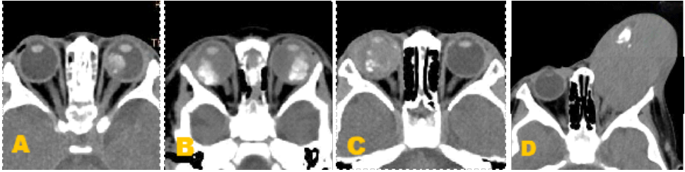
(A)眼内期CT表现:眼球内肿块小,钙化少,眼环完整,无视神经增粗或眶内侵犯。 (B)青光眼期CT表现:肿块内钙化增多,玻璃体密度增高,可见散在钙化点,视神经仍无明显增粗,偶可见前房变浅征象。(C)眼外期CT表现:眼球内大片钙化灶,玻璃体密度显著增高,可伴眼球增大或变形。(D)全身转移期CT表现:眼球壁破坏,肿块不规则增大,肿瘤侵犯眼眶软组织或颅内。
(A) Intraocular CT findings: small mass in the eye, minimal calcification, intact orbital rim, no enlargement of the optic nerve or invasion into the orbit. (B) Glaucomatous phase CT findings: increased calcification within the mass, increased vitreous density, scattered calcification points visible, no significant enlargement of the optic nerve, occasional signs of shallower anterior chamber. (C) Extraocular CT findings: large areas of calcification within the eye,significantly increased vitreous density, may be accompanied by globe enlargement or deformity. (D) Systemic metastatic phase CT findings: destruction of the eye wall, irregular enlargement of the mass, tumor invasion into orbital soft tissues or intracranial space.
3 讨论
3.1 RB的典型临床进程表现
3.2 RB的影像学诊断
3.3 研究结果
3.4 临床启示
声明
利益冲突
所有作者均声明不存在利益冲突。开放获取声明
基金
参考文献
1. 1.高晓曼. 视网膜母细胞瘤:儿童眼内的“恶魔”[C]//广东省肿瘤康复学会. 2025年“《健康大湾区》-科普引领健康”论坛曁第2期健康科普作品征集活动作品集. 建德市第二人民医院眼科, 2025: 75-77. DOI: 10.26914/c.cnkihy.2025.011480. Gao XM. Retinoblastoma: the "demon" in children's eyes[C]//Guangdong Anti-Cancer Association for Rehabilitation. Proceedings of 2025 "Healthy Greater Bay Area - Popular Science Leading Health" Forum and the 2nd Health Science Popularization Works Collection Activity. Department of Ophthalmology, Jiande Second People's Hospital, 2025: 75-77. DOI: 10.26914/c.cnkihy.2025.011480.
5. 崔雪皓, 李筱荣. 视网膜母细胞瘤诊断和治疗的研究进展[J]. 眼科新进展, 2022, 42(08): 634-638. DOI: 10.13389/j.cnki.rao.2022.0130. Cui XH, LI XR. Research Progress in Diagnosis and Treatment of Retinoblastoma[J]. Recent Adv Ophthalmol, 2022, 42(8): 634-638. DOI: 10.13389/j.cnki.rao.2022.0130.
9. 崔若棣, 马常友, 杨光勇, 等. 儿童视网膜母细胞瘤CT、MRI影 像特征及临床应用价值[J]. 中国CT和MRI杂志, 2022, 20(06): 23-25. DOI: 10.3969/j.issn.1672-5131.2022.06.008.
Cui RD, Ma CY, Yang GY, et al. CT and MRI features of retinoblastoma in children and their clinical application value[J]. Chin J CT MRI, 2022, 20(6): 23-25. DOI: 10.3969/j.issn.1672-5131.2022.06.008.
17. 首都医科大学眼部肿瘤临床诊疗与研究中心, 中华医学会放射学分会头颈学组, 中华医学会放射学分会儿科学组, 等. 视网膜母细胞瘤影像检查与诊断及选择性眼动脉化疗专家共识[J]. 中华放射学杂志, 2021, 55(5): 470-477. DOI: 10.3760/cma.j.cn112149-20201214-01306. Clinical Diagnosis and Treatment Center for Ocular Tumors, Capital Medical University, Head and Neck Radiology Group, Chinese Society of Radiology, Pediatric Radiology Group, Chinese Society of Radiology, et al. Expert consensus on imaging examination and diagnosis of retinoblastoma and selective ophthalmic artery chemotherapy[J]. Chin J Radiol, 2021, 55(5): 470-477. DOI: 10.3760/cma.j.cn112149-20201214-01306.
19. 王亮, 周珉, 盛茂, 等. 儿童视网膜母细胞瘤的影像诊断[J]. 江苏医药, 2011, 37(8): 934-936. DOI: 10.19460/j.cnki.0253-3685.2011.08.026. Wang L, Zhou M, Sheng M, et al. Imaging diagnosis of retinoblastoma in children[J]. Jiangsu Med J, 2011, 37(8): 934-936. DOI: 10.19460/j.cnki.0253-3685.2011.08.026.
20. 孙丰源. 黑色素瘤及顺磁性病变的影像特征和鉴别诊断[C]//中国超声医学工程学会. 中国超声医学工程学会第十三届全国眼科超声医学学术会议论文汇编. 天津医科大学眼科医院, 2024: 13. DOI: 10.26914/c.cnkihy.2024.044680. Sun FY. Imaging Features and Differential Diagnosis of Melanoma and Paramagnetic Lesions [C]//Chinese Society of Ultrasound in Medicine. Proceedings of the 13th National Conference on Ophthalmic Ultrasound Medicine. Tianjin Medical University Eye Hospital, 2024: 13. DOI: 10.26914/c.cnkihy.2024.044680.
21. 陈向东, 刘悦, 彭清华, 等. Coats病伴眼内钙化灶形成1例[J]. 临床眼科杂志, 2016, 24(5): 465-466. DOI: 10.3969/j.issn.1006-8422.2016.05.028. Chen XD, Liu Y, Peng QH, et al. Coats disease with calcification in the eye: a case report[J]. J Clin Ophthalmol, 2016, 24(5): 465-466. DOI: 10.3969/j.issn.1006-8422.2016.05.028.
22. 马京琪, 黄东生, 张谊, 等. 244例儿童视网膜母细胞瘤临床特点[J]. 眼科, 2011, 20(2): 113-115. Ma JQ, Huang DS, Zhang Y, et al. Study on the clinical characteristics of 244 retinoblastoma cases[J]. Ophthalmol China, 2011, 20(2): 113-115.
23. 刘强, 耿左军, 贾歆, 等. CT在视网膜母细胞瘤钙化研究中的价值[J]. 实用医学影像杂志, 2020(1). DOI: 10.16106/j.cnki.cn14-1281/r.2020.01.002. Liu Q, Geng ZJ, Jia X, et al. Value of CT in the Study of Retinoblastoma Calcification[J]. Journal of Practical Medical Imaging, 2020, 21(1). DOI: 10.16106/j.cn14-1281/r.2020.01.002.
24. 李婷, 鲜军舫. 眼眶和颅脑磁共振成像促进视网膜母细胞瘤精准诊疗:视网膜母细胞瘤影像检查与诊断专家共识解读[J]. 磁共振成像, 2021, 12(11): 74-79. DOI: 10.12015/issn.1674-8034.2021.11.016. Li T, Xian JF. Orbital and Cranial Magnetic Resonance Imaging Promotes Precision Diagnosis and Treatment of Retinoblastoma: Interpretation of the Expert Consensus on Imaging Examination and Diagnosis of Retinoblastoma[J]. Magnetic Resonance Imag, 2021, 12(11): 74-79. DOI: 10.12015/issn.1674-8034.2021.11.016.