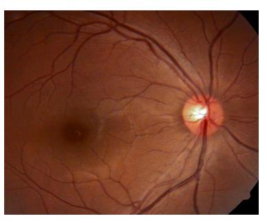
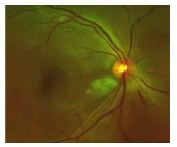
Figure 1 Color fundus of the right eye showed enhanced arterial reflection and slightly tortuous veins (oneset of 13 d)
图2 左眼眼底彩照见颞上方视网膜大片火焰状出血,棉绒斑,静脉迂曲,黄斑水肿(发病13 d)
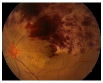
Figure 2 Color fundus of the left eye showed large flaming hemorrhage, cotton velvet spot, and tortuous veins of the supratemporal retina , macular edema (oneset of 13 d)
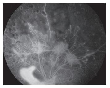
图3 左眼眼底荧光血管造影见颞上方网膜大片荧光遮蔽,微血管瘤,未见明显无灌注区(发病13 d)
Figure 3 Fluorescein fundus angiography of the left eye showed large fluorescent occlusion, microhemangioma, and no non-perfusion area of the supratemporal retina (oneset of 13 d)
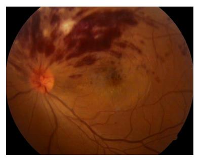
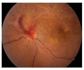
Figure 4 Color fundus of the left eye showed large flaming hemorrhage and tortuous veins of the supratemporal retina, exudation of the macula, the neovascularization of the optic disc (oneset of 36 d)
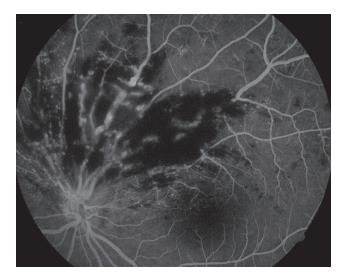
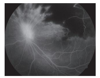
图5 左眼眼底荧光血管造影检查见左眼视盘荧光渗漏,颞上视网膜大片无灌注区(发病36 d)
Figure 5 Fluorescein fundus angiography of the left eye showed high fluorescence of the optic disc,non-perfusion area of the supratemporal retina (oneset of 36 d)
图6 左眼眼底彩照见视盘新生血管增殖膜(发病75 d)
Figure 6 Color fundus of the left eye showed the neovascularization membrane of the optic disc (oneset of 75 d)
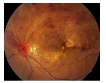
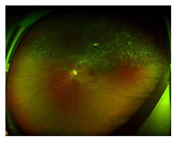
图7 左眼眼底彩照见颞上方视网膜血管闭塞,呈白线状,网膜部分出血吸收,散在激光斑(发病75 d)
Figure 7 Color fundus of the left eye showed the vessels were occluded and the retinal hemorrhage was partly absorbed by the supratemporal retina (oneset of 75 d)
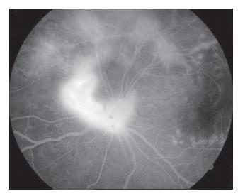
图8 左眼眼底荧光血管造影见视盘荧光渗漏(发病103 d)
Figure 8 Fluorescein fundus angiography of the left eye showed fluorescence leakage of optic disc (oneset of 103 d)
图9 左眼眼底荧光血管造影见颞上方网膜可见新生异常血管网(发病103 d)
Figure 9 Fluorescein fundus angiography of the left eye showed abnormal neovascularization network of the supratemporal retina (oneset of 103 d)
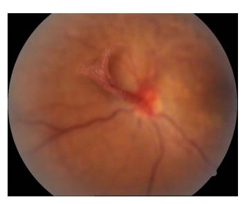
图10 左眼眼底彩照见视盘形成新生血管膜(发病138 d)
Figure 10 Color fundus of the left eye showed the optic disc forms a neovascularization membrane (oneset of 138 d)
图11 左眼眼底彩照见视盘新生血管膜表面血管旺盛(发病138 d)
Figure 11 Color fundus of the left eye showed the optic disc neovascularization membrane with vigorous surface vessels (oneset of 138 d)
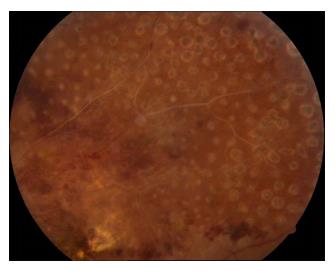
图12 左眼眼底彩照见视盘新生血管膜机化萎缩,表面无血管(发病10年)
Figure 12 Color fundus of the left eye showed the neovascularization membrane of the optic disc were mechanized and atrophied, and there were no vessels on the surface (oneset of 10 years)
Figure 13 Color fundus of the right eye showed enhanced arterial reflectance, tortuous veins, scattered cotton-wool spots of the Inferior temporal and superior temporal retina. There was superficial hemorrhage in the subnasal and lateral retinas and some rigid exudation in the supranasal retinas (oneset of 10 years)
1. 山东省自然科学基金青年项目 (ZR2020QH149)。 This work was supported by the Shandong Natural Science Foundation Youth Program, China (ZR2020QH149)
参考文献
1. 张桂, 刘军, 李中凯. 全视网膜激光光凝联合玻璃体腔注射雷珠
单抗治疗新生血管性青光眼效果观察[ J]. 中国实用医刊, 2017,
44 (23): 64-66.
ZHANG G , LIU J , LI Zk. Effect of pan retinal
photocoagulation combined with intravitreal injection of ranibizumab
on neo-vascular glaucoma[ J]. Chinese Journal of Practical Medicine,
2017, 44(23): 64-66.
2. 海鸥, 刘芳, 高丹宇. 玻璃体腔注射康柏西普联合Ahmed青光眼
引流阀植入和视网膜光凝治疗新生血管性青光眼[ J]. 中华眼
视光学与视觉科学杂志, 2019, 21(11): 838-841.
HAI O, LIU F, GAO DY. Intravitreal injection of conbercept
combined with Ahemd glaucoma valve implants and panretinal
photocoagulation treatment for neovascular glaucoma[ J]. Chinese
Journal of Optometry Ophthalmology and Visual Science, 2019,
21(11): 838-841
3. 王海波. 抗VEGF药物联合PRP治疗新生血管性青光眼的效
果[ J]. 中国医药指南, 2019, 17(22): 72.
WANG HB. Anti-VEGF medicine with PRP for neovascular
glaucoma[ J]. Guide of China Medicine, 2019, 17(22): 72.
4. 王惠英, 李臻, 徐蔚, 等. 视网膜激光光凝后糖尿病大鼠眼玻璃体
中色素上皮衍生因子的表达[ J]. 中华眼底病杂志, 2006, 22(1):
52-53.
WANG HY, LI Z, XU W, et al. Expression of pigment
epithelium-derived factors in vitreous of diabetic rats after retinal laser
photocoagulation[ J]. Chinese Journal of Ocular Fundus Diseases,
2006, 22(1): 52-53.
5. 秦海峰, 徐国旭, 张敬法. 缺氧和炎症在视网膜静脉阻塞继发黄
斑水肿中的作用[ J]. 国际眼科纵览, 2022, 46(2): 173-178.
QIN HF, XU GX, ZHANG JF. Roles of hypoxia and
inflammation in the pathogenesis of macular edema secondary to
retinal vein occlusion[ J]. International Review of Ophthalmology,
2022, 46(2): 173-178.
6. 王雅芬, 郭长梅. 巨噬细胞的异质性在视网膜新生血管中的作
用[ J]. 国际眼科纵览, 2018, 42(2): 119-124.
WAN YF, GUO CM. The role of heterogeneity of
macrophages in retinal neovascularization[ J]. International Review of
Opthalmology, 2018, 42(2): 119-124.
7. Campbell M, Doyle SL. Current perspectives on established and novel
therapies for pathological neovascularization in retinal disease[ J].
Biochem Pharmacol, 2019, 164: 321-325.
8. Alizadeh E, Mammadzada P, André H. The different facades of retinal
and choroidal endothelial cells in response to hypoxia[ J]. Int J Mol
Sci, 2018, 19(12): 3846.
9. Hayreh SS. Ocular vascular occlusive disorders: natural history of visual
outcome[ J]. Prog Retin Eye Res, 2014, 41: 1-25.
11. 赵明威, 苗恒. 视网膜静脉阻塞诊疗重在全病程管理[ J]. 中华眼
科杂志, 2020, 56(4): 246-249.
ZHAO MW, MIAO H. Whole course management is the key
to retinal vein occlusion[ J]. Chinese Journal of Ophthalmology, 2020,
56(4): 246-249.
12. Stenner AM, Frederiksen KH, Grauslund J. Is there still a role of
macular laser treatment in branch retinal vein occlusion in the era of
intravitreal injections?[ J]. Acta Ophthalmol, 2020, 98(1): 9-21.
13. 陈露璐, 陈有信. 2019年《EURETINA视网膜静脉阻塞诊疗指
南》解读[ J]. 中华实验眼科杂志, 2020, 38(1): 60-63.
CHEN LL, CHEN YX. Interpretation of the 2019 Guidelines
for the Management of retinal vein occlusion by the EURETINA[ J].
Chinese Journal Of Experimental Ophthalmology, 2020, 38(1): 60-63.
15. Csernok E, Bossuyt X. Investigations in systemic vasculitis. The role of
the laboratory[ J]. Best Pract Res Clin Rheumatol, 2018, 32(1): 52-62.
16. Moisseiev E, Sagiv O, Lazar M. Intense exercise causing central retinal
vein occlusion in a young patient: case report and review of the
literature[ J]. Case Rep Ophthalmol, 2014, 5(1): 116-120.
17. Vieira MJ, Campos A, do Carmo A, et al. Thrombophilic risk factors for
retinal vein occlusion[ J]. Sci Rep, 2019, 9(1): 18972.
18. Kolar P. Risk factors for central and branch retinal vein occlusion: a
meta-analysis of published clinical data[ J]. J Ophthalmol, 2014, 2014:
724780.
20. Rogers S, McIntosh RL, Cheung N, et al. The prevalence of retinal vein
occlusion: pooled data from population studies from the United States,
Europe, Asia, and Australia[J]. Ophthalmology, 2010, 117(2): 313-319.e1.
'%20fill='white'%20fill-opacity='0.01'/%3e%3cmask%20id='mask0_3477_29692'%20style='mask-type:luminance'%20maskUnits='userSpaceOnUse'%20x='0'%20y='0'%20width='16'%20height='16'%3e%3crect%20id='&%23232;&%23146;&%23153;&%23231;&%23137;&%23136;_2'%20x='16'%20width='16'%20height='16'%20transform='rotate(90%2016%200)'%20fill='white'/%3e%3c/mask%3e%3cg%20mask='url(%23mask0_3477_29692)'%3e%3cpath%20id='&%23232;&%23183;&%23175;&%23229;&%23190;&%23132;'%20d='M14%205L8%2011L2%205'%20stroke='%23333333'%20stroke-width='1.5'%20stroke-linecap='round'%20stroke-linejoin='round'/%3e%3c/g%3e%3c/g%3e%3c/svg%3e)