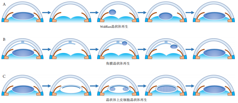
(A) Wolffiffiffian lens regeneration: the representative model is the newts, where after removal of the crystalline lens, the PECs on the dorsal side of the iris undergo dedifferentiation to form LECs and achieve lens regeneration. (B) Corneal lens regeneration: the representative model is the Xenopus, where after removal of the crystalline lens, corneal-derived cells (stem cells or transient amplify cells presented in the corneal stroma) differentiate into LECs and achieve lens regeneration. (C) Lens epithelial cell lens regeneration: In mammals, the preserved lens capsule and cells are essential when the lens contents are removed, the cells will proliferate and differentiate to produce lens fibers filling the closed capsule cavity to achieve lens regeneration.
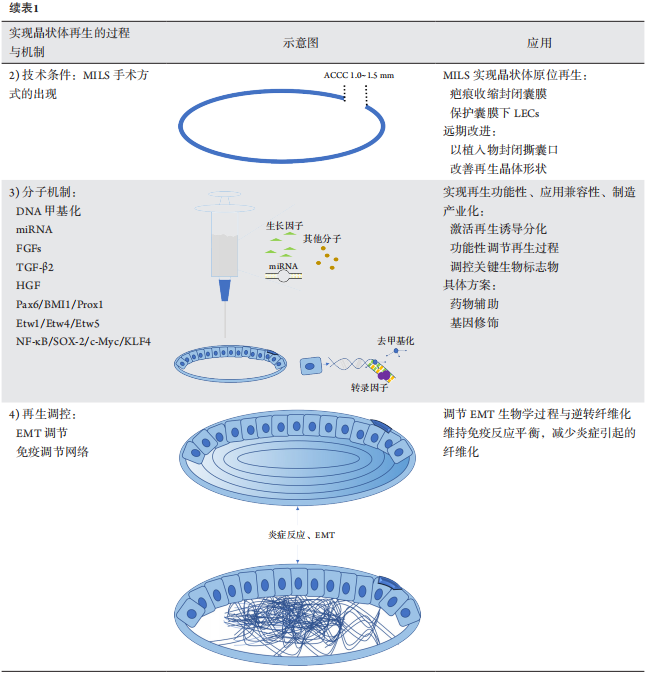
MILS操作在取出晶状体内容物的过程中,保护了前囊下和赤道处的LECs。水分离的操作要求尽量轻柔,可以使用黏弹性剂代替平衡盐溶液(balanced salt solution,BSS),以降低晶状体前囊下上皮细胞脱落的风险。此外,水分离要求尽量充分,幼年哺乳动物和人类先天性白内障患儿的晶状体核柔软但黏性大,充分的水分离有助于避免将超声乳化手柄和灌注抽吸手柄过度插入囊袋,保护前囊下细胞层的完整性。
图2 晶状体微创手术
Figure 2 Minimally invasive lens-content removal surgery
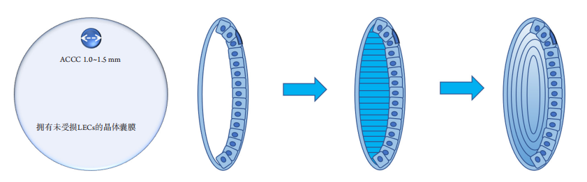
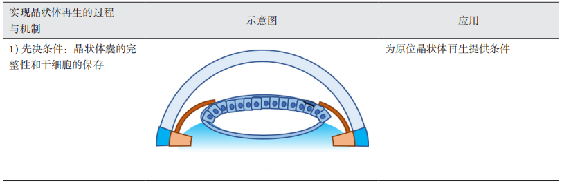
During the procedure, a periphery capsulorhexis opening of 1–1.5 mm in diameter will be made, the lens content and/or cortical opacities will be removed with a 0.9 mm phacoemulsification probe. With the reservation of the lens stem cells and the intact capsule membrane, the closed capsule accommodates the lens fibers. Intact lens will regenerate if microenvironment is proper. ACCC: anterior continuous curvilinear capsulorhexis.
其他与晶状体发育相关的重要转录因子还有核因子-κB(nuclear factor kappa-B,NF-κB)[55]、Sox2、c-Myc和Klf4[56]等。此外,DNase-seq(DNase I hypersensitive sites sequencing)、ATAC-seq(Assay for Transposase-Accessible Chromatin using sequencing)等表观遗传学新技术有利于发现新的调控晶状体分化的转录因子或DNA结合区,如gatad1和NF1[57]等。
1. Tzahor E, Poss KD. Cardiac regeneration strategies: staying young at
heart[ J]. Science, 2017, 356(6342): 1035-1039.
2. Jorstad NL, Wilken MS, Grimes WN, et al. Stimulation of functional
neuronal regeneration from Müller glia in adult mice[ J]. Nature, 2017,
548(7665): 103-107.
3. Zhang Y, Kim MS, Jia B, et al. Hypothalamic stem cells control ageing
speed partly through exosomal miRNAs[ J]. Nature, 2017, 548(7665):
52-57.
4. Bassat E, Mutlak YE, Genzelinakh A, et al. The extracellular matrix
protein agrin promotes heart regeneration in mice[ J]. Nature, 2017,
547(7662): 179-184.
5. Otsuki L, Brand AH. Cell cycle heterogeneity directs the timing
of neural stem cell activation from quiescence[ J]. Science, 2018,
360(6384): 99-102.
6. Perico L, Morigi M, Rota C, et al. Human mesenchymal stromal
cells transplanted into mice stimulate renal tubular cells and enhance
mitochondrial function[ J]. Nat Commun, 2017, 8(1): 983.
7. Leeman DS, Hebestreit K, Ruetz T, et al. Lysosome activation clears
aggregates and enhances quiescent neural stem cell activation during
aging[ J]. Science, 2018, 359(6381): 1277-1283.
8. Karin M, Clevers H. Reparative inflammation takes charge of tissue
regeneration[ J]. Nature, 2016, 529(7586): 307-315.
9. Kinoshita S, Koizumi N, Ueno M, et al. Injection of cultured cells
with a rock inhibitor for bullous keratopathy[ J]. N Engl J Med, 2018,
378(11): 995-1003.
10. Laha B, Stafford BK, Huberman AD. Regenerating optic pathways from
the eye to the brain[ J]. Science, 2017, 356(6342): 1031-1034.
11. Servick K. Stem cell approach for cataracts challenged[ J]. Science,
2017, 356(6345): 1318-1319.
12. van der Flier LG, Clevers H. Stem cells, self-renewal, and differentiation
in the intestinal epithelium[ J]. Annu Rev Physiol, 2009, 71: 241-260.
13. Donati G, Watt FM. Stem cell heterogeneity and plasticity in
epithelia[ J]. Cell Stem Cell, 2015, 16(5): 465-476.
14. Wang EX, Jiang X. Stem cells from trabecular meshwork cells can
secrete extracellular matrix[ J]. Biochem Biophys Res Commun, 2020,
523(2): 522-526.
15. Koizumi N, Okumura N, Ueno M, et al. New therapeutic modality for
corneal endothelial disease using Rho-associated kinase inhibitor eye
drops[ J]. Cornea, 2014, 33 Suppl 11: S25-S31.
17. Yao K, Qiu S, Wang YV, et al. Restoration of vision after de novo
genesis of rod photoreceptors in mammalian retinas[ J]. Nature, 2018,
560(7719): 484-488.
18. Ueki Y, Wilken MS, Cox KE, et al. Transgenic expression of the
proneural transcription factor Ascl1 in Müller glia stimulates retinal
regeneration in young mice[ J]. Proc Natl Acad Sci U S A, 2015,
112(44): 13717-13722.
19. Marote A, Teixeira FG, Mendes-Pinheiro B, et al. MSCs-derived
exosomes: cell-secreted nanovesicles with regenerative potential[ J].
Front Pharmacol, 2016, 7: 231.
20. Vannella KM, Wynn TA. Mechanisms of organ injury and repair by
macrophages[ J]. Annu Rev Physiol, 2017, 79: 593-617.
21. Tang J, Liu S, Han Y, et al. Surface modification of intraocular lenses
via photodynamic coating for safe and effective PCO prevention[ J]. J
Mater Chem B, 2021, 9(6): 1546-1556.
22. Huang H, Zhu S, Liu D, et al. Antiproliferative drug-loaded multi-
functionalized intraocular lens for reducing posterior capsular
opacification[ J]. J Biomater Sci Polym Ed, 2021, 32(6): 735-748.
23. Qin C, Liu S, Wen S, et al. Enhanced PCO prevention of drug eluting
IOLs via endocytosis and autophagy effects of a PAMAM dendrimer[ J].
J Mater Chem B, 2021, 9(3): 793-800.
24. Brockes JP, Kumar A. Comparative aspects of animal regeneration[ J].
Annu Rev Cell Dev Biol, 2008, 24: 525-549.
25. Malloch EL, Perry KJ, Fukui L, et al. Gene expression profiles of lens
regeneration and development in Xenopus laevis[ J]. Dev Dyn, 2009,
238(9): 2340-2356.
26. Logan CM, Bowen CJ, Menko AS. Induction of immune surveillance of
the dysmorphogenic lens[ J]. Sci Rep, 2017, 7(1): 16235.
27. Ursell PG, Dhariwal M, O'Boyle D, et al. 5-year incidence of YAG
capsulotomy and PCO after cataract surgery with single-piece
monofocal intraocular lenses: a real-world evidence study of 20,763
eyes[ J]. Eye (Lond), 2020, 34(5): 960-968.
28. Spierer A, Desatnik H, Blumenthal M. Refractive status in children
after long-term follow up of cataract surgery with intraocular lens
implantation[ J]. J Pediatr Ophthalmol Strabismus, 1999, 36(1): 25-29.
30. Aurora AB, Olson EN. Immune modulation of stem cells and
regeneration[ J]. Cell Stem Cell, 2014, 15(1): 14-25.
31. Arnoux V, Nassour M, L'Helgoualc'h A, et al. Erk5 controls Slug
expression and keratinocyte activation during wound healing[ J]. Mol
Biol Cell, 2008, 19(11): 4738-4749.
32. Shaw TJ, Martin P. Wound repair: a showcase for cell plasticity and
migration[ J]. Curr Opin Cell Biol, 2016, 42: 29-37.
33. Zhao Y, Zheng D, Cvekl A. Profiling of chromatin accessibility and
identification of general cis-regulatory mechanisms that control two
ocular lens differentiation pathways[ J]. Epigenetics Chromatin, 2019,
12(1): 27.
34. Maki N, Suetsugu-Maki R, Tarui H, et al. Expression of stem cell
pluripotency factors during regeneration in newts[ J]. Dev Dyn, 2009,
238(6): 1613-1616.
35. Sun Y, Rong X, Li D, et al. NF-κB/cartilage acidic protein 1 promotes
ultraviolet B irradiation-induced apoptosis of human lens epithelial
cells[ J]. DNA Cell Biol, 2020, 39(4): 513-521.
36. Garg A, Hannan A, Wang Q, et al. Etv transcription factors functionally
diverge from their upstream FGF signaling in lens development[ J].
Elife, 2020, 9: 51915.
37. Aryal S, Viet J, Weatherbee BAT, et al. The cataract-linked RNA-binding
protein Celf1 post-transcriptionally controls the spatiotemporal
expression of the key homeodomain transcription factors Pax6 and
Prox1 in lens development[ J]. Hum Genet, 2020, 139(12): 1541-1554.
38. Huang X, Wang Y, Zhang P, et al. A HGF-derived peptide suppresses
EMT in human lens epithelial cells via the TGF-β/Smad and Akt/
mTOR signaling pathways[ J]. Mol Med Rep, 2020, 22(1): 551-558.
40. McAvoy JW, Chamberlain CG. Fibroblast growth factor (FGF)
induces different responses in lens epithelial cells depending on its
concentration[ J]. Development, 1989, 107(2): 221-228.
41. Chen X, Xiao W, Chen W, et al. MicroRNA-26a and -26b inhibit lens
fibrosis and cataract by negatively regulating Jagged-1/Notch signaling
pathway[ J]. Cell Death Differ, 2017, 24(8): 1431-1442.
42. Dirks RP, Klok EJ, van Genesen ST, et al. The sequence of regulatory
events controlling the expression of the gamma D-crystallin gene during
fibroblast growth factor-mediated rat lens fiber cell differentiation[ J].
Dev Biol, 1996, 173(1): 14-25.
43. Lu Y, Brommer B, Tian X, et al. Reprogramming to recover youthful
epigenetic information and restore vision[ J]. Nature, 2020, 588(7836):
124-129.
44. Wang Y, Guan H. The Role of DNA methylation in lens development
and cataract formation[ J]. Cell Mol Neurobiol, 2017, 37(6): 979-984.
45. Sevilla A, Papatsenko D, Mazloom AR, et al. An Esrrb and Nanog cell
fate regulatory module controlled by feed forward loop interactions[ J].
Front Cell Dev Biol, 2021, 9: 630067.
46. Li R, Li B, Cao Y, et al. Long non-coding RNA Mir22hg-derived miR-
22-3p promotes skeletal muscle differentiation and regeneration by
inhibiting HDAC4[ J]. Mol Ther Nucleic Acids, 2021, 24: 200-211.
47. Peng S, Shi S, Tao G, et al. JKAMP inhibits the osteogenic capacity of
adipose-derived stem cells in diabetic osteoporosis by modulating the
Wnt signaling pathway through intragenic DNA methylation[ J]. Stem
Cell Res Ther, 2021, 12(1): 120.
48. Khurana I, Al-Hasani K, Maxwell S, et al. DNA methylation status
correlates with adult β-cell regeneration capacity[ J]. NPJ Regen Med,
2021, 6(1): 7.
49. Wu X, Liu Z, Zhang X, et al. Proteomics analysis and proteogenomic
characterization of different physiopathological human lenses[ J]. BMC
Ophthalmol, 2017, 17(1): 253.
50. Huang Y, Xie L. Expression of transcription factors and crystallin
proteins during rat lens regeneration[ J]. Mol Vis, 2010, 16: 341-352.
51. Lin L, Lin Q, Li J, et al. ROCK inhibitor modified intraocular lens as an
approach for inhibiting the proliferation and migration of lens epithelial
cells and posterior capsule opacification[ J]. Biomater Sci, 2019, 7(10):
4208-4217.
52. Han Y, Tang J, Xia J, et al. Anti-adhesive and antiproliferative synergistic
surface modification of intraocular lens for reduced posterior capsular
opacification[ J]. Int J Nanomedicine, 2019, 14: 9047-9061.
53. Inanc M, Tekin K, Erol YO, et al. The ultrastructural alterations in the
lens capsule and epithelium in eyes with traumatic white cataract[ J].
Int Ophthalmol, 2019, 39(1): 47-53.
54. Jiang J, Shihan MH, Wang Y, et al. Lens epithelial cells initiate
an inflammatory response following cataract surgery[ J]. Invest
Ophthalmol Vis Sci, 2018, 59(12): 4986-4997.
55. Tan X, Zhu Y, Chen C, et al. Sprouty2 suppresses epithelial-
mesenchymal transition of human lens epithelial cells through
blockade of Smad2 and ERK1/2 pathways[ J]. PLoS One, 2016, 11(7):
e0159275.
56. Gwon A , Gruber L. Engineering the cr ystalline lens w ith a
biodegradable or non-degradable scaffold[ J]. Exp Eye Res, 2010,
91(2): 220-228.
57. Hurvitz LM. YAG anterior capsulectomy and lysis of posterior
synechiae after cataract surgery[ J]. Ophthalmic Surg, 1992, 23(2):
103-107.
58. Martínez Toldos JJ, Artola Roig A, Chipont Benabent E. Total anterior
capsule closure after silicone intraocular lens implantation[ J]. J
Cataract Refract Surg, 1996, 22(2): 269-271.
59. Shammas HJ. Relaxing the fibrosed capsulorhexis rim to correct
induced hyperopia after phacoemulsification[ J]. J Cataract Refract
Surg, 1995, 21(2): 228-229.
60. Nagamoto T, Tanaka N, Fujiwara T. Inhibition of posterior capsule
opacification by a capsular adhesion-preventing ring[ J]. Arch
Ophthalmol, 2009, 127(4): 471-474.
61. Gwon A . Lens regeneration in mammals: a rev iew[ J]. Sur v
Ophthalmol, 2006, 51(1): 51-62.
62. Anchan RM, Lachke SA, Gerami-Naini B, et al. Pax6- and Six3-
mediated induction of lens cell fate in mouse and human ES cells[ J].
PLoS One, 2014, 9(12): e115106.
63. Han C, Li J, Wang C, et al. Wnt5a contributes to the differentiation
of human embryonic stem cells into lentoid bodies through the
noncanonical Wnt/JNK signaling pathway[ J]. Invest Ophthalmol Vis
Sci, 2018, 59(8): 3449-3460.
64. Lin H, Ouyang H, Zhu J, et al. Lens regeneration using endogenous
stem cells with gain of visual function[ J]. Nature, 2016, 531(7594):
323-328.
65. Andjeli? S, Dra?lar K, Lumi X, et al. Morphological and proliferative
studies on ex vivo cultured human anterior lens epithelial cells -
relevance to capsular opacification[ J]. Acta Ophthalmol, 2015, 93(6):
e499-e506.
66. 柳夏林,张新愉,刘奕志,等. 兔眼晶状体再生模型的建立及观
察[ J]. 眼科学报, 2002, 18(4): 230-234.
LIU Xialin, ZHANG Xinyu, LIU Yizhi, et al. To establish and observe
the experimental lens regeneration model in rabbits[ J]. Yan Ke Xue
Bao, 2002, 18(4): 230-234.
68. Zhou KJ, Li YN, Huang FR , et al. In vivo observation of lens
regeneration in rat using ultra-long scan depth optical coherence
tomography[ J]. Invest Ophthalmol Vis Sci, 2016, 57(15): 6615-6623.
69. 刘晓敏, 代云海, 谢立信. 哺乳动物晶状体再生的研究进展[ J].
中华眼科杂志, 2019, 55(7): 549-553.
LIU Xiaomin, DAI Yunhai, XIE Lixin. Advances and clinical application
of lens regeneration in mammal[ J]. Chinese Journal of Ophthalmology,
2019, 55(7): 549-553.
70. Perry KJ, Hamilton PW, Sonam S, et al. The role of sensory innervation
in cornea-lens regeneration[ J]. Dev Dyn, 2019, 248(7): 530-544.
71. Perry KJ, Thomas AG, Henry JJ. Expression of pluripotency factors in
larval epithelia of the frog Xenopus: evidence for the presence of cornea
epithelial stem cells[ J]. Dev Biol, 2013, 374(2): 281-294.
72. Tsonis PA. Lens regeneration[M]//Encyclopedia of the eye. Oxford:
Academic Press, 2010: 557-564.
73. Filoni S, Bosco L, Cioni C. Reconstruction of the crystalline lens from
fragments of capsular membrane and epithelium in larvae of Rana
esculenta[ J]. Acta Embryol Exp (Palermo), 1977, (1): 41-49.
74. Ledwon JK, Turin SY, Gosain AK, et al. The expression of fgfr3 in the
zebrafish head[ J]. Gene Expr Patterns, 2018, 29: 32-38.
79. Wan J, Goldman D. Retina regeneration in zebrafish[ J]. Curr Opin
Genet Dev, 2016, 40: 41-47.
80. Sukhija J, Kaur S. Nature nurtures: lens regeneration, a breakthrough in
ophthalmology[ J]. Ann Eye Sci, 2017, 2: 17.
81. Henry JJ, Hamilton PW. Diverse evolutionary origins and mechanisms
of lens regeneration[ J]. Mol Biol Evol, 2018, 35(7): 1563-1575.
82. Jiang S, Tian G, Li X, et al. Research progress on stem cell therapies
for articular cartilage regeneration[ J]. Stem Cells Int, 2021, 2021:
8882505.
83. Gonzalez G, Sasamoto Y, Ksander BR, et al. Limbal stem cells: identity,
developmental origin, and therapeutic potential[ J]. Wiley Interdiscip
Rev Dev Biol, 2018, 7(2): 10.1002/wdev.303.
84. Eichstadt S, Barriga M, Ponakala A, et al. Phase 1/2a clinical trial
of gene-corrected autologous cell therapy for recessive dystrophic
epidermolysis bullosa[ J]. JCI Insight, 2019, 4(19): 130554.
85. Dietz AB, Dozois EJ, Fletcher JG, et al. Autologous mesenchymal stem
cells, applied in a bioabsorbable matrix, for treatment of perianal fistulas
in patients with Crohn's disease[ J]. Gastroenterology, 2017, 153(1):
59-62.e2.
86. Stern JH, Tian Y, Funderburgh J, et al. Regenerating eye tissues to
preserve and restore vision[ J]. Cell Stem Cell, 2018, 22(6): 834-849.
87. Xia H, Li X, Gao W, et al. Tissue repair and regeneration with
endogenous stem cells[ J]. Nat Rev Mater, 2018, 3(7): 174-193.
88. GBD 2019 Blindness and Vision Impairment Collaborators; Vision
Loss Expert Group of the Global Burden of Disease Study. Causes of
blindness and vision impairment in 2020 and trends over 30 years,
and prevalence of avoidable blindness in relation to VISION 2020: the
Right to Sight: an analysis for the Global Burden of Disease Study[ J].
Lancet Glob Health, 2021, 9(2): e144-e160.
89. GBD 2019 Blindness and Vision Impairment Collaborators; Vision
Loss Expert Group of the Global Burden of Disease Study. Trends in
prevalence of blindness and distance and near vision impairment over
30 years: an analysis for the Global Burden of Disease Study[ J]. Lancet
Glob Health, 2021, 9(2): e130-e143.
'%20fill='white'%20fill-opacity='0.01'/%3e%3cmask%20id='mask0_3477_29692'%20style='mask-type:luminance'%20maskUnits='userSpaceOnUse'%20x='0'%20y='0'%20width='16'%20height='16'%3e%3crect%20id='&%23232;&%23146;&%23153;&%23231;&%23137;&%23136;_2'%20x='16'%20width='16'%20height='16'%20transform='rotate(90%2016%200)'%20fill='white'/%3e%3c/mask%3e%3cg%20mask='url(%23mask0_3477_29692)'%3e%3cpath%20id='&%23232;&%23183;&%23175;&%23229;&%23190;&%23132;'%20d='M14%205L8%2011L2%205'%20stroke='%23333333'%20stroke-width='1.5'%20stroke-linecap='round'%20stroke-linejoin='round'/%3e%3c/g%3e%3c/g%3e%3c/svg%3e)