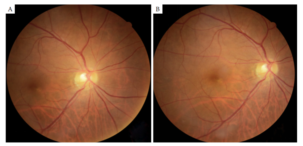
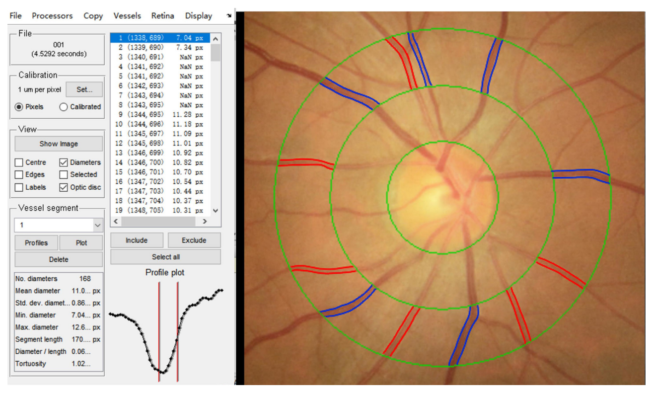
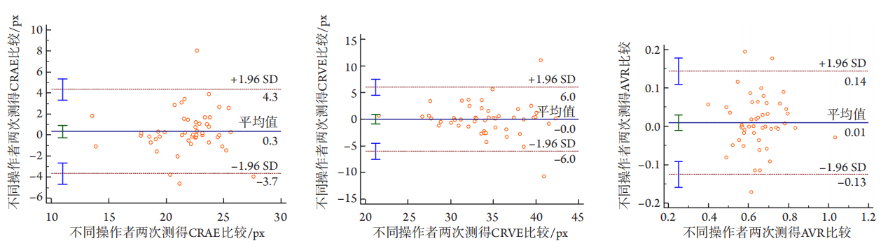
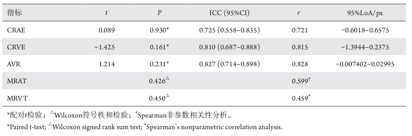
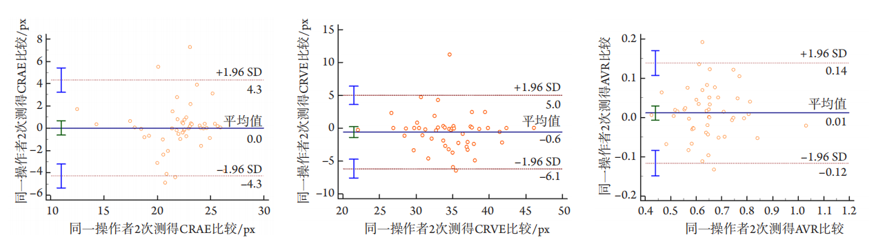
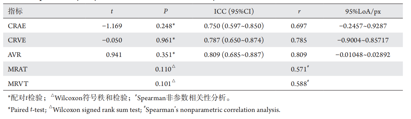
ARIA软件先通过小波变换的阈值分割技术进行血管分割,后采用基于图像的算法提取血管的中心线,通过曲线拟合确定血管方向,并搜索垂直于血管的二阶导数零点,最终分析其平均管径及迂曲度。静态图像中血管分析首先依赖于对血管的准确定位,该算法的视网膜血管分割技术较其他无监督分割方法具有明显的速度优势,处理2 160×1 440像素图像大约需要3~7s,且保证了较高的准确度。当使用公开的视网膜图像数据库DRIVE(Digital Retinal Image for Vessel Extraction)进行验证时,血管分割的真阳性率为70.27%,假阳性率为2.83%,准确率为93.71%。应用于图像分辨率更高的公开视网膜图像数据库REVIEW(REtinal Vessel Image set for Estimation of Widths)时,该算法输出的血管直径与其提供的3个独立的观察者的手动测量结果呈现良好的一致性。 在该软件准确度良好的基础上,本研究评估了ARIA软件应用于我国糖尿病患者的视网膜血管管径测量及迂曲度的再现性和重复性。已有研究[2]报道了糖尿病患者的视网膜动静脉管径及迂曲度的变化可早于微血管瘤等眼底体征,因此ARIA软件在糖尿病患者中的应用前景主要在于识别DR的早期改变,故本研究对象为D R分期为0~3期的患者,结果表明:操作者内和操作者间的CRAE、CRVE、AVR、MR AT、MRVT的差异均无统计学意义,2组数据间相关性好,操作者内优于操作者间,静脉优于动脉。而Bland-Altman法作为图形分析法,可以直观地从集中趋势、离散趋势、同步变化程度多角度评价结果的一致性,弥补了t检验及相关分析的不足。但在使用中应注意检验样本的数据行为,避免错误使用。在本研究中,同一操作者2次测量所得和不同操作者的测量所得CRAE、CRVE、AVR的Bland-Altman一致性分析图示均不超过4个点位于95%LoA外,且95%LoA在临床可接受的范围,可以认为操作者内和操作者间的数据是一致的、可相互替代的。为后续将ARIA软件有效推广应用于DR的诊疗奠定基础。
1. 王金洒. 视网膜血管分割与基于视盘定位的动静脉管径测
量[D]. 武汉: 华中师范大学, 2018.
WANG Jinsa. Retinal vessel segmentation and diameter measurement
of arteriovenous vessel based on OD location[D]. Wuhan: Central
China Normal University, 2018.
2. Perez-Rovira A, MacGillivray T, Trucco E, et al. VAMPIRE: vessel
assessment and measurement platform for images of the REtina[ J].
Annu Int Conf IEEE Eng Med Biol Soc, 2011, 2011: 3391-3394.
3. 王思远. 眼底图像中血管管径的测量方法及其软件实现[D]. 长
沙: 湖南大学, 2018.
WANG Siyuan. Measurement method of vessel width in retinal images
4. Ghasemi Falavarjani K, Al-Sheikh M, Darvizeh F, et al. Retinal
vessel calibre measurements by optical coherence tomography
angiography[ J]. Br J Ophthalmol, 2017, 101(7): 989-992.
5. 李春艳. 早期糖尿病视网膜病变血管形态学改变及其相关机制
的研究[D]. 呼和浩特: 内蒙古医科大学, 2016.
LI Chunyan. Assess morphological changes of retinal vascular and its
related mechanism in early diabetic retinopathy[D]. Hohhot: Inner
Mongolia Medical University, 2016.
6. 李春艳, 徐力, 王春燕, 等. IVAN软件测量视网膜管径的重复性
和再现性评价[ J]. 中华实验眼科杂志, 2017, 35(7): 634-639.
LI Chunyan, XU Li, WANG Chunyan, et al. Repeatability and
reproducibility of IVAN software measuring retinal vascular caliber[ J].
Chinese Journal of Ophthalmology, 2017, 35(7): 634-639.
7. Stanton AV, Mullaney P, Mee F, et al. A method of quantifying retinal
microvascular alterations associated with blood pressure and age[ J]. J
Hypertens, 1995, 13(1): 41-48.
8. Knudtson MD, Lee KE, Hubbard LD, et al. Revised formulas for
summarizing retinal vessel diameters[ J]. Curr Eye Res, 2003, 27(3):
143-149.
9. 中华医学会眼科学会眼底病学组. 我国糖尿病视网膜病变筛
查的图像采集及阅片指南(2017年)[ J]. 中华眼科杂志, 2017,
53(12): 890-896.
Fundus Disease Group in Chinese Academy of Ophthalmology.
Guidelines for image acquisition and interpretation of diabetic
retinopathy screening in China (2017)[ J]. Chinese Journal of
Ophthalmology, 2017, 53(12): 890-896.
10. Scanlon PH, Malhotra R, Greenwood RH, et al. Comparison of two
reference standards in validating two field mydriatic digital photography
as a method of screening for diabetic retinopathy[ J]. Br J Ophthalmol,
2003, 87(10): 1258-1263.
11. 中华医学会眼科学会眼底病学组. 我国糖尿病视网膜病变临床
诊疗指南(2014年)[ J]. 中华眼科杂志, 2014, 50(11): 851-865.
Fundus Disease Group in Chinese Academy of Ophthalmology.
Guidelines for clinical diagnosis and treatment of diabetic retinopathy
in China (2014)[ J]. Chinese Journal of Ophthalmology, 2014, 50(11):
851-865.
12. Gabir MM, Hanson RL, Dabelea D, et al. The 1997 American
Diabetes Association and 1999 World Health Organization criteria for
hyperglycemia in the diagnosis and prediction of diabetes[ J]. Diabetes
Care, 2000, 23(8): 1108-1112.
13. 李淑婷, 吴强. 远程医疗在糖尿病视网膜病变筛查项目中应用
的价值及前景[ J]. 国际眼科杂志, 2021, 21(2): 257-261.
LI Shuting, WU Qiang. Research advances in telemedicine program
for diabetic retinopathy screening[ J]. International Eye Science, 2021,
21(2): 257-261.
14. Bankhead P, Scholfield CN, McGeown JG, et al. Fast retinal vessel
detection and measurement using wavelets and edge location
refinement[ J]. PLoS One, 2012, 7(3): e32435.
15. Gopinath B, Liew G, Lewis JR, et al. Associations between dietary
flavonoids and retinal microvasculature in older adults[ J]. Eur J Nutr,
2020, 59(7): 3093-3101.
16. Invernizzi A, Schiuma M, Parrulli S, et al. Retinal vessels modifications
in acute and post-COVID-19[ J]. Sci Rep, 2021, 11(1): 19373.
17. Simkiene J, Pranskuniene Z, Vitkauskiene A, et al. Ocular microvascular
changes in patients with sepsis: a prospective observational study[ J].
Ann Intensive Care, 2020, 10(1): 38.
18. Cheung CY, Ikram MK, Chen C, et al. Imaging retina to study dementia
and stroke[ J]. Prog Retin Eye Res, 2017, 57: 89-107.
19. Phan K, Mitchell P, Liew G, et al. Associations between retinal
arteriolar and venular calibre with the prevalence of impaired fasting
glucose and diabetes mellitus: A cross-sectional study[ J]. PLoS One,
2018, 13(5): e0189627.
20. Ponto KA, Werner DJ, Wiedemer L, et al. Retinal vessel metrics:
normative data and their use in systemic hypertension: results from the
Gutenberg Health Study[ J]. J Hypertens, 2017, 35(8): 1635-1645.
21. Lockhart CJ, McCann AJ, Pinnock RA, et al. Multimodal functional
and anatomic imaging identifies preclinical microvascular abnormalities
in type 1 diabetes mellitus[ J]. Am J Physiol Heart Circ Physiol, 2014,
307(12): H1729-H1736.
22. Jenkins AJ, Joglekar MV, Hardikar AA, et al. Biomarkers in diabetic
retinopathy[ J]. Rev Diabet Stud, 2015, 12(1-2): 159-195.
'%20fill='white'%20fill-opacity='0.01'/%3e%3cmask%20id='mask0_3477_29692'%20style='mask-type:luminance'%20maskUnits='userSpaceOnUse'%20x='0'%20y='0'%20width='16'%20height='16'%3e%3crect%20id='&%23232;&%23146;&%23153;&%23231;&%23137;&%23136;_2'%20x='16'%20width='16'%20height='16'%20transform='rotate(90%2016%200)'%20fill='white'/%3e%3c/mask%3e%3cg%20mask='url(%23mask0_3477_29692)'%3e%3cpath%20id='&%23232;&%23183;&%23175;&%23229;&%23190;&%23132;'%20d='M14%205L8%2011L2%205'%20stroke='%23333333'%20stroke-width='1.5'%20stroke-linecap='round'%20stroke-linejoin='round'/%3e%3c/g%3e%3c/g%3e%3c/svg%3e)