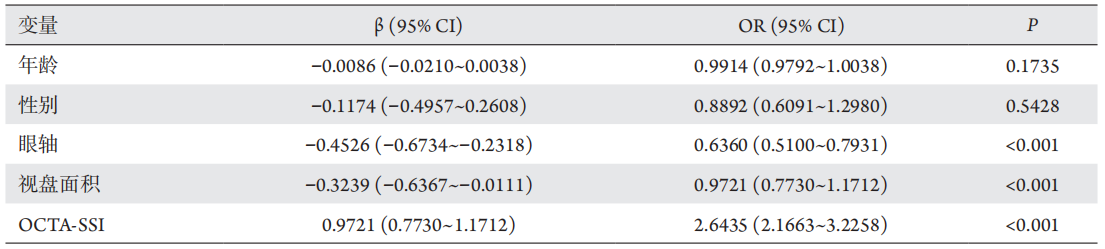
在使用OCTA检查过程中,研究者[10-11]发现视网膜或视盘血流密度不仅与疾病的种类、年龄和眼轴等有关,也与OCTA扫描信号强度(scanning signal intensity,SSI)有关[12-14]。SSI是一个关于成像质量的重要参数,在血流密度的临床应用或研究中,为保证图片的质量,常常要求OCTA- SSI要至少达到一定的强度才能视为有效扫描(如蔡司OCTA的SSI为0~10,一般要求SSI至少为7)[14],但对于这些符合质量要求的OCTA图像,SSI是否仍然会对视网膜或视盘血流产生常影响以及可能产生什么样的影响,目前少有报道。
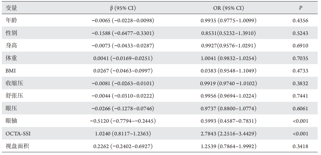
受试者均接受了视力、眼压、裂隙灯、眼轴和OCTA检查。最佳矫正视力使用国际标准视力表检查,眼压采用自动眼压计连续测量3次取其平均值。使用眼球生物测量仪Lenstar测量眼轴。用蔡司OCT(Cirrus 5000 HD-OCT)的200×200扫描获取视盘面积,同时记录受试者的身高、体重和血压指标,并计算BMI(body mass index,BMI)。
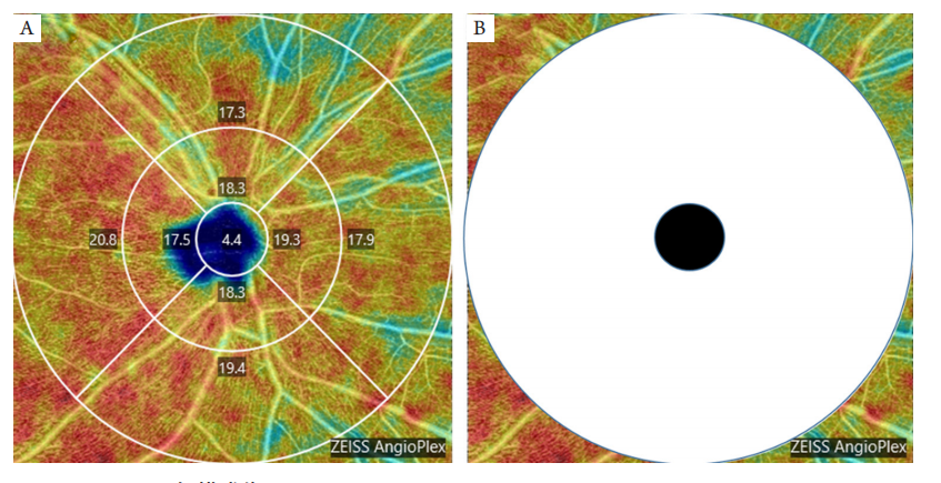
(A) Vessel density of ETDRS chart in optic disc using the 6 mm×6 mm scanning model of OCTA. (B)The black circle represents the central circle of ETDRS in Figure 1A (diameter: 1mm), and the white annulus area represents the area around the optic disc (length of the full circle – length of the central circle)/(area of the full circle – area of the central circle).
1. 广东省基础与应用基础研究基金 (2019B1515120011);佛山市科技局项目 (1920001000811)。 This work was
supported by the Guangdong Basic and Applied Basic Research Foundation (2019B1515120011) and the Science and Technology Planning Project of Foshan
City (1920001000811), China.
参考文献
1. Hayreh SS. The blood supply of the optic nerve head and the evaluation
of it - myth and reality[ J]. Prog Retin Eye Res, 2001, 20(5): 563-593.
2. Hayreh SS. Blood supply of the optic nerve head[ J]. Ophthalmologica,
1996, 210(5): 285-295.
3. Liu L, Jia Y, Takusagawa HL, et al. Optical coherence tomography
angiography of the peripapillary retina in glaucoma[ J]. JAMA
Ophthalmol, 2015, 133(9): 1045-1052.
4. Zhang S, Wu C, Liu L, et al. Optical coherence tomography angiography
of the peripapillary retina in primary angle-closure glaucoma[ J]. Am J
Ophthalmol, 2017, 182: 194-200.
5. Balducci N, Cascavilla ML, Ciardella A, et al. Peripapillary vessel
density changes in Leber’s hereditary optic neuropathy: a new
biomarker[ J]. Clin Exp Ophthalmol, 2018, 46(9): 1055-1062.
6. Kay MD. Color Doppler imaging in disorders of the orbit, retina, and
optic nerve[ J]. Semin Ophthalmol, 1995, 10(3): 242-250.
8. Zhang Y, Zhang B, Fan M, et al. The vascular densities of the macula
and optic disc in normal eyes from children by optical coherence
tomography angiography[ J]. Graefes Arch Clin Exp Ophthalmol, 2020,
258(2): 437-444.
9. Wylegala A . Principles of OCTA and applications in clinical
neurology[ J]. Curr Neurol Neurosci Rep, 2018, 18(12): 96.
10. Sampson DM, Gong P, An D, et al. Axial length variation impacts
on superficial retinal vessel density and foveal avascular zone area
measurements using optical coherence tomography angiography[ J].
Invest Ophthalmol Vis Sci, 2017, 58(7): 3065-3072.
11. Jo YH, Sung KR, Shin JW. Effects of age on peripapillary and macular
vessel density determined using optical coherence tomography
angiography in healthy eyes[ J]. Invest Ophthalmol Vis Sci, 2019,
60(10): 3492-3498.
12. Lim HB, Kim YW, Nam KY, et al. Signal strength as an important factor
in the analysis of peripapillary microvascular density using optical
coherence tomography angiography[ J]. Sci Rep, 2019, 9(1): 16299.
13. Lim HB, Kim YW, Kim JM, et al. The importance of signal strength in
quantitative assessment of retinal vessel density using optical coherence
tomography angiography[ J]. Sci Rep, 2018, 8(1): 12897.
14. Chen CL, Ishikawa H, Wollstein G, et al. Histogram matching extends
acceptable signal strength range on optical coherence tomography
images[ J]. Invest Ophthalmol Vis Sci, 2015, 56(6): 3810-3819.
15. Lee TH, Lim HB, Nam KY, et al. Factors affecting repeatability of assessment
of the retinal microvasculature using optical coherence tomography
angiography in healthy subjects[J]. Sci Rep, 2019, 9(1): 16291.
17. You QS, Chan JCH, Ng ALK, et al. Macular vessel density measured
with optical coherence tomography angiography and its associations
in a large population-based study[ J]. Invest Ophthalmol Vis Sci, 2019,
60(14): 4830-4837.
18. Spaide RF, Fujimoto JG, Waheed NK. Image artifacts in optical coherence
tomography angiography[J]. Retina, 2015, 35(11): 2163-2180.
19. Zhang X, Iverson SM, Tan O, et al. Effect of signal intensity on
measurement of ganglion cell complex and retinal nerve fiber layer
scans in Fourier-domain optical coherence tomography[ J]. Transl Vis
Sci Technol, 2015, 4(5): 7.
20. She X, Guo J, Liu X, et al. Reliability of vessel density measurements
in the peripapillary retina and correlation with retinal nerve fiber layer
thickness in healthy subjects using optical coherence tomography
angiography[ J]. Ophthalmologica, 2018, 240(4): 183-190.
21. Rao HL, Pradhan ZS, Weinreb RN, et al. Determinants of peripapillary
and macular vessel densities measured by optical coherence
tomography angiography in normal eyes[ J]. J Glaucoma, 2017, 26(5):
491-497.
22. Llanas S, Linderman RE, Chen FK , et al. Assessing the use of
incorrectly scaled optical coherence tomography angiography images
in peer-reviewed studies: a systematic review[ J]. JAMA Ophthalmol,
2019, Epub ahead of print.
23. Zhang Q, Zhang A, Lee CS, et al. Projection artifact removal improves
visualization and quantitation of macular neovascularization imaged
by optical coherence tomography angiography[ J]. Ophthalmol Retina,
2017, 1(2): 124-136.
'%20fill='white'%20fill-opacity='0.01'/%3e%3cmask%20id='mask0_3477_29692'%20style='mask-type:luminance'%20maskUnits='userSpaceOnUse'%20x='0'%20y='0'%20width='16'%20height='16'%3e%3crect%20id='&%23232;&%23146;&%23153;&%23231;&%23137;&%23136;_2'%20x='16'%20width='16'%20height='16'%20transform='rotate(90%2016%200)'%20fill='white'/%3e%3c/mask%3e%3cg%20mask='url(%23mask0_3477_29692)'%3e%3cpath%20id='&%23232;&%23183;&%23175;&%23229;&%23190;&%23132;'%20d='M14%205L8%2011L2%205'%20stroke='%23333333'%20stroke-width='1.5'%20stroke-linecap='round'%20stroke-linejoin='round'/%3e%3c/g%3e%3c/g%3e%3c/svg%3e)