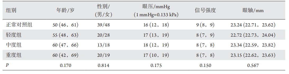
HUMPHREY视野计(德国Carl Zeis公司)检查视野。采用30-2标准程序,III号光标。全部试验组对象视野结果满足以下条件:1)青光眼Hemifield测试在正常范围外;2)固定损失率<20%、假阳性率<15%;3)至少2次连续可靠测试证实的模式标准偏差(pattern standard deviation,PSD)与青光眼乳头神经纤维层丢失一致。
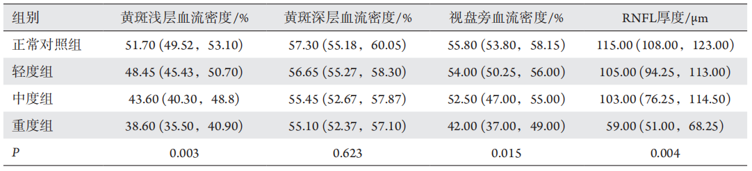
1 )设备:Optovue公司的Avanti RT Vue XR OCT进行眼底黄斑区和视盘的定量检测。2 )扫描模式:在OCTA检查模式中,选择黄斑区血管高清成像6 mm × 6 mm模式(纳入范围:以中心凹为中心,边长为6 mm × 6 mm的正方形)及视乳头4.5 mm×4.5 mm模式[9](纳入范围:以视乳头为中心,边长为4.5 mm × 4.5 mm的正方形),自动追踪扫描模式完成横向及纵向扫描(OCTA信号强度≤6予以排除)。
1.3 统计学处理
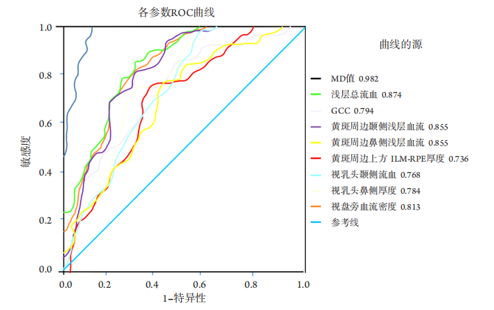
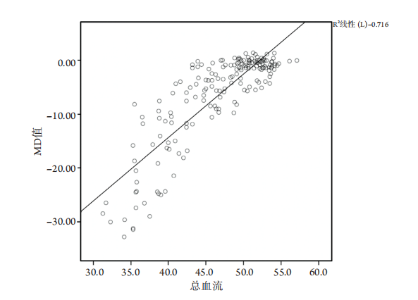
所有数据使用SPSS24.0统计学软件分析。样、本资料经正态性检验,推断总体不服从正态分布(P <0.05),以中位数(1/4位数,3/4位数)表示,用KrusKal-WallissH进行多个独立样本比较、用Friedman M检验进行多个相关样本比较、用q检验进行相关样本间两两比较,采用卡方检验进行性别的统计分析。受试者工作曲线(receiver operating characteristic,ROC)及曲线下面积(area under the curve,AUC)用以反映OCTA的各项参数鉴别CPACG的能力,相关性采用Spearman等级相关系数、多元线性回归分析。以P <0.05为差异具有统计学意义。
1. 国家自然科学基金 (81970789)。 This work was supported by the National Natural Science Foundation of China (81970789).
参考文献
1. Pollack IP. Chronic angle-closure glaucoma; diagnosis and treatment
in patients with angles that appear open[ J]. Arch Ophthalmol, 1971,
85(6): 676-689.
2. Kim DY, Fingler J, Zawadzki R J, et al. Optical imaging of the
chorioretinal vasculature in the living human eye[ J]. Proc Natl Acad
Sci U S A, 2013, 110(35): 14354-14359.
3. Uzun S, Pehlivan E. Vascular density in retina and choriocapillaris as
measured by optical coherence tomography angiography[ J]. Am J
Ophthalmol, 2016, 169: 290.
4. Chansangpetch S, Lin SC. Optical coherence tomography angiography
in glaucoma care[ J]. Curr Eye Res, 2018, 43(9):1067-1082.
5. Kashani AH, Chen CL, Gahm JK, et al. Optical coherence tomography
angiography: a comprehensive review of current methods and clinical
applications[ J]. Prog Retin Eye Res, 2017, 60: 66-100.
6. Faridi A, Jia Y, Gao SS, et al. Sensitivity and specificity of OCT
angiography to detect choroidal neovascularization[ J]. Ophthalmol
Retina, 2017, 1(4): 294-303.
7. Suh MH, Zangwill LM, Manalastas PI, et al. Deep retinal layer
microvasculature dropout detected by the optical coherence
tomography angiography in glaucoma[ J]. Ophthalmology, 2016,
123(12): 2509-2518.
8. Lee EJ, Lee SH, Kim JA, et al. Parapapillary deep-layer microvasculature
dropout in glaucoma: topographic association with glaucomatous
damage[ J]. Invest Ophthalmol Vis Sci, 2017, 58(7): 3004-3010.
9. Miguel AIM, Silva AB, Azevedo LF. Diagnostic performance of optical
coherence tomography angiography in glaucoma: a systematic review
and meta-analysis[ J]. Br J Ophthalmol, 2019, 103(11): 1677-1684.
10. Hood DC, Raza AS, de Moraes CG, et al. Glaucomatous damage of the
macula[ J]. Prog Retin Eye Res, 2013, 32: 1-21.
11. Triolo G, Rabiolo A, Shemonski ND, et al. Optical coherence
tomography angiography macular and peripapillary vessel perfusion
density in healthy subjects, glaucoma suspects, and glaucoma
patients[ J]. Invest Ophthalmol Vis Sci, 2017, 58(13): 5713-5722.
12. Richter GM, Chang R, Situ B, et al. Diagnostic performance of macular
versus peripapillary vessel parameters by optical coherence tomography
angiography for glaucoma[ J]. Transl Vis Sci Technol, 2018, 7(6): 21
13. Shin JW, Lee J, Kwon J, et al. Relationship between macular vessel
density and central visual field sensitivity at different glaucoma
stages[ J]. Br J Ophthalmol, 2019, 103(12): 1827-1833.
14. Akil H, Chopra V, Al-Sheikh M, et al. Swept-source OCT angiography
imaging of the macular capillary network in glaucoma[ J]. Br J
Ophthalmol, 2018, 102(4): 515-519.
15. Takusagawa HL, Liu L, Ma KN, et al. Projection-resolved optical
coherence tomography angiography of macular retinal circulation in
glaucoma[ J]. Ophthalmology, 2017, 124(11): 1589-1599.
16. Yarmohammadi A, Zangwill LM, Diniz-Filho A, et al. Optical
coherence tomography angiography vessel density in healthy, glaucoma
suspect, and glaucoma eyes[ J]. Invest Ophthalmol Vis Sci, 2016, 57(9):
OCT451-OCT459.
17. Jo YH, Sung KR, Yun SC. The relationship between peripapillary
vascular density and visual field sensitivity in primary open-angle and
angle-closure glaucoma[ J]. Invest Ophthalmol Vis Sci, 2018, 59(15):
5862-5867.
18. Jia Y, Wei E, Wang X, et al. Optical coherence tomography angiography
of optic disc perfusion in glaucoma[ J]. Ophthalmology, 2014, 121(7):
1322-1332.
19. Lommatzsch C, Rothaus K, Koch JM, et al. Vessel density in OCT
angiography permits differentiation between normal and glaucomatous
optic nerve heads[ J]. Int J Ophthalmol, 2018, 11(5): 835-843.
20. Rao HL, Kadambi SV, Weinreb RN, et al. Diagnostic ability of
peripapillary vessel density measurements of optical coherence
tomography angiography in primary open-angle and angle-closure
glaucoma[ J]. Br J Ophthalmol, 2017, 101(8): 1066-1070.
21. Moghimi S, Bowd C, Zangwill LM, et al. Measurement floors and
dynamic ranges of OCT and OCT angiography in glaucoma[ J].
Ophthalmology, 2019, 126(7): 980-988.
22. Lu P, Xiao H, Liang C, et al. Quantitative analysis of microvasculature
in macular and peripapillary regions in early primary open-angle
glaucoma[ J]. Curr Eye Res, 2020, 45(5): 629-635.
'%20fill='white'%20fill-opacity='0.01'/%3e%3cmask%20id='mask0_3477_29692'%20style='mask-type:luminance'%20maskUnits='userSpaceOnUse'%20x='0'%20y='0'%20width='16'%20height='16'%3e%3crect%20id='&%23232;&%23146;&%23153;&%23231;&%23137;&%23136;_2'%20x='16'%20width='16'%20height='16'%20transform='rotate(90%2016%200)'%20fill='white'/%3e%3c/mask%3e%3cg%20mask='url(%23mask0_3477_29692)'%3e%3cpath%20id='&%23232;&%23183;&%23175;&%23229;&%23190;&%23132;'%20d='M14%205L8%2011L2%205'%20stroke='%23333333'%20stroke-width='1.5'%20stroke-linecap='round'%20stroke-linejoin='round'/%3e%3c/g%3e%3c/g%3e%3c/svg%3e)