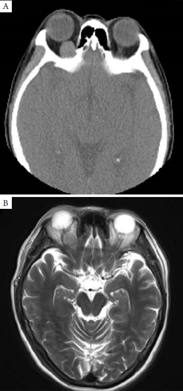
(A) Orbital CT shows that the tumor is quasi-circular with homogenous density and clear boundaries; (B) Orbital MRI shows that the tumor is quasi-circular, well-circumscribed and with homogenous intensity. Note the medial rectus and optic nerve is compressed.
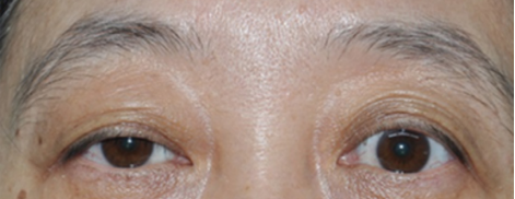
入院后完善相关辅助检查,经患者同意后于2018年3月1日在全身麻醉下行右眼眼眶内肿物切除。术中见肿物位于肌锥内,紧邻上直肌、提上睑肌和视神经。病理大体检查:灰白灰红色组织1块,质地较韧,肿物破裂,大小18 mm×17 mm×8 mm,切面灰白色实性。病理检查显微镜下结构:肿瘤细胞呈巢状或围绕血管呈条索状分布,大小一致,圆形或多角形,界限不清,细胞质丰富,嗜伊红性,颗粒感,胞核圆形或卵圆形位于中央,染色质细小,可见核仁结构,间质内含有丰富扩张的毛细血管结构,周围有散在淋巴细胞浸润(图3 )。免疫组织化学检查结果(图4 )示:嗜铬素A(chromogranin A,CgA)(+)、神经元特异性烯醇化酶(neuron specific enolase,NSE)(+)、Ki-67散在(+)、p53少量胞核(+)、CD31、CD34血管壁(+)、波形蛋白血管壁(+)、广谱型细胞角蛋白(cytokeratin CK)(?)、突触素(synaptophysin,Syn)(?)。病理诊断:符合右眼眶副神经节瘤。术后1周出院,眼部检查右眼上睑完全下垂,其他未见明显异常,嘱其定期复诊。1年后复查,右眼上睑下垂症状消失,随访3年,肿瘤无复发。
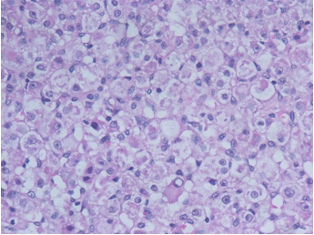
图3 眼眶副神经节瘤HE染色结果(×400)
Figure 3 HE staining of orbital paraganglioma (×400)
肿瘤细胞的细胞质丰富,嗜伊红性,颗粒感,胞核圆形或卵圆形位于中央,染色质细小,可见核仁结构。
The cytoplasm of tumor cell is rich, eosinophilic, and granular, and the nuclei are round or oval in the center and the nucleolus are clear.
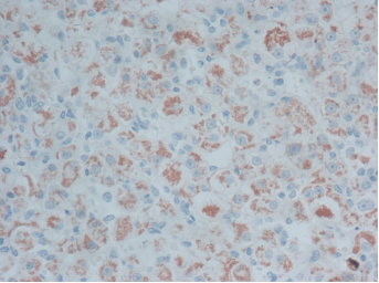
图4 免疫组织化学CgA染色结果(×400)示:肿瘤细胞的细胞质阳性
Figure 4 IHC staining of CgA (×400): the cytoplasm of the tumour cell is positive
1. Delellis RA, Lloyd RV, Heitz PU, et al. World Health Organization
classification of tumors. Pathology and genetics, tumors of endocrine organs[M]. Lyon: IARC Press, 2004: 162-163.
2. Huang N, Rayess HM, Svider PF. et al. Orbital paraganglioma: a
systematic review[ J]. J Neurol Surg B Skull Base, 2018, 79(4):
407-412.
3. Fisher ER, Hazard JB. Nonchromaffin paraganglioma of the orbit[ J].
Cancer, 1952, 5(3): 521-524.
4. Salinas-La R osa CM. Orbital paraganglioma and succinate
dehydrogenase staining for genetic testing triage and prognosis[ J].
Ocul Oncol Pathol, 2015, 2(1): 36-39.
5. 文阳, 王华, 王伯胤. 肾上腺外副神经节瘤的临床特点与CT诊断
价值[ J]. 中国临床医学影像杂志, 2009, 20(4): 286-289.
WEN Y, WANG H, WANG BY. Clinical features and CT
diagnostic value of extradrenal paraganglioma[ J]. Journal of China
Clinic Medical Imaging, 2009, 20(4): 286-289.
6. 王关顺, 刘云霞, 廖承德, 等. 副神经节瘤的CT和MRI表现[ J]. 中
国临床医学影像杂志, 2014, 25(8): 542-546.
WANG GS, LIU YX, LIAO CD, et al. CT and MRI
findings of paraganglioma[ J]. Journal of China Clinic Medical Imaging,
2014, 25(8): 542-546.
7. Zhu B, Yan J. Orbital paraganglioma[ J]. J Craniofac Surg, 2019, 30(6):
e503-e506.
8. Makhdoomi R, Nayil K, Santosh V, et al. Orbital paraganglioma—
a case report and review of the literature[ J]. Clin Neuropathol, 2010,
29(2): 100-104.
9. Archer KF, Hurwitz JJ, Balogh JM, et al. Orbital nonchromaffin
paraganglioma. A case report and review of the literature[ J].
Ophthalmology, 1989, 96(11): 1659-1666.
'%20fill='white'%20fill-opacity='0.01'/%3e%3cmask%20id='mask0_3477_29692'%20style='mask-type:luminance'%20maskUnits='userSpaceOnUse'%20x='0'%20y='0'%20width='16'%20height='16'%3e%3crect%20id='&%23232;&%23146;&%23153;&%23231;&%23137;&%23136;_2'%20x='16'%20width='16'%20height='16'%20transform='rotate(90%2016%200)'%20fill='white'/%3e%3c/mask%3e%3cg%20mask='url(%23mask0_3477_29692)'%3e%3cpath%20id='&%23232;&%23183;&%23175;&%23229;&%23190;&%23132;'%20d='M14%205L8%2011L2%205'%20stroke='%23333333'%20stroke-width='1.5'%20stroke-linecap='round'%20stroke-linejoin='round'/%3e%3c/g%3e%3c/g%3e%3c/svg%3e)