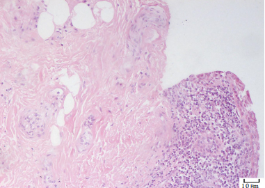
Figure 2 The skin tissue and conjunctival tissue showed severe chronic inflammation, some areas with acute inflammation,papillomatous hyperplasia, hyperkeratosis and incomplete keratosis (HE, ×200)
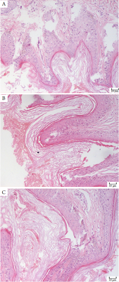
Figure 3 Skin tissues of (A) right temporal, (B) right mandibular, (C) r ight neck : papillar y and verr ucous hyperplasia could be seen in epidermis, sebaceous gland hyperplasia could be seen in dermis, no hair, which was considered to be more consistent with sebaceous nevus(HE, ×200)
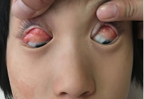
Figure 4 General image of the patient showed angulation deformity in the outer 1/3 of the upper eyelid of the right eye;there was yellowish white hyperplasia in the upper bulbar conjunctiva and fornix conjunctiva, located 1 mm above the upper limbus of the cornea; an angle deformity in the outer 1/3 of the upper eyelid of the left eye, eyelash inverted to the cornea, full-thickness defect in the central part of the upper eyelid, yellow and white hyperplasia in the conjunctiva and fornix of the upper temporal side, located 2 mm above the upper temporal corneal limbus
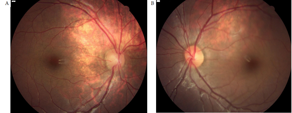
图5 眼底像示双眼视盘颞上方可见不规则形黄白色病灶,右眼大小约6个视盘直径,左眼大小约3个视盘直径
Figure 5 Fundus image showed irregular yellowish white lesions above the temporal of optic disc in both eyes; the right eye was about 6 optic disc diameters, and the left eye was about 3 optic disc diameters
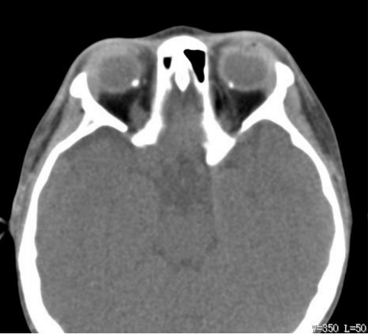
Figure 6 CT examination of the patient’s orbit showed dense shadows on the nasal side of the posterior wall of bilateral eyeballs,short strip dense shadows on the right superior ocular vein, enlarged eyelid of bilateral lacrimal glands, right sigmoid sinus and maxillary sinusitis, and multiple strip soft tissue density shadows under the skin of right maxillofacial area
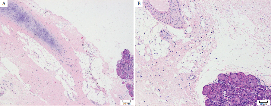
图7 患者病变组织病理组织学表现(HE染色)
Figure 7 Histopathological findings of the patient (HE staining)
(A) Fibrous connective and adipose, cartilage and lacrimal gland, consistent with the pathological changes of steatoadenoma (×100); (B)Fibrous connective tissue and adipose tissue with lacrimal glands, consistent with the pathological changes of steatoadenoma (×200).
1. 国家自然科学青年基金 (81800864)。 This work was supported by the National Natural Science Youth Foundation of China
(81800864).
参考文献
1. Pan C, Zhou X, Hong A, et al. Identification of KRAS mutation in a
patient with linear nevus sebaceous syndrome: a case report[ J]. BMC
Med Genomics, 2020, 13(1): 188.
2. Wang H, Qian Y, Wu B, et al. KRAS G12D mosaic mutation in a
Chinese linear nevus sebaceous syndrome infant[ J]. BMC Med Genet,
2015, 16: 101.
3. 张红, 刘兵, 王玉颖, 等. 线状皮脂腺痣综合征1例[ J]. 临床皮肤
科杂志, 2004, 33(11): 679-681.
ZHANG H, LIU B, WANG YY, et al. A case of linear
sebaceous nevus syndrome[ J]. Journal of Clinical Dermatology, 2004,
33(11): 679-681.
4. Kuroda Y, Ohashi I, Enomoto Y, et al. A postzygotic NRAS mutation
in a patient with Schimmelpenning syndrome[ J]. Am J Med Genet A,
2015, 167A(9): 2223-2225.
5. Groesser L, Herschberger E, Ruetten A, et al. Postzygotic HRAS
and KRAS mutations cause nevus sebaceous and Schimmelpenning
syndrome[ J]. Nat Genet, 2012, 44(7): 783-787.
6. Sun BK, Saggini A, Sarin KY, et al. Mosaic activating RAS mutations in
nevus sebaceus and nevus sebaceus syndrome[ J]. J Invest Dermatol,
2013, 133(3): 824-827.
7. 颜美荣, 王连元, 周一鸣. 眼睑皮脂腺痣伴眶颅骨畸形1例[ J]. 眼
科新进展, 2007, 27(6): 480.
YAN MR, WANG LY, ZHOU YM. A case of eyelid
sebaceous nevus with orbital skull deformity[ J]. Recent Advances in
Ophthalmology, 2007, 27(6): 480.
8. Yu KC, Lalwani AK. Inner ear malformations and hearing loss in linear
nevus sebaceous syndrome[ J]. Int J Pediatr Otorhinolaryngol, 2000,
56: 211-216.
9. Lihua J, Feng G, Shanshan M, et al. Somatic KRAS mutation in an
infant with linear nevus sebaceous syndrome associated with lymphatic
malformations: A case report and literature review[ J]. Medicine
(Baltimore), 2017, 96(47): e8016.
10. Eisen DB, Michael DJ. Sebaceous lesions and their associated
syndromes: part II[ J]. J Am Acad Dermatol, 2009, 61: 563-578, quiz
579-580.
11. 曹媛, 丁侠, 林明, 等. 线性皮脂腺痣综合征及其眼部表现[ J]. 临
床眼科杂志, 2019, 27(2): 142-144.
CAO Y, DING X, LIN M, et al. Linear sebaceous nevus
syndrome and its ocular manifestations[ J]. Journal of Clinical
Ophthalmology, 2019, 27(2): 142-144.
12. 章征, 顼晓琳, 李栋军, 等. 线状皮脂腺痣综合征眼部临床
特征及手术整复效果分析[ J]. 中华眼科杂志, 2020, 56(11):
846-852.
ZHANF Z, XU XL, LI DJ, et al. Clinical features of
linear sebaceous nevus syndrome and analysis of the effect of surgical
restoration[ J]. Chinese Journal of Ophthalmology, 2020, 56(11):
846-852.
13. Kausar A, Zafar SN, Altaf S, et al. Ophthalmic manifestations of linear
nevus sebaceous/organoid nevus syndrome[ J]. J Coll Physicians Surg
Pak, 2015, 25(3): 220-222.
14. Caputo R, Tadini G. Atlas of gerodermatoses[M]. London: Taylor &
Francis, 2004: 192-193.
15. 佟柏楠, 肖骏. 皮脂腺痣综合征合并脉络膜骨瘤一例[ J]. 中华眼
底病杂志, 2017, 33(3): 306.
TONG BN, XIAO J. A case of sebaceous nevus syndrome with
choroidal osteoma[ J]. Chinese Journal of Ocular Fundus Diseases,
2017, 33(3): 306.
16. Lambert HM, Sipperley JO, Shore JW, et al. Linear nevus sebaceous
syndrome[ J]. Ophthalmology, 1987, 94(3): 278-282.
'%20fill='white'%20fill-opacity='0.01'/%3e%3cmask%20id='mask0_3477_29692'%20style='mask-type:luminance'%20maskUnits='userSpaceOnUse'%20x='0'%20y='0'%20width='16'%20height='16'%3e%3crect%20id='&%23232;&%23146;&%23153;&%23231;&%23137;&%23136;_2'%20x='16'%20width='16'%20height='16'%20transform='rotate(90%2016%200)'%20fill='white'/%3e%3c/mask%3e%3cg%20mask='url(%23mask0_3477_29692)'%3e%3cpath%20id='&%23232;&%23183;&%23175;&%23229;&%23190;&%23132;'%20d='M14%205L8%2011L2%205'%20stroke='%23333333'%20stroke-width='1.5'%20stroke-linecap='round'%20stroke-linejoin='round'/%3e%3c/g%3e%3c/g%3e%3c/svg%3e)