本研究已获得佛山市第二人民医院医学伦理委员会审核批准,同时将试验方法、可能存在的风险及应对方案告知患者,并在患者同意下签署知情同意书。检查仪器:Corvis ST(Oculus,德国)、A型超声角膜测厚仪(A-scan Plus,美国)、Lenstar LS 900(Haag-Streit,瑞士)。
1.2 方法
1.2.1 Corvis ST角膜生物力学分析仪检查
嘱近视患者下颌部置于下颌托,额头紧贴额托,嘱受检者眨眼数次后睁开双眼并注视中央红点固视目标;将Corvis ST 检查仪测压头对准角膜顶点后进行自动识别,均匀向角膜施加空气脉冲压力,完成1次操作;重复检查5次,两次测量之间间隔2~5 min,在5次获取的CCT中取图像质量最好的一次并将CCT纳入本研究。
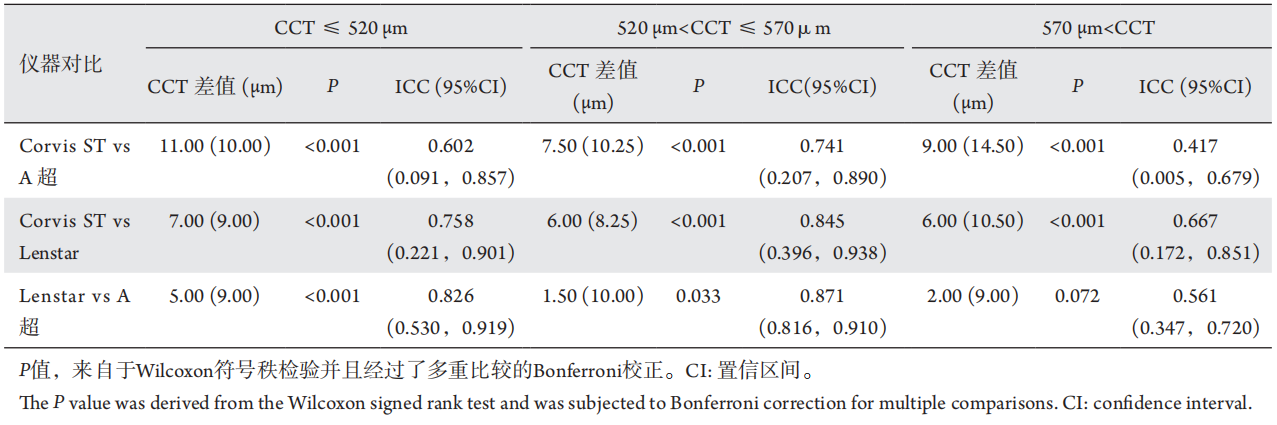
Table 1 Comparison of difffference of CCT measured by three instruments in each group
2.1.2 相关性
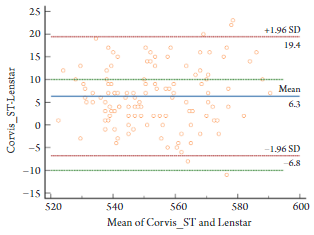
Corvis ST、A超、Lenstar测量值的正态性检验分别为P=0.032、P<0.001、P=0.916,3组数据不完全合正态分布,采用Spearman分析法。CorvisST与A超、Lenstar间呈高度正相关(r=0.841,P<0.001;r=0.832,P<0.001);Lenstar与A超呈高度正相关性(r=0.905,P<0.001)。 表1各分组三种仪器测量CCT的差异性比较 Table 1 Comparison of difference of CCT measured by three instruments in each group
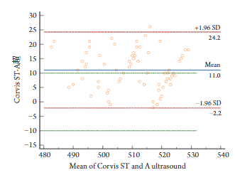
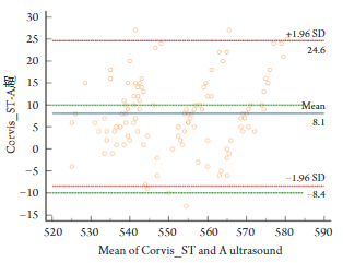
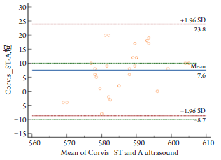
图5厚角膜组Corvis ST与A超的Bland-Altman一致性分析 Figure 5 Bland Altman consistency analysis of Corvis ST and A ultrasound in the thick cornea group
图5 厚角膜组 Corvis ST与A超的Bland-Altman一致性分析
Figure 5 Bland Altman consistency analysis of Corvis ST and A ultrasound in the thick cornea group
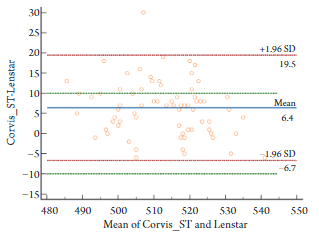
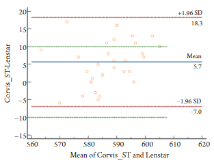
图6 厚角膜组Corvis ST与Lenstar的Bland-Altman一致性分析 Figure 6 Bland Altman consistency analysis of Corvis ST and Lenstar in the thick cornea group
3 讨论
在角膜屈光手术研究领域,角膜厚度是一个重要的监测指标[6-7],也是屈光手术设计方案及矫正屈光度数的必要参数[8],因此角膜厚度测量成为术前检查必要步骤。目前对角膜厚度测量的仪器繁多,包括角膜接触式的A型角膜测厚仪、非接触式的Lenstar LS 900测厚仪以及最新引进的Corvis ST超高速Scheimpflug动态成像仪,但在角膜厚度测量上也存在差异。临床研究更倾向于寻找简易便捷、精准度较高且无创的角膜厚度测量方法。Hon等[9]、祖培培等[10]的研究表明:Corvis ST的检测值具有较高的可重复性。本研究主要通过将Corvis ST与A超、Lenstar两种仪器在角膜厚度测量上的对比,探究Corvis ST与后两者的差异性、相关性与一致性,研究其使用特点,为屈光术前检查提供新的参考。
在角膜厚度测量上,传统的A型超声角膜测厚仪被公认为测量的“金标准”,其原理是通过测量超声波穿过角膜所需时间来计算角膜厚度,优点在于其测量的精准度高、可重复性好,在Iskander等[11]的研究中已经被证实。但其无法进行对角膜厚度进行多点测量,同时,由于需要接触角膜,一定程度上增加了角膜感染的概率。其次,测量值也会受到操作者与受检者的影响[12]。随着越来越多无创方法的出现,超声测厚仪逐渐被取代。Lenstar LS 900是一种非接触式光学仪器[13],以光学低相干反射为原理使其理论上具有较好的精确性和分辨率[14]。同时Tappeiner等[15]、黄磊等[16]的研究也表明Lenstar具有检测时间短、重复性高的特点,因此本文与该仪器作对比有临床意义。
1. Wong YL, Saw SM. Epidemiology of pathologic myopia in asia and worldwide[J]. Asia Pac J Ophthalmol (Phila), 2016, 5(6): 394-402.
2. Ambrosio JRR, Ramos I, Luz A, et al. Dynamic ultra-high-speed Scheimpflug imaging for assessing corneal biomechanical properties[J]. RevBras Oftalmol, 2013, 72(2): 99-102.
3. Lee H, Kang DSY, Ha BJ, et al. Biomechanical properties of the cornea using a dynamic scheimpflug analyzer in healthy eyes[J]. Yonsei Med J, 2018, 59(9): 1115-1122.
4. Hon Y, Wan K, Chen GZ, et al. Diurnal variation of corneal tangent modulus in normal Chinese[J]. Cornea, 2016, 35(12): 1600-1604.
5. Hamed-Azzam S, Briscoe D, Tomkins O, et al. Evaluation of intraocular pressure according to corneal thickness before and after excimer laser corneal ablation for myopia[J]. Int Ophthalmol, 2013, 33(4): 349-354.
6. Viswanathan D, Goldberg I, Graham SL. Relationship of change in central corneal thickness to visual field progression in eyes with glaucoma[J]. Graefes Arch Clin Exp Ophthalmol, 2013, 251(6): 1593-1599.
7. Christensen A, Narváez J, Zimmerman G. Comparison of central corneal thickness measurements by ultrasound pachymetry, konan noncontact optical pachymetry, and orbscan pachymetry[J]. Cornea, 2008, 27(8): 862-865.
8. Hon Y, Lam AK. Corneal deformation measurement using Scheimpflug noncontact tonometry[J]. Optom Vis Sci, 2013, 90(1): e1-e8.
10. Study on repeatability and consistency of Corvis ST measured by corneal biomechanical intraocular pressure analyzer[J]. Chinese Journal of Optometry Ophthalmology and Visual Science, 2013, 15(5): 261-265.
11. Iskander NG, Anderson Penno E, Peters NT, et al. Accuracy of Orbscan pachymetry measurements and DHG ultrasound pachymetry in primary laser in situ keratomileusis and LASIK enhancement procedures[J]. J Cataract Refract Surg, 2001, 27(5): 681-685.
13. A comparative study of three instruments for measuring central corneal thickness[J]. Chinese Journal of Optometry & Ophthalmology, 2009, 11(5): 364-367.
14. Bjelo? Ron?evi? M, Bu?i? M, Cima I, et al. Intraobserver and interobserver repeatability of ocular components measurement in cataract eyes using a new optical low coherence reflectometer[J]. Graefes Arch Clin Exp Ophthalmol, 2011, 249(1): 83-87.
16. Clinical application progress of Lenstar LS900[J]. International Eye Science, 2012, 12(11): 2123-2125.
17. Tappeiner C, Rohrer K, Frueh BE, et al. Clinical comparison of biometry using the non-contact optical low coherence reflectometer (Lenstar LS 900) and contact ultrasound biometer (Tomey AL-3000) in cataract eyes[J]. Br J Ophthalmol, 2010, 94(5): 666-667.
21. Accuracy evaluation of corneal biomechanical analyzer in measuring central corneal thickness and intraocular pressure in patients with myopia[J]. Chinese Journal of Experimental Ophthalmology, 2016, 34(4): 340-344.
23. Clinical observation of corneal thickness and intraocular pressure measured by Corvis ST and Pentacam combined diagnostic system[J]. Journal of Clinical Ophthalmology, 2020, 28(4): 341-346.
24. Yu A, Zhao W, Savini G, et al. Evaluation of central corneal thickness using corneal dynamic scheimpflug analyzer Corvis ST and comparison with Pentacam rotating scheimpflug system and ultrasound pachymetry in normal eyes[J]. J Ophthalmol, 2015, 2015: 767012.
25. Smedowski A, Weglarz B, Tarnawska D, et al. Comparison of three intraocular pressure measurement methods including biomechanical properties of the cornea[J]. Invest Ophthalmol Vis Sci, 2014, 55(2): 666-673.
27. Clinical study of Corvis ST in measuring central corneal thickness and intraocular pressure in glaucoma[J]. Chongqing Medicine, 2020, 49(21): 3556-3560.
28. Niimi J, Tan B, Chang J, et al. Diurnal pattern of tear osmolarity and its relationship to corneal thickness and deswelling[J]. Cornea, 2013, 32(10): 1305-1310.
'%20fill='white'%20fill-opacity='0.01'/%3e%3cmask%20id='mask0_3477_29692'%20style='mask-type:luminance'%20maskUnits='userSpaceOnUse'%20x='0'%20y='0'%20width='16'%20height='16'%3e%3crect%20id='&%23232;&%23146;&%23153;&%23231;&%23137;&%23136;_2'%20x='16'%20width='16'%20height='16'%20transform='rotate(90%2016%200)'%20fill='white'/%3e%3c/mask%3e%3cg%20mask='url(%23mask0_3477_29692)'%3e%3cpath%20id='&%23232;&%23183;&%23175;&%23229;&%23190;&%23132;'%20d='M14%205L8%2011L2%205'%20stroke='%23333333'%20stroke-width='1.5'%20stroke-linecap='round'%20stroke-linejoin='round'/%3e%3c/g%3e%3c/g%3e%3c/svg%3e)