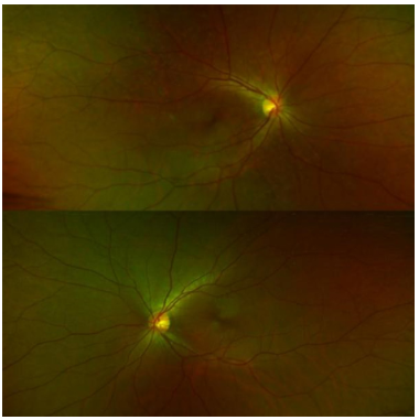
Figure 1 Binocular scanning laser fundus, the optic disc boundary of the right eye is clear, with mild edema, and retinal blood vessels can be deformed, dimly visible sunken reflections, no obvious abnormality is found in the left eye
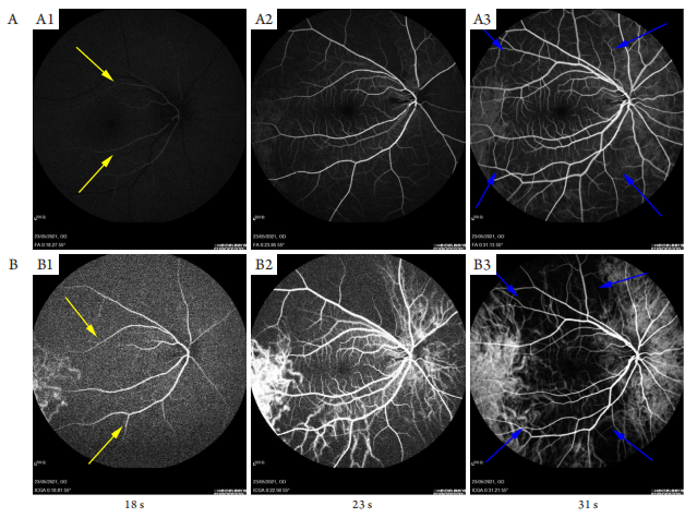
(A) FFA; (B) ICGA. Arterial phase (A1, B1) for 18 s, “for-ward” phenomenon of retinal artery (yellow arrows), arteriovenous phase (A2, B2) for 23 s, venous phase (A3, B3) for 31 s. The filling time of arteries and veins was delayed, and the circulation time was significantly delayed. FFA and ICGA showed that the choroidal filling in the posterior pole was slow, affffecting the macular area, and it was still not filled after 31 s, as shown by the blue arrows in figure (A3, B3). The choroidal circulation time is prolonged, the choroidal background fluorescence is weakened in the early stage and is in an uneven and slow filling state. The choroidal blood vessels first fill locally from the temporal side of the macula at the posterior pole, showing a “watershed” like change with a clear boundary between the central area of the macula and the surrounding area and a dark vertical shape.
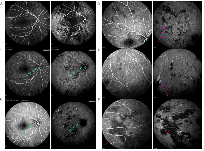
(A) 0:40 s; (B) 6:21 s; (C) 18:57 s. Contrast and late fundus peripheral image (D: upper, E: lower, F: nasal side). Orange arrow shows that the retinal vascular wall can be accompanied by light to moderate staining; blue arrow shows optic disc edema with unclear boundary; purple arrows show focal hyperfluorescence scattered in the fundus at the late stage of angiography, and several hyperfluorescence rays can be seen at the ends of some arterioles; red arrows show partial filling defect of retinal background fluorescence.
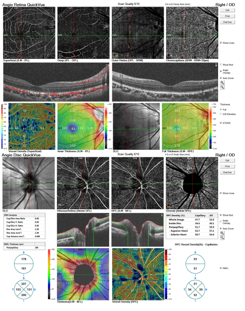
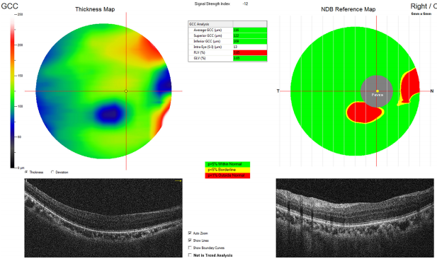
Figure 4 Right eye Angio OCT. Superficial retinal blood flow density around macular fovea of right eye decrease, oct of optic disc shows that the blood flow density of temporal side and inferior nose of optic papilla is relatively positive The RNFL of optic disc is thickened obviously in the supratemporal and infratemporal regions
1. 山东省医药卫生科技发展计划项目 (202007021030)。This work was supported by the Shandong Medical and Health Science and Technology Development Plan Project, China (202007021030)
参考文献
1. Schmidt-Erfurth U, Garcia-Arumi J, Bandello F, et al. Guidelines for the management of diabetic macular edema by the European Society of Retina Specialists (EURETINA)[J]. Ophthalmologica, 2017, 237(4): 185-222.
2. Saito K, Akiyama H, Mukai R . Aliteration of optical coherence tomography angiography in a patient w ith ocular ischemic
syndrome[ J]. Retin Cases Brief Rep, 2021, 15(5): 588-592.
5. 庄岩, 刘春军, 杨明勇. 眼动脉及其分支栓塞:严重的面部注射填充术并发症[ J]. 中国美容整形外科杂志, 2015, 26(5): 308-310.
ZHUANG Yan, LIU Chunjun, YANG Mingyong. Ophthalmic artery and its branches embolism: serious complications of facial injection and filling surgery[ J]. Chinese Journal of Aesthetic and Plastic Surgery, 2015, 26(5): 308-310.
6. Kahn M, Green WR , Knox DL, et al. Ocular features of carotid
occlusive disease[ J]. Retina, 1986, 6(4): 239-252.
7. 杨秀芬, 李红阳, 赵露, 等. 眼缺血综合征的临床及影像学特点分
析[ J]. 山东大学耳鼻喉眼学报, 2019, 33(4): 119-123.
YANG Xiufen, LI Hongyang, ZHAO Lu, et al. Clinical and imaging
characteristics in patients with ocular ischemic syndrome[ J]. Journal
of Otolaryngology and Ophthalmology of Shandong University, 2019,
33(4): 119-123.
8. Taylor GI, Shoukath S, Gascoigne A, et al. The functional anatomy of the ophthalmic angiosome and its implications in blindness as a complication of cosmetic facial filler procedures[J]. Plast Reconstr Surg, 2020, 146(4): 745.
9. 王露萍, 黄映湘, 王艳玲. 眼缺血综合征研究进展[ J]. 山东大学耳鼻喉眼学报, 2020, 34(4): 23-27.
WANG Luping, HUANG Yingxiang, WANG Yanling. Recent ocular ischemic syndrome advances[ J]. Journal of Otolaryngology and Ophthalmology of Shandong University, 2020, 34(4): 23-27.
10. 高景恒, 袁继龙, 朱利娜, 等. 面部注射填充突发失明、脑血栓并发症的文献复习[J]. 中国美容整形外科杂志, 2017, 28(9): 513-516.
GAO Jingheng, YUAN Jilong, ZHU Lina, et al. Literature review of sudden blindness and cerebral thrombosis complications after facial injection filling[ J]. Chinese Journal of Aesthetic and Plastic Surgery,2017, 28(9): 513-516.
11. Kim YK, Jung C, Woo SJ, et al. Cerebral angiographic findings of cosmetic facial filler-related ophthalmic and retinal artery occlusion[ J]. J Korean Med Sci, 2015, 30(12): 1847-1855.
12. Wang K, Rong X, Dang J, et al. Severe vascular complications caused by facial autologous fat grafting: a critical review[ J]. Ann Plast Surg, 2021,86(3S Suppl 2): S208-S219.
13. 陈竹林, 黄光, 赵涵, 等. 面部注射美容手术致失明和肢体偏瘫的临床分析[J]. 中国神经免疫学和神经病学杂志, 2017, 24(6): 438-440.
CHEN Zhulin, HUANG Guang, ZHAO Han, et al. Clinical analysis of blindness and hemiplegia caused by facial injection cosmetic surgery[ J]. Chinese Journal of Neuroimmunology and Neurology,2017, 24(6): 438-440.
14. Moon HS, Song SW, Park TH, et al. Treatment of liponecrotic pseudocysts following autologous fat transfer with minimally invasive combination therapy[J]. Plast Reconstr Surg Glob Open, 2018, 6(8S): 8-9.
15. An TT, Colon-Acevedo B, Mettu P, et al. An anatomical analysis of the supratrochlear artery: considerations in facial filler injections and preventing vision loss[ J]. Aesthet Surg J, 2017, 37(2): 203-208.
16. 狄宇, 叶俊杰. 眼缺血综合征二例[ J]. 中华眼科杂志, 2019, 55(6):454-457.
DI Yu, YE Junjie. Two cases of ocular ischemia syndrome[ J]. Chinese Journal of Ophthalmology, 2019, 55(6): 454-457.
17. 万锦麟, 鲁峰. 颗粒脂肪移植后严重并发症的文献分析[ J]. 中华整形外科杂志, 2015, 31(3): 237-238.
WAN Jinlin, LU Feng. Literature analysis of severe complications after granular fat transplantation[ J]. Chinese Journal of Plastic Surgery,2015, 31(3): 237-238.
18. Park KH, Kim YK, Woo SJ, et al. Latrogenic occlusion of the ophthalmic artery after cosmetic facial filler injections: A national survey by the Korean Retina Society[J]. JAMA Ophthalmol, 2014, 132(6): 714-723.
19. Eleznay K, Carruthers JD, Humphrey S, et al. Avoiding and treating blindness from fillers: a review of the world literature[ J]. Dermatol Surg, 2015, 41(10): 1097-1117.
20. Utsugi N, Takahashi K, Kishi S. Choroidal vascular occlusion in internal carotid artery obstruction[ J]. Retina, 2004, 24(6): 915-919.
21. Mineda K , Kuno S, Kato H, et al. Chronic inflammation and progressive Calcification as a result of fat necrosis: the worst outcome in fat grafting[ J]. Plast Reconstr Surg, 2014, 133(5): 1064-1072.
'%20fill='white'%20fill-opacity='0.01'/%3e%3cmask%20id='mask0_3477_29692'%20style='mask-type:luminance'%20maskUnits='userSpaceOnUse'%20x='0'%20y='0'%20width='16'%20height='16'%3e%3crect%20id='&%23232;&%23146;&%23153;&%23231;&%23137;&%23136;_2'%20x='16'%20width='16'%20height='16'%20transform='rotate(90%2016%200)'%20fill='white'/%3e%3c/mask%3e%3cg%20mask='url(%23mask0_3477_29692)'%3e%3cpath%20id='&%23232;&%23183;&%23175;&%23229;&%23190;&%23132;'%20d='M14%205L8%2011L2%205'%20stroke='%23333333'%20stroke-width='1.5'%20stroke-linecap='round'%20stroke-linejoin='round'/%3e%3c/g%3e%3c/g%3e%3c/svg%3e)