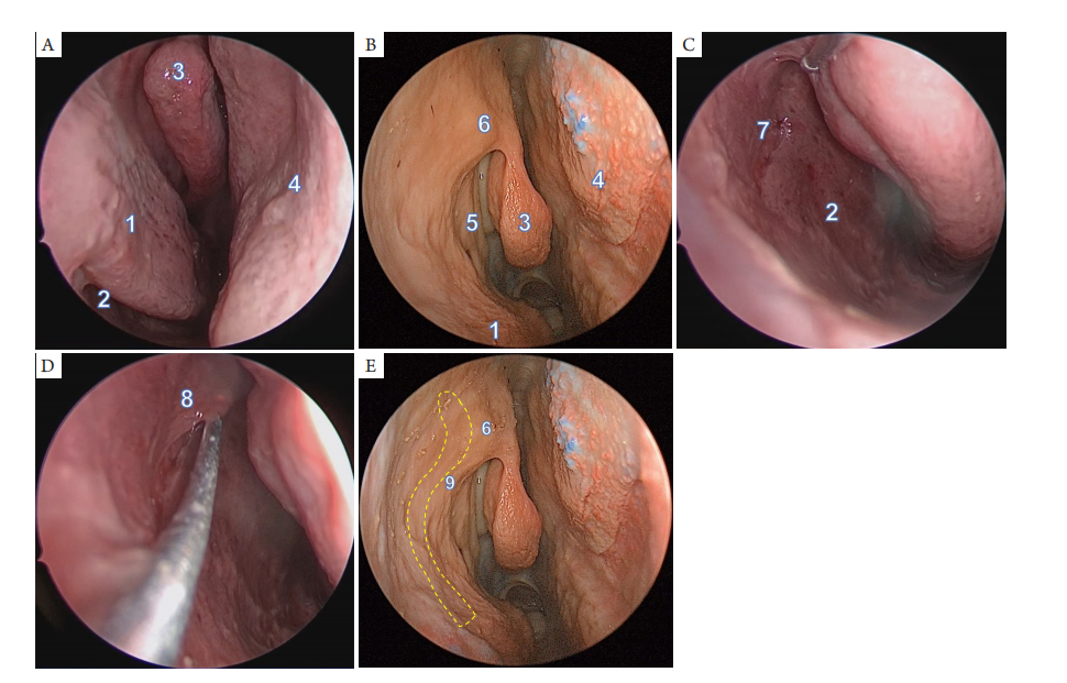
(A,B) Endoscopic views of the right side of nasal cavity; (C,D) Exposure of the opening of the right nasolacrimal duct on the later wall of inferior meatus; (E) The projection of the nasolacrimal duct and lacrimal sac on the lateral nasal wall (Yellow dash line). 1: inferior turbinate; 2: inferior meatus; 3: middle turbinate; 4: nasal septum; 5: uncinate process; 6: nasal mound; 7: opening of nasolacrimal duct; 8: Hasner valve; 9: frontal process of maxilla.
图2 鼻内镜下显露鼻泪管-泪囊
Figure 2 Exposure of nasolacrimal duct and lacrimal sac under nasal endoscopy
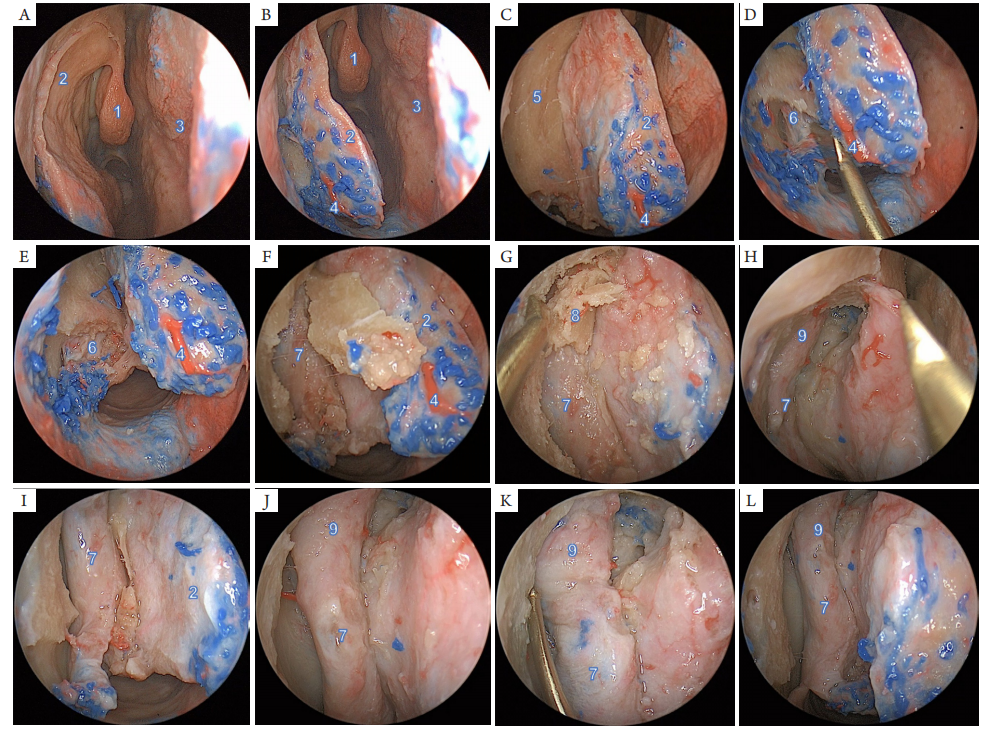
(A-C) An arc-shaped longitudinal incision was made on the lateral wall of the right nasal cavity, starting from the attachment point of theanterior end of the middle turbinate, forward and upward to the level above the nasal mound, and then turning forward and downward to the front of the anterior end of the inferior turbinate. Then elevator was used to separate the mucosal flap from the bony surface, and the head of the bony inferior turbinate was seen. (D-F) The connection of the mucosal flap at the axilla of the inferior turbinate was separated, and the opening of the nasolacrimal duct was located. Then the anterior part of the bony inferior turbinate was removed in front of the opening of the nasolacrimal duct, and the bony lateral nasal wall on the surface of the nasolacrimal duct was removed upward. (G-I) After the nasolacrimal duct was exposed, the bone of the frontal process of maxilla was removed upward with the nasolacrimal duct as a landmark, and then the medial wall of the lacrimal sac was exposed. The nasolacrimal duct was freed from the bottom up. ( J-L) After dissecting the connecting tissue around the membranous part of nasolacrimal duct, the nasolacrimal duct and lacrimal sac were fully freed. 1: middle turbinate; 2: mucosal flap on the nasal lateral wall; 3: nasal septum; 4: lateral nasal branch of anterior ethmoidal artery; 5: bony lateral nasal wall; 6: the inferior meatus opening of the bony part of nasolacrimal duct; 7: membrane part of nasolacrimal duct; 8: The inner bony wall of lacrimal sac; 9: lacrimal sac.
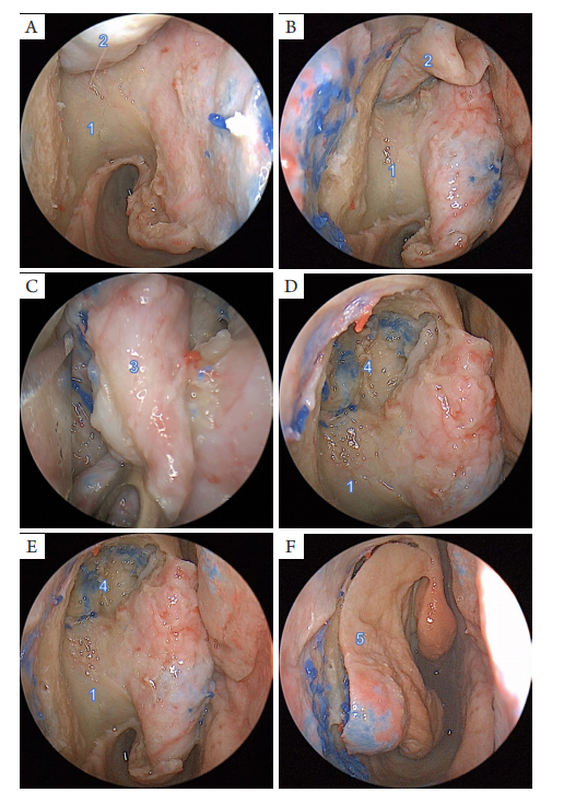
(A-D) After dissecting the connection between the opening of the right nasolacrimal duct and the mucosa of the inferior meatus, the membranous part of the nasolacrimal duct was lifted from the bony canal from the bottom up. The lateral part of the upper lacrimal sac was revealed. Then the lacrimal sac and the medial orbital tissue was separated from anteriorly to posteriorly, and finally the lacrimal sac and nasolacrimal duct were resected completely. (E) Endoscopic views showing structures of the lacrimal fossa, the bony part of nasolacrimal duct after removal of nasolacrimal duct and lacrimal sac. (F) Reposition of the mucosal flap on the nasal lateral wall. 1: bony part of nasolacrimal duct; 2: membrane part of nasolacrimal duct; 3: lacrimal sac; 4: lacrimal fossa; 5: mucosal flap on the nasal lateral wall.
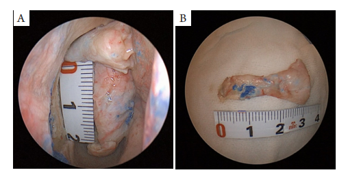
图4 骨性鼻泪管(A)和膜性鼻泪管及泪囊(B)的测量
Figure 4 Measurement of the bony part of nasolacrimal duct (A) and the membrane part of nasolacrimal duct and the lacrimal sac (B)
1. 张速勤, 贾沛靓, 唐海红, 等. 泪囊鼻内解剖研究及临床应用. 中华耳鼻咽喉头颈外科杂志, 2006, 41(7): 506-509.
ZHANG Suqin, JIA Peiliang, TANG Haihong, et al. Endonasal anatomy of lacrimal sac and its clinical significance in dacryocystorhinostomy[ J]. Chinese Journal of Otorhinolaryngology Head and Neck Surgery, 2006, 41(7): 506-509.
2. Wormald PJ, Kew J, Van Hasselt A. Intranasal anatomy of the nasolacrimal sac in endoscopic dacryocystorhinostomy[ J]. Otolaryngol Head Neck Surg, 2000, 123(3): 307-310.
3. Rebeiz EE, Shapshay SM, Bowlds JH, et al. Anatomic guidelines for dacryocystorhinostomy[ J]. Laryngoscope, 1992, 102(10): 1181-1184.
4. El-Sawy T, Frank SJ, Hanna E, et al. Multidisciplinary management of lacrimal sac/nasolacrimal duct carcinomas[ J]. Ophthalmic Plast Reconstr Surg, 2013, 29(6): 454-457.
5. Parmar DN, Rose GE. Management of lacrimal sac tumours[ J]. Eye (Lond), 2003, 17(5): 599-606.
6. Ramberg I, Toft PB, Heegaard S. Carcinomas of the lacrimal drainage system[ J]. Surv Ophthalmol, 2020, 65(6): 691-707.
7. Reshef ER , Bleier BS, Freitag SK . The endoscopic transnasal approach to orbital tumors: A review[ J]. Semin Ophthalmol, 2021, 36(4): 232-240.
8. Heindl LM, Jünemann AG, Kruse FE, et al. Tumors of the lacrimal drainage system[ J]. Orbit, 2010, 29(5): 298-306.
9. 赵云, 惠靖雯, 杨丽红, 等. 原发性泪道肿物64例临床组织病理学分析. 中华眼科杂志, 2020, (05): 364-369.
ZHAO Yun, HUI Jingwen, YANG Lihong , et al. Clinical and pathological analysis of 64 patients with primary neoplasms of the lacrimal drainage system[ J]. Chinese Journal of Ophthalmology, 2020, 56(5): 364-369.
10. 蒋永强, 王彬, 李晓华, 等. 泪囊原发性恶性肿瘤22例临床病理学分析[ J]. 中华眼外伤职业眼病杂志, 2019, 41(2): 81-84.
JIANG Yongqiang, WANG Bin, LI Xiaohua, et al. Clinicopathological analysis of 22 cases with primary malignant tumors of the lacrimal sac[ J]. Chinese Journal of Ocular Trauma and Occupational Eye Disease, 2019, 41(2): 81-84.
11. Singh S, Ali MJ. Primary malignant epithelial tumors of the lacrimal drainage system: A major review[ J]. Orbit, 2021, 40(3): 179-192.
12. Krishna Y, Coupland SE. Lacrimal sac tumors—A review[ J]. Asia Pac J Ophthalmol (Phila), 2017, 6(2): 173-178.
13. Alam MS, Mukherjee B, Krishnakumar S. Clinical profile and management outcomes of lacrimal drainage system malignancies[ J]. Orbit, 2022, 41(4): 429-436.
15. Chang CH, Ku WN, Kung WH, et al. Navigation-assisted endoscopic surgery of lacrimal sac tumor[ J]. Taiwan J Ophthalmol, 2020, 10(2): 141-143.
16. Rajesh Raju G, Sandeep S. Lacrimal Sac Rhinosporidiosis and surgical management by transnasal endoscopic excision: A case series[ J]. Laryngoscope, 2018, 128(12): 2693-2696.
17. Curragh DS, Psaltis AJ, Tan NC, et al. Prelacrimal approach for nasolacrimal duct excision in the management of lacrimal system tumours[ J]. Orbit, 2019, 38(4): 308-312.
18. Curragh DS, James C, Selva D. Swinging inferior turbinate approach to the nasolacrimal duct[ J]. Orbit, 2020, 39(2): 112-117.
19. Villaret AB, Lombardi D, Schreiber A, et al. Oncocytic carcinoma of the nasolacrimal duct treated by transnasal endoscopic resection[ J]. Head Neck, 2013, 35(1): E24-E27.
20. 中华医学会眼科学分会眼整形眼眶病学组. 中国内镜泪囊鼻腔吻合术治疗慢性泪囊炎专家共识(2020年)[ J]. 中华眼科杂志,2020, 56(11): 820-823.
Ophthalmology Orbital Disease Group of Chinese Medical Association Ophthalmology Branch. Expert consensus on the treatment of chronic dacryocystitis with endoscopic dacryocystorhinostomy in China[ J]. Chinese Journal of Ophthalmology, 2020, 56(11): 820-823.
21. Rajak SN, Psaltis AJ. Anatomical considerations in endoscopic lacrimal surgery[ J]. Ann Anat, 2019, 224: 28-32.
'%20fill='white'%20fill-opacity='0.01'/%3e%3cmask%20id='mask0_3477_29692'%20style='mask-type:luminance'%20maskUnits='userSpaceOnUse'%20x='0'%20y='0'%20width='16'%20height='16'%3e%3crect%20id='&%23232;&%23146;&%23153;&%23231;&%23137;&%23136;_2'%20x='16'%20width='16'%20height='16'%20transform='rotate(90%2016%200)'%20fill='white'/%3e%3c/mask%3e%3cg%20mask='url(%23mask0_3477_29692)'%3e%3cpath%20id='&%23232;&%23183;&%23175;&%23229;&%23190;&%23132;'%20d='M14%205L8%2011L2%205'%20stroke='%23333333'%20stroke-width='1.5'%20stroke-linecap='round'%20stroke-linejoin='round'/%3e%3c/g%3e%3c/g%3e%3c/svg%3e)