Corneal biomechanics after rigid gas permeable contact lens wear in keratoconus eyes
阅读量:1463
DOI:doi: 10.3978/j.issn.1000-4432.2016.05.03
发布日期:2024-11-29
作者:
Fereshteh Shokrollahzadeh ,Hassan Hashemi ,Ebrahim Jafarzadehpur ,Ali Mirzajani ,Mehdi Khabazkhoob ,Soheila Asgari
展开更多 '%20fill='white'%20fill-opacity='0.01'/%3e%3cmask%20id='mask0_3477_29692'%20style='mask-type:luminance'%20maskUnits='userSpaceOnUse'%20x='0'%20y='0'%20width='16'%20height='16'%3e%3crect%20id='&%23232;&%23146;&%23153;&%23231;&%23137;&%23136;_2'%20x='16'%20width='16'%20height='16'%20transform='rotate(90%2016%200)'%20fill='white'/%3e%3c/mask%3e%3cg%20mask='url(%23mask0_3477_29692)'%3e%3cpath%20id='&%23232;&%23183;&%23175;&%23229;&%23190;&%23132;'%20d='M14%205L8%2011L2%205'%20stroke='%23333333'%20stroke-width='1.5'%20stroke-linecap='round'%20stroke-linejoin='round'/%3e%3c/g%3e%3c/g%3e%3c/svg%3e)
关键词
Keratoconus
corneal biomechanics
rigid gas permeable lens (RGP lens)
摘要
Background: Evaluation of corneal biomechanical properties 3 months after using rigid gas permeable
(RGP) contact lenses in keratoconus.
Methods: In this prospective trial study, cases were 32 keratoconic eyes with no history of RGP lens
wear. All eyes were examined with the Ocular Response Analyzer (ORA) and the Corneal Visualization
Scheimpflug Technology (CORVIS-ST) to measure corneal hysteresis (CH), corneal resistance factor
(CRF), deformation amplitude (DA), applanation velocity (AV) 1 and 2, applanation length (AL) 1 and 2, and
peak distance before and 3 months after fitting aspheric RGP lenses. The effect of the correlation between
contralateral eyes and maximum keratometry were controlled for in the analysis. Results were compared
using repeated measures analysis of covariance.
Results: At 3 months, neither the increases in mean CH (0.14±2.77 mmHg, P=0.789), CRF (0.41±4.35 mmHg, P=0.612), AV1 (0.03±0.17 m/s, P=0.301), AV2 (0.11±0.59 m/s, P=0.299), AL1 (0.44±1.56 m/s, P=0.118),
AL2 (1.16±5.06 m/s, P=0.211), and peak distance (0.19±1.29 m/s, P=0.409), nor the decrease in mean DA
(0.03±0.17 mm, P=0.402) was statistically significant.
Conclusions: Results in our series of patients indicated that 3 months of RGP lens wear had no significant
impact on corneal biomechanics, and perhaps non progression of keratoconus. Therefore, RGP lenses can be
regarded safe and appropriate in keratoconic patients.
全文
Introduction
Corneal biomechanical properties provide important
indicators when evaluating treatment results and monitoring
progression of disease such as keratoconus (1,2), and can be
helpful for the detection of keratoconus even before common
topographic signs of disease develop (3). Current devices
available for the measurement of corneal biomechanical
properties include the Ocular Response Analyzer (ORA;
Reichert, USA) and the Corneal Visualization Scheimpflug
Technology (CORVIS-ST; Oculus Optikgeräte GmbH,
Germany). The ORA measures corneal hysteresis (CH)
and the corneal resistance factor (CRF) (4). CORVIS-ST
shows corneal deformations in response to air puff
pressures. Measured indices with this device, including the
deformation amplitude (DA), the velocity of the first and
second applanation velocity (AV1 and AV2), the length of the first and second applanation (AL1 and AL2), and the
peak distance can complete ORA results to assess structural
changes, stability or no change in the corneal status, and the
appropriateness of the treatment method (5).
Methods
The target of this before-after study was patients with
mild to severe keratoconus referring to the Contact Lens
Clinic. All patients had already had a complete ophthalmic
evaluation by a cornea sub-specialist, based on which the
diagnosis of keratoconus was made and confirmed through
imaging. The Ethics Committee of Iran University of
Medical Sciences approved the study. Objectives and
methods of the study were explained to patients, and they
all signed informed consents before participation in the
study.
After enrollment, patients had complete visual examinations including the measurement of visual acuity using a Snellen chart (Nidek-34605-6004-LCD Chart) at 4 meters, objective refraction with a streak retinoscope (HEINE BETA 200, Germany) and an auto refractometer (Nidek, Japan), and subjective refraction with a trial lens set and frame. The Pentacam (Oculus Optikgeräte GmbH, Germany) was used to measure and record maximum keratometry and the central corneal thickness.
The appropriate aspherical RGP (Lens Gostar, Tehran, Iran) lens was prescribed using the diagnostic lens fitting method (8). On slit-lamp examination, good tear exchange was verified by observing the fluorescein pattern with mild apical clearance over the corneal cone and slight edge and midperipheral clearance (9). The ORA and CORVIS-ST were used to measure indices related to the biomechanical properties of the cornea. Testing with ORA was done first, and measurements with CORVIS were done after at least 1 hour. Both were done in the same room by the same technician. Before testing, patients’ blinking pattern was noted if necessary. Both devices were checked and calibrated before use.
Recorded biomechanical indices included CH and CRF, as measured with the ORA and DA, AV1, AV2, AL1, AL2 and peak distance, as measured with the CORVIS-ST. All examinations were repeated 3 months after daily use of the RGP lens.
The results were compared using repeated measure analysis of variance. P values less than 0.05 were considered significant.
Results
In this before-after study, 32 eyes of 19 patients with
keratoconus who met the inclusion criteria were enrolled.
Patients were 7 women and 12 men, with an age range
of 19–35 years (27.9±4.30 years). Based on Rabinowitz/
McDonnell criteria (10), 15 eyes were in the early stage of
keratoconus, 3 were in the moderate stage, and 14 were
categorized as having advanced keratoconus. Mean spherical
equivalent refraction was −4.13±3.16 diopter (D) and mean
LogMAR visual acuity was 0.81±0.46 without contact lenses
and 0.01±0.02 with lenses.
Mean maximum keratometry reading was 53.34±6.93 D at baseline and 53.87±6.97 D at 3 months (P=0.368) and mean central corneal thickness was 455.76±33 µm and 452.33±47.32 µm, respectively (P=0.80).
Mean maximum keratometry reading was 53.34±6.93 D at baseline and 53.87±6.97 D at 3 months (P=0.368) and mean central corneal thickness was 455.76±33 µm and 452.33±47.32 µm, respectively (P=0.80).
At 3 months, neither the increases in mean CH (0.14±
2.77 mmHg), CRF (0.41±4.35 mmHg), AV1 (0.03±0.17 m/s),
AV2 (0.11±0.59 m/s), AL1 (0.44±1.56 m/s), AL2 (1.16±5.06 m/s),
and peak distance (0.19±1.29 m/s), nor the decrease in mean
DA (0.03±0.17 mm) was statistically significant (all P>0.05).Table 1 shows ORA and CORVIS-ST indices at baseline and
3 months after RGP lens wear.
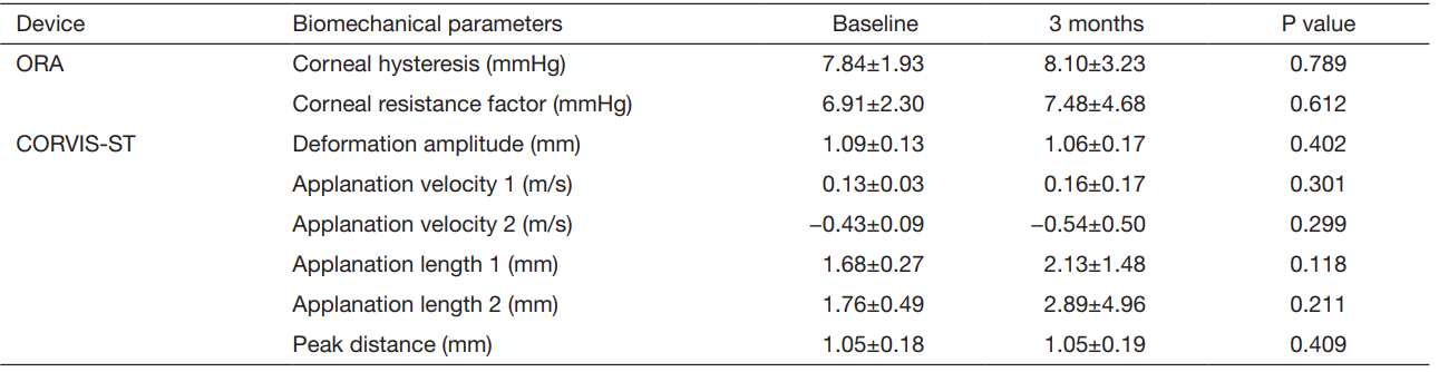 ORA, Ocular Response Analyzer; CORVIS-ST, Corneal Visualization Scheimpflug Technology.
Table 1 Descriptive statistics indices of the ORA and CORVIS-ST parameters in the studied keratoconus patients at baseline and at 3 months after wearing rigid gas permeable contact lenses
Discussion
This study showed that corneal biomechanical properties
were stable after 3 months RGP lens wear. The ORA has
been used in a number of studies with contrasting results; some believe its measurements in keratoconus add little
value to the routine workup (11-13).
The ORA lacks
the ability to measure dynamic deformation in real time,
and certain indices such as velocity (14). A study in 2014
introduced the CORVIS as an appropriate method for
measuring corneal biomechanical parameters. In their
study, two groups of normal and keratoconic corneas were
compared, and DA was reported as the best parameter with
a sensitivity of 81.7%. Results indicated increased DA,
AV1, and AV2, and decreased AL1 and AL2 in keratoconus
patients. Moreover, DA was introduced as a reliable
parameter for corneal biomechanical evaluation with
emphasis on the necessity of more studies in this regard (14).
In another study, DA was the most repeatable parameter,
and it appeared to be higher in thinner corneas, which could
be important in light of progressive thinning in keratoconus
patients (15).
Comparison of the results of the two devices in normal
and keratoconic individuals showed decreased elasticity
and resistance of the keratoconic corneas as measured with
the ORA (16) and an increase in DA and velocity (14,15) as measured with the CORVIS in line with the structural
weakness of the cornea.
RGP lens wear is an efficient method for improving
visual acuity. To our knowledge, our study is the first
to examine the effect of RGP lens wear on corneal
biomechanics, and 3 months after fitting RGP lenses in
keratoconus patients. We found no significant change in
any of the studied variables including the central corneal
thickness and maximum keratometry reading (Table 1). This
is in favor of the safety of RGP lenses for the treatment
of keratoconus patients, or even a halting effect on the
progression of the disease. Further studies with appropriate controls are required to elucidate this mater.
In conclusion, prescribing RGP lenses is a safe
approach with minimal short-term effects on the corneal
biomechanics. This finding is important for practitioners
and patients on account of concerns about lens-induced
changes in the corneal surface or structure. Further studies
with longer follow-up periods are suggested to obtain longterm
results.
基金
暂无基金信息
参考文献
1. Garcia-Porta N, Fernandes P, Queiros A, et al. Corneal biomechanical properties in different ocular conditions and new measurement techniques. ISRN Ophthalmol 2014;2014:724546.
2. Piñero DP, Alio JL, Barraquer RI, et al. Corneal biomechanics, refraction, and corneal aberrometry in keratoconus: an integrated study. Invest Ophthalmol Vis Sci 2010;51:1948-55.
3. Kozobolis V, Sideroudi H, Giarmoukakis A, et al. Corneal biomechanical properties and anterior segment parameters in forme fruste keratoconus. Eur J Ophthalmol 2012;22:920-30.
4. Luce DA. Determining in vivo biomechanical properties of the cornea with an ocular response analyzer. J Cataract Refract Surg 2005;31:156-62.
5. Ambrósio RJ Jr, Caldas DL, Ramos IC, et al. Corneal biomechanical assessment using dynamic ultra high-speed Scheimpflug technology noncontact tonometry (UHSST
NCT): preliminary results. In Proceedings of the American Society of Cataract and Refractive Surgery, the American Society of Ophthalmic Administrators (ASCRSASOA
'11); 2011 March; San Diego, California, USA.
6. Lawless M, Coster DJ, Phillips AJ, et al. Keratoconus: diagnosis and management. Aust N Z J Ophthalmol 1989;17:33-60.
7. Jinabhai A, Radhakrishnan H, O'Donnell C. Corneal changes after suspending contact lens wear in early pellucid marginal corneal degeneration and moderate keratoconus. Eye Contact Lens 2011;37:99-105.
8. Bennett ES, Weissman BA. Clinical Contact Lens Practice. Philadelphia: Lippincott Williams & Wilkins, 2005.
9. Herranz RM, Zarzuelo GR, Herráez VJ. Contact Lens Correction of Regular and Irregular Astigmatism. In: Goggin M, editor. Astigmatism - Optics, Physiology and Management. Croatia: Intechopen, 2012:157-80.
10. Sedghipour MR, Sadigh AL, Motlagh BF. Revisiting corneal topography for the diagnosis of keratoconus: use of Rabinowitz's KISA% index. Clin Ophthalmol 2012;6:181-4.
11. Terai N, Raiskup F, Haustein M, et al. Identification of biomechanical properties of the cornea: the ocular response analyzer. Curr Eye Res 2012;37:553-62.
12. Wolffsohn JS, Safeen S, Shah S, et al. Changes of corneal biomechanics with keratoconus. Cornea 2012;31:849-54.
13. Moshirfar M, Edmonds JN, Behunin NL, et al. Corneal biomechanics in iatrogenic ectasia and keratoconus: A review of the literature. Oman J Ophthalmol 2013;6:12-7.
14. Tian L, Huang YF, Wang LQ, et al. Corneal biomechanical assessment using corneal visualization scheimpflug technology in keratoconic and normal eyes. J Ophthalmol 2014;2014:147516.
15. Hon Y, Lam AK. Corneal deformation measurement using Scheimpflug noncontact tonometry. Optom Vis Sci 2013;90:e1-8.
16. Galletti JG, Pförtner T, Bonthoux FF. Improved keratoconus detection by ocular response analyzer testing after consideration of corneal thickness as a confounding factor. J Refract Surg 2012;28:202-8.
相关文章
Honghe Xia;Riping Zhang;Alvin L. Young;Mingzhi Zhang,Long term changes of posterior corneal elevation after myopic laser in situ keratomileusisYali Du;Lixia Sun;Mingzhi Zhang,The early change of corneal vertical coma and trefoil in 2.8-mm superior incision cataract surgeryHassan Hashemi;Nahid Ashraf;Ebrahim Jafarzadehpur;Alireza Hedayatfar;Soheila Asgari,Subclinical inflammatory response after accelerated corneal cross-linking