Overhanging glaucoma ff ltration bleb related to cataract surgery
阅读量:1476
DOI:doi: 10.3978/j.issn.1000-4432.2016.08.03
发布日期:2024-12-01
作者:
Haisheng Zheng ,Jingjing Huang
展开更多 '%20fill='white'%20fill-opacity='0.01'/%3e%3cmask%20id='mask0_3477_29692'%20style='mask-type:luminance'%20maskUnits='userSpaceOnUse'%20x='0'%20y='0'%20width='16'%20height='16'%3e%3crect%20id='&%23232;&%23146;&%23153;&%23231;&%23137;&%23136;_2'%20x='16'%20width='16'%20height='16'%20transform='rotate(90%2016%200)'%20fill='white'/%3e%3c/mask%3e%3cg%20mask='url(%23mask0_3477_29692)'%3e%3cpath%20id='&%23232;&%23183;&%23175;&%23229;&%23190;&%23132;'%20d='M14%205L8%2011L2%205'%20stroke='%23333333'%20stroke-width='1.5'%20stroke-linecap='round'%20stroke-linejoin='round'/%3e%3c/g%3e%3c/g%3e%3c/svg%3e)
关键词
Overhanging ff ltration bleb
trabeculectomy
cataract surgery
摘要
A 74-year-old man presented with a three-year history of foreign body sensation in the right eye
after cataract surgery. He underwent uneventful trabeculectomy with mitomycin C (MMC) in the right eye
seven years ago. Slit-lamp examination revealed a large avascular filltration bleb overhanging on the cornea
with a thin base connected to the conjunctiva. Preoperative ultrasound biomicroscopy (UBM) impressions
were confirmed by leakage of aqueous from the incision intraoperatively. Surgical dissection and revision
of the bleb was performed with satisfactory outcome. Histopathologic evaluation showed proliferation of
fibrous tissue under the conjunctival epithelia with irregular cystoids change. The current case may be the
first report of a post-trabeculectomy overhanging filtration bleb related to cataract surgery. The possible
mechanism may be related to microleakage of the surgical wound after phacoemulsiff cation which initiated
the healing and scarring process.
全文
Case presentation
A 74-year-old man presented with a three-year history
of foreign body sensation in the right eye after cataract
surgery. He underwent uneventful trabeculectomy with
mitomycin C (MMC) in the right eye for advanced stage
primary open angle glaucoma (POAG) seven years ago,
and phacoemulsiff cation with intraocular lens implantation
for senile cataract three years ago. From then on, he felt
uncomfortable in the right eye with mild vision loss. On
examination, his best-corrected visual acuity (BCVA) was
20/40. Intraocular pressure (IOP) was 18 mmHg without
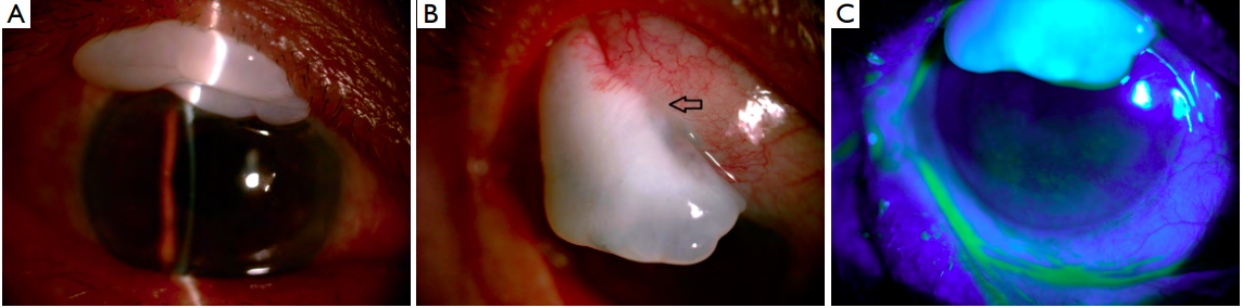
glaucoma medications. Slit-lamp examination revealed a
large avascular filtration bleb overhanging on the cornea
with a thin base connected to the conjunctiva (Figure 1A,B).
No leakage from the filtration bleb could be detected
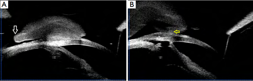
(Figure 1C). Ultrasound biomicroscopy (UBM) confirmed
that the bleb was connected to the conjunctiva through a
thin base with partial free edge (Figure 2A), and the inner
path of the ff ltration bleb was patent (Figure 2B).
 The patient underwent surgical dissection of the
overhanging bleb and scar tissues on the cornea. Leakage of
aqueous could be seen from the base of the bleb. Revision
of the bleb using superior conjunctival flap was performed.
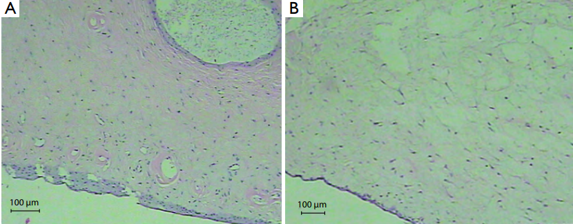
Histopathologic evaluation showed proliferation of fibrous
tissue under the conjunctival epithelia with irregular
cystoids change (Figure 3A,B).
The patient underwent surgical dissection of the
overhanging bleb and scar tissues on the cornea. Leakage of
aqueous could be seen from the base of the bleb. Revision
of the bleb using superior conjunctival flap was performed.
Histopathologic evaluation showed proliferation of fibrous
tissue under the conjunctival epithelia with irregular
cystoids change (Figure 3A,B).
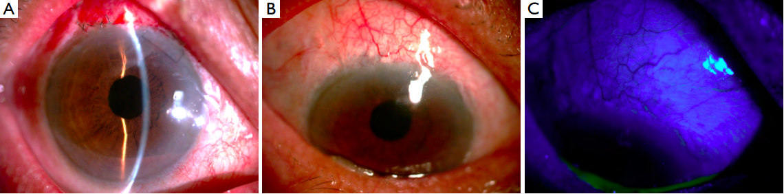
On the first post-operative day, BCVA was 20/40 and IOP was 15 mmHg. The bleb was flat and diffuse (Figure 4A). Six months later, BCVA increased to 20/25, while the IOP was stable and the bleb was functioning well (Figure 4B,C).
(A). The bleb was wellmaintained six months after surgery (B) without leakage (C).
Figure 1 Slit-lamp examination revealed a large avascular filtration bleb overhanging on the cornea
(A) with a thin base connected to the
conjunctiva (B). No leakage could be seen (C). Black arrow: thin base connected to the conjunctiva.
Figure 2 Ultrasound biomicroscopy showed that the bleb was connected to the conjunctiva through a thin base with a partial free edge
(A).
The inner passage of the ff ltration bleb was patent (B). White arrow: partial free edge. Yellow arrow: inner passage of the ff ltration bleb.
Figure 3 Histopathologic evaluation showed proliferation of ff brous tissue under the conjunctival epithelia (A) with irregular cystoids (B).
On the first post-operative day, BCVA was 20/40 and IOP was 15 mmHg. The bleb was flat and diffuse (Figure 4A). Six months later, BCVA increased to 20/25, while the IOP was stable and the bleb was functioning well (Figure 4B,C).
Figure 4 Post-operative slit-lamp examination.
(A). The bleb was wellmaintained six months after surgery (B) without leakage (C).
Discussion
Large overhanging blebs are an uncommon complication
of glaucoma filtration surgery. Though the pathogenesis
is unknown, it may be related to the use of MMC. Its
shape varies, especially after application of MMC (1). In
the current case, the occurrence of such bleb appeared to
be related to cataract surgery instead, evidenced by the
following two signs. First, foreign body sensation and
uncomfortable feeling appeared after the cataract surgery. Second, for the two-side phacoemulsification on the right
eye, the side incision was on the superior peripheral cornea
near the ff ltration bleb, which might be interrelated.
It has been reported that the size of functioning filtration bleb decreased and IOP increased after phacoemulsiff cation (2). There has also been a report of an inadvertent bleb developing after phacoemulsification (3). However, the current case may be the ff rst report of a post-trabeculectomy overhanging filtration bleb related to cataract surgery. The possible mechanism may be related to microleakage of the surgical wound of the side incision after phacoemulsiff cation which initiated the healing and scarring process (3). Thus, the morphology of the bleb is similar to a conjunctival granuloma with a thin base connected to the bleb.
Excision and revision of overhanging blebs are indicated when they are symptomatic or leaking. However, complications such as bleb leakage or failure occurred occasionally (4). Use of indocyanine green during excision or bleb revision guided by anterior segment optical coherence tomography has been reported with good results (5). Anis et al. reported sutureless revision with the aid of corneal contact lenses in six cases of overhanging filtering blebs (4). Success was achieved in five, while one patient had bleb leak requiring suture repair (4). In the current case, preoperative UBM impressions were confirmed by leakage of aqueous from the incision intraoperatively. Therefore, suture revision with superior conjunctival flap was performed with satisfactory outcome.
基金
1. This work is supported by the Natural Science Foundation of Guangdong Province in China (grant No. 2015A030313052)
参考文献
1. Kapoor KG, Syed MF. Dramatic dysesthetic overhanging bleb. Int Ophthalmol 2011;31:403-4.
2. Rebolleda G, Muñoz-Negrete FJ. Phacoemulsiff cation in eyes with functioning ff ltering blebs: a prospective study. Ophthalmology 2002;109:2248-55.
3. Zetterström C. Filtration bleb after phacoemulsiff cation. Acta Ophthalmol Scand 2003;81:188-90.
4. Anis S, Ritch R, Shihadeh W, et al. Sutureless revision of overhanging ff ltering blebs. Arch Ophthalmol 2006;124:1317-20.
5. Kojima S, Inoue T, Kawaji T, et al. Filtration bleb revision guided by 3-dimensional anterior segment optical coherence tomography. J Glaucoma 2014;23:312-5.
相关文章
张佳晴;谢潇杭;林灏文;谈旭华;刘臻臻;丘晓樟;靳光明;陈晓云;韩晓彤;罗莉霞;刘奕志,Near visual acuity of high myopes after cataract surgery: a real-world analysisYali Du;Lixia Sun;Mingzhi Zhang,The early change of corneal vertical coma and trefoil in 2.8-mm superior incision cataract surgeryYong Yao;Ming-Zhi Zhang;Vishal Jhanji,Surgically induced scleral staphyloma