Comparison of Visual Quality after Implantation of Big Bag and Akreos Adapt Intraocular Lenses in Patients with High Myopia
'%20fill='white'%20fill-opacity='0.01'/%3e%3cmask%20id='mask0_3477_29692'%20style='mask-type:luminance'%20maskUnits='userSpaceOnUse'%20x='0'%20y='0'%20width='16'%20height='16'%3e%3crect%20id='&%23232;&%23146;&%23153;&%23231;&%23137;&%23136;_2'%20x='16'%20width='16'%20height='16'%20transform='rotate(90%2016%200)'%20fill='white'/%3e%3c/mask%3e%3cg%20mask='url(%23mask0_3477_29692)'%3e%3cpath%20id='&%23232;&%23183;&%23175;&%23229;&%23190;&%23132;'%20d='M14%205L8%2011L2%205'%20stroke='%23333333'%20stroke-width='1.5'%20stroke-linecap='round'%20stroke-linejoin='round'/%3e%3c/g%3e%3c/g%3e%3c/svg%3e)
关键词
摘要
Purpose:To compare vision quality following phacoemulsifi- cation cataract extraction and implantation of a Big Bag or Akreos Adapt intraocular lens(IOL)in patients diagnosed with high myopia complicated with cataract.
Methods:This was a randomized prospective control study. The patients with high myopia complicated with cataract,with axial length ≥28 mm,and comeal astigmatism ≤1D were enrolled and randomly divided into the Big Bag and Akreos Adapt IOL groups.All patients underwent phacoemulsification cataract extraction and lens implantation. At 3 months after surgery, intraocular high-order aberration was measured by a Tracey-iTrace wavefront aberrometer at a pupil diameter of 5 mm in an absolutely dark room and statistically compared between two groups.The images of the anterior segment of eyes were photographed with a Scheimpflug camera using Pentacam three-dimensional anterior segment analyzer.The tilt and decentration of the IOL were calculated by Image-pro plus 6.0 imaging analysis software and statistically compared between two groups.
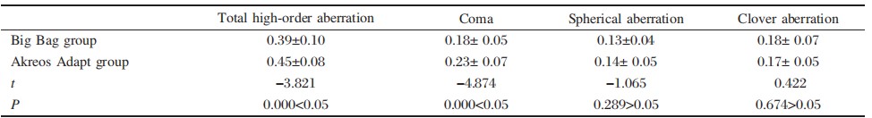
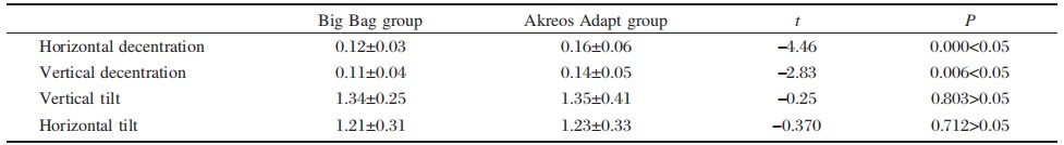
Results: In total,127 patients(127 eyes),including 52 males and 75 females,were enrolled in this study.The total high-order aberration and coma in the Akreos Adapt group(59 eyes) were significantly higher compared with those in the Big Bag (P<0.05). The clover and spherical aberration did not differ between the two groups(P>0.05).The horizontal and vertical decentration were significantly smaller in the Big Bag lens group than in the Akreos Adapt group(both P<0.05), whereas the tilt of IOL did not significantly differ between the two groups(P>0.05).
Conclusion: Both Big Bag and Akreos Adapt IOLs possess relatively good intraocular stability implanted in patients with high myopia. Compared with the Akreos Adapt IOL, the Big Bag IOL presents with smaller intraocular high-order aberration. Coma is the major difference between the two groups.
全文
Introduction
In China,the proportion of the elderly myopic population with an axial length ≥27.0 mm and≥ 28.5 mm is significantly higher compared with the 1.0%and 0.1%,respectively,found in western cou ntries¹ IOL implantation after phacoemulsification cataract extraction has been considered as a conventional procedure of cataract surgery.Along with the introduction of low-degree IOL,the postoperative visual acuity is improved in patients suffering from high myopia accompanied with cataract.However, the lens position is unstable because these IOLs are not specifically designed for patients with high myopia².The Big Bag IOL is specifically designed for patients diagnosed with high myopia and this lens in a bag has been confirmed to show good stability³. The Akreos Adapt IOL is designed with four haptics and possesses good stability in the lens capsule⁴, which is consistent with the opinion that an IOL with a multi-haptic design has greater stability than a twohaptic IOL⁵. However,the best type of IOL for providing high stability,reducing postoperative high-order aberration,and improving visual quality has not been established.
This prospective control study provides a statistical comparison of the high-order aberration,decentration,and tilt of two types of IOLs implanted in patients with high myopia complicated with cataract following phacoemulsification cataract surgery.
Materials and methods
Patient selection
The patients undergoing phacoemulsification combined with IOL implantation in Guangzhou Red Cross Hospital between February 2010 and May 2014 were enrolled in this study.Inclusion criteria: Aged 55-70 years,with axial length ≥28 mm,with high myopia complicated with cataract.Exclusion criteria:Having diseases of macular hemorrhage and hiatus and retinal detachment determined by preo perative Goldmann three-mirror lens or B-ultrasound examination.Retinal laser photocoagulation was pe rformed to treat retinal degeneration within two weeks before surgery.Patients with alternative di seases,such as uveitis,glaucoma,corneal diseases, and diabetes,were excluded.This clinical study was approved by the Ethics Committee of our hospital.
Study group
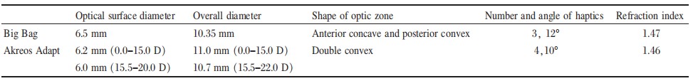
All patients were randomly assigned into two groups.After phacoemulsification cataract extraction,implantation of three-loop(Big Bag,Zeiss, Germany) and four-loop (AkreosAdapt, BauschLomb, U.S.) IOLs was performed. Both types of IOLs are made of hydrophilic polyacrylate, as illustrated incording to the SRK-T formula.IOL degree was selected by -2.0 D -3.0 D of the postoperative theoretical value. Only one eye was included for those undergoing surgery on both eyes.
Surgical approach
All surgeries were conducted by one single physician.Prior to operation,tropicamide eye drops were administered for full mydriasis and alcaine for topical anesthesia.A transparent incision 3.2 mm in diameter was made in the upper cornea,and continuous curvilinear capsulorhexis was conducted with a diameter ≥5 mm.An INFINITI phacoemulsification instrument(ALCON,U.S.)was employed to perform phacoemulsification and cataract extraction. The lens nucleus was emulsified and aspirated out, the residual lens cortex was removed with suction, the IOL was implanted into the capsular bag,and the remnant viscoelastic agent was eliminated.Tobradex eye drops(tobramycin and dexamethasone) were delivered 4 times/day for consecutive 2 weeks postoperatively.
Observational index
All patients were followed up for 3 months.Co nventional ocular examinations,including visual ac uity and intraocular pressure measurement,were pe rformed before and after surgery.Intraoperative and postoperative complications were carefully observed.
At postoperative 3 months,intraocular high-order aberration was measured by a Tracey-iTrace wavefront aberrometer in an absolutely dark room.The measurement was conducted three times for each eye and the highest quality image was selected.Intraocular high-order aberration,spherical aberration,coma,and clover aberration at a pupil diameter of 5 mm were statistically analyzed between the two groups.
The tilt and decentration ofIOL were measured by using a Pentacam Scheimpflug system(Oculus,Germany)at postoperative 3 months.Prior to examination,all patients were administered compound tropicamide eye drops for mydriasis until the pupil diameter was enlarged to approximately 8 mm.The images of 50 positions were collected and adjusted for the brightness and contrast ratio until the profile of the IOL was distinct.The images at 0°and 180° scan were chosen for imaging analysis by Image-pro plus 6.0 image analysis software.The vertical and horizontal decentration,and tilt ofthe IOL were calculated and statistically compared between the two groups⁴.
Statistical analysis
The SPSS 13.0 software package was utilized for data analysis.The chi-square test and t-test were adopted for statistical comparisons.P<0.05 was considered as statistically significant.
Results
General data
In total,127 patients (127 eyes),including 52 males and 75 females,were divided into the Big Bag(68 eyes)and Akreos Adapt(59 eyes)groups. Preoperative data of the patients did not significantly differ between the two groups.
The patients in the Big Bag lens group were aged 63.9±4.4 years on average and the mean axial length was 31.5±1.7 mm.The patients in the Akreos Adapt group were aged 62.6±4.5 years and had an average axial length of 31.0±1.9)mm.The differences b etween the two groups were not statistically significant(t=1.7,P>0.05 for both parameters).
Intraocular high-order aberration
The total high-order aberration was significantly lower in the Big Bag group than in the Akreos Adapt,and the main difference was coma(Table 2).
IOL tilt and decentration
Both horizontal and vertical decentration were si gnificantly smaller in the Big Bag lens group than in the Akreos Adapt group,whereas the tilt did not significantly differ between two groups,as illustrated in Table 3.
Intraoperative and postoperative complications
No related complications were observed during the surgery in both groups.All IOLs were successfully implanted into the capsular bag without posterior capsular rupture.During subsequent follow-up,no retinal detachment or other complications were noted in either group.
Discussion
A patient with high myopia presents with a long eye axis,thin scleral wall,and abnormal lens zonule, which makes surgery more difficult and causes more surgical complications.Currently,cataract extraction followed by IOL implantation is regarded as the co nventional treatment for cataract.However,IOL i mplantation can not be performed in a timely manner for patients with high myopia complicated with cataract due to the lack of low-degree IOLs.Although low-or negative-degree foldable IOLs have been i ntroduced to resolve this problem,the lens shape is not specifically designed for patients with high m yopia,which is likely to lead to adverse effects such as after-cataract and lens instability².
Cataract surgery has evolved from vision-restoring surgery into a refraction operation.Therefore,wide spread attention has been placed upon enhancing the visual quality of patients with high myopia after u ndergoing cataract surgery.Postoperative visual acuity is closely correlated with the axial length and the severity of fundus changes.Hence,those patients with macular hiatus,lacquer crack,or hemorrhage, which severely affect postoperative visual acuity, were excluded from this clinical trial.
Postoperative visual acuity was not utilized as the main observational parameter.Wavefront aberration acts as one of the factors evaluating the visual quality of human eyes.The aberration of phakic eyes was significantly increased due to the following causes. First,the optics characteristics differ between an IOL and the human lens.Second,the relative relationship between an IOL and the cornea is likely to alter. Third,the surgery exerts an effect upon the cornea⁶ . Along with the improvement of phacoemulsification technique,the incidence of corneal aberration induced by surgical procedures is extremely low. Hence,IOL implantation is the primary cause of increased aberration after surgery.
The IOL position exerts an evident effect on corneal defocus and different types of astigmatism⁷ . Continuous curvilinear capsulorhexis and IOL implantation are the most vital factors that guarantee postoperative IOL stability.In this study,all surgical procedures were completed by one single physician to avoid errors.The IOL was successfully implanted into the capsular bag in all patients and no posterior capsular rupture or alternative complications were noted.However,the occurrence of IOL tilt and decentration was still inevitable.Hayashi et al⁸.sugges ted that IOL tilt and decentration tended to occur even though the IOL was well inserted into the capsular bag.
The diameter of the capsular bag varies among i ndividuals and increases along with the axial elongation⁹.Consequently,the diameter of the capsular bag is relatively long for patients diagnosed with high myopia.The optical surface and overall diameters of the Akreos Adapt IOL tend to alter according to the IOL degree.The overall diameter of 0-15D IOL is up to ll mm,and 10.7 mm for 15.5-20.0D IOL. Therefore,a large capsular bag is required for IOL implantation to achieve good stability.The Akreos Adapt IOL is designed with four haptics to generate an acting force on two lines that vertically intersect at the center of the posterior capsular membrane. This widens the capsular bag as much as possible, allowing for direct contact between the IOL and the capsular bag,and making the lens well centered within the capsular bag.
Wesendahl et al¹.have demonstrated that the i ncluded angle between IOL haptics and optical su rface should be >10°,which allows for direct contact between the IOL optical surface and the posterior capsular membrane.The angle between the haptics and optics part is 12°,and the optics zone is designed as concave anteriorly and convex posteriorly,
which increases the contact between the IOL and the posterior capsular membrane,supports the vitreous body backwards,and decreases the movement of the vitreous body.The overall diameter of the Big Bag IOL is merely 10.35 mm.It is designed with three ear-shaped haptics,which allow for 240°contact b etween the haptics and capsular bag.The Big Bag IOL is better suited for the capsular bag of patients with high myopia,as it more evenly supports the posterior capsular membrane,decreases posterior capsular membrane hernia,and reduces the incidence ofIOL decentration and tilt.
In this study,the postoperative decentration and tilt of the IOLs were relatively small in both groups. The IOL tilt did not significantly differ between two groups.Both the horizontal and vertical decentration ofIOL in the Akreos Adapt group was higher compared with those in the Big bag group.This could be explained by the fact that the Big Bag IOL is designed with a symmetrical structure with one center and even stress is imposed on the capsular bag. However,the Akreos Adapt IOL has a symmetrical structure with one axis,so slight decentration might be induced by gravity or alternative factors.
Postoperative IOL tilt exerts significant effects u pon spherical aberration and total aberration¹¹.The i ntraocular spherical aberration after surgery did not significantly differ between two groups,consistent with the results of the IOL tilt.Intraocular spherical aberration was correlated with refraction index of IOL and shape of optical surface.Both types of IOLs share similar IOL refraction indexes(Big Bag=1.47, Akreos Adapt=1.46).However,the Big Bag lens is designed as concave anteriorly and convex poster iorly,whereas the Akreos Adapt lens is double co nvex in shape.Certain studies have suggested that a planoconcave IOL yields the minimal spherical aberration,whereas a double convex lens generates relatively large spherical aberration¹².However, spherical aberration in a lens that is concave anteriorly and convex posteriorly has rarely been reported. The two types of IOLs in this study did not significantly differ in terms of intraocular spherical aberr ation.The coma tended to increase after the incidence of IOL tilt and decentration³.Hence,we suggest that the postoperative intraocular coma is larger for the Akeros Adapt IOL than for the Big Bag lens, as a result ofIOL decentration.
The optical surface diameter of the Akeros Adapt IOL with 0-15D was 6.2 mm,whereas it was 6.5 mm for the Big Bag IOL.Both IOLs had a relatively large optical surface among the IOL products available.This not only decreases the incidence of glare induced by a large pupil,but also facilitates postoperative observation and treatment of the peripheral retina in patients with high myopia.Implantation e ither the Big Bag or the Akreos Adapt IOLs will provide good stability within the capsular bag.
Compared with Akreos Adapt lens,however,the Big Bag IOL yields less decentration,smaller intraocular high-order aberration,and better visual quality.
Conflicts of interests
The authors declare no conflicts of interest.
基金
参考文献
1. Sanders DR,Retzlaff JA,Kraff MC,et al.Comparison of the SRK/T formula and other theoretical and regression formulas. J Cataract Refract Surg, 1990, 16: 341-346.
2. Sundelin K,Friberg-Riad Y,Ostberg A,et al.Posterior capsule opacification with AcrySof and poly(methyl methacrylate)intraocular lenses.Comparative study with a 3-year follow-up.J Cataract Refract Surg,2001,27:1586- 1590.
3. Chen QY,Duan,ZB,Luo ZW.Stability of different in- traocular lens implanted in sac of high myopia.Interna- tional Jounal of Ophthalmology,2012;01:49-51.
4. Xing XJ,Tang X,Song H.Comparison of tilt and decen- tration of four different kinds of aspheric intraocular lens- es implantation.Chinese Journal of Ophthalmology,2010, 04:332-336.
5. Spath U,Liekfeld A,Hartmann C,et al.Evaluation of pos- terior capsule opacification after implantation of the Akreos Disc and Akreos Fit acrylic intraocular lenses - pilot studies.Klin Monbl Augenheilkd,2003,220:695- 698.
6. Shi D,Dong YJ,Zhang JS.Application of wavefront aber- ration technology in cataract and intraocular lens.2006, 04:362-367.
7. Kozaki J,Takahashi F.Theoretical analysis of image de- focus with intraocular lens decentration.J Cataract Refract Surg,1995,21:552-555.
8. Hayashi K,Harada M,Hayashi H,et al.Decentration and tilt of polymethyl methacrylate,silicone,and acrylic soft intraocular lenses.Ophthalmology,1997,104:793-798.
9. Schmidbauer JM,Peng Q,Apple DJ,et al.Rates and caus- es of intraoperative removal of foldable and rigid intraoc- ular lenses: clinicopathological analysis of 100 cases.J Cataract Refract Surg,2002, 28:1223-1228.
10. Wesendahl TA,Hunold W,Auffarth GU,et al.Area of contact of the artificial lens and posterior capsule.System- atic study of various haptic parameters.Ophthalmology, 1994,91:680-684.
11. Taketani F,Matuura T,Yukawa E,et al.Influence of in- traocular lens tilt and decenration on wavefront aberra- tions.J Cataract Refract Surg,2004,30:2158-2162.
12. Uchio E,Ohno S,Kusakawa T.Spherical aberration and glare disability with intraocular lenses of different optical design.J Cataract Refract Surg,1995,21:690-696.
13. Dietze HH,Cox MJLimitations of correcting spherical aberration with aspheric intraocular lenses.J Refract Surg, 2005,21:S541-546.