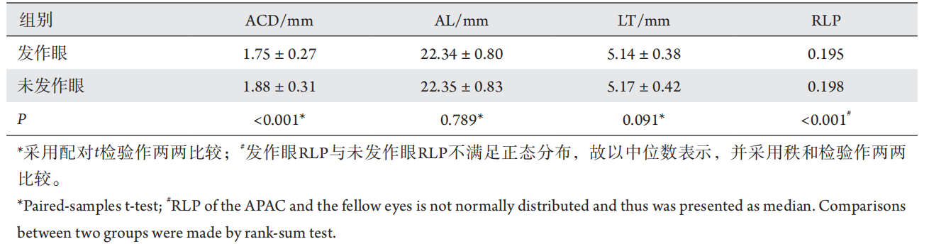
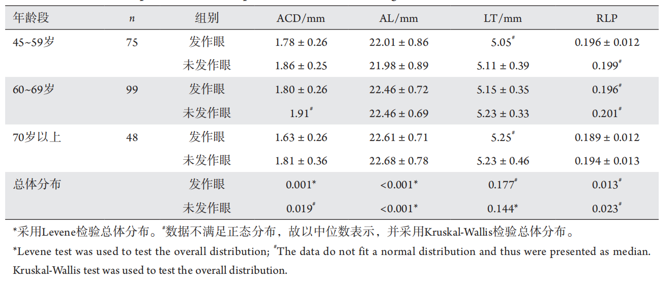
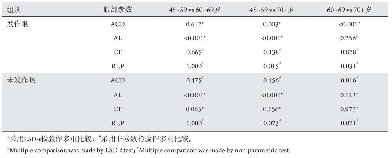
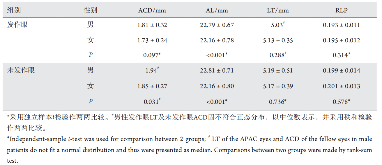
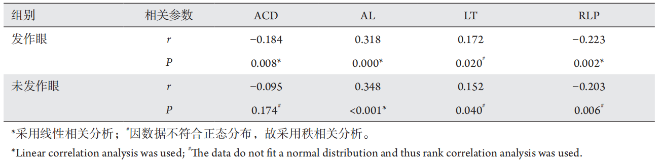
Objective: To compare the ocular biometric parameters between the acute primary angle closure (APAC) eyes and the fellow eyes as well as the risk factors associated with APAC. Methods: From January 2008 to March 2020,222 monocular APAC patients over 45 years old from the Glaucoma Department of Zhongshan Ophthalmic Center, Sun Yat-sen University were retrospectively studied. Patients with binocular attack, previous attack in the fellow eyes, and secondary factors such as lens-induced and traumatic glaucoma were excluded. Ocular biometric parameters including axial length (AL) and lens thickness (LT) were measured with A-scan ultrasound, while the anterior chamber depth (ACD) was measured by ultrasonic biological microscope. AL, ACD, LT and relative lens position (RLP) were compared between the APAC and the fellows eyes. Results: The average age of onset was (62.57±9.14) years. The ACD was (1.75±0.27) and (1.88±0.31) mm, AL was (22.34±0.80) and (22.35±0.83) mm,LT was (5.14±0.38) and (5.17±0.42) mm, and the RLP was 0.195 and 0.198 for the APAC and the fellow eyes,respectively. Compared with the fellow eyes, the ACD of the APAC eyes was shallower, and the RLP was more anterior (both P<0.001), while the differences of AL and LT were not statistically significant (both P>0.05).Furthermore, AL of patients with a younger age of onset (aged 45 to 59 years) was shorter than that of those with an older age of onset (aged 60 to 69 or over 70 years); patients with an onset age of over 70 years have shallower ACD and more anterior RLP, all statistically significant (P<0.05). In addition, correlation analysis indicated that younger onset age was significantly correlated to shorter axial length of APAC eyes (P<0.001). Conclusion:APAC eyes had shallower ACD and more anterior RLP. Shorter AL and female were associated with APAC attack between individuals. Shallower ACD, thicker lens and more anterior RLP are potential risk factors for APAC among aged population.
广东省科技计划项目 (2017A020215092,2019A070701006)。 This work was supported by Guangdong Science and
Technology Project, China (2017A020215092, 2019A070701006).
References
Sun X, Dai Y, Chen Y, et al. Primary angle closure glaucoma: What we
know and what we don't know[ J]. Prog Retin Eye Res, 2017, 57: 26-45.
Wang L, Huang W, Huang S, et al. Ten-year incidence of primary angle
closure in elderly Chinese: The Liwan Eye Study[ J]. Br J Ophthalmol,
2019, 103(3): 355-360.
Marchini G, Pagliarusco A, Toscano A, et al. Ultrasound biomicroscopic
and conventional ultrasonographic study of ocular dimensions in primary
angle-closure glaucoma[ J]. Ophthalmology, 1998, 105(11): 2091-2098.
Ang LP, Aung T, Chew PT. Acute primary angle closure in an Asian
population: Long-term outcome of the fellow eye after prophylactic
laser peripheral iridotomy[ J]. Ophthalmology, 2000, 107(11):
2092-2096.
Sng CCA, Aquino MCD, Liao J, et al. Pretreatment anterior segment
imaging during acute primary angle closure: Insights into angle
closure mechanisms in the acute phase[ J]. Ophthalmology, 2014,
121(1): 119-125.
Jonas JB, Iribarren R, Nangia V, et al. Lens position and age: The
Central India Eye and Medical Study[ J]. Invest Ophthalmol Vis Sci,
2015, 56(9): 5309-5314.
Jonas JB, Nangia V, Gupta R, et al. Lens thickness and associated
factors[ J]. Clin Exp Ophthalmol, 2012, 40(6): 583-590.
Jonas JB, Nangia V, Gupta R, et al. Anterior chamber depth and its
associations with ocular and general parameters in adults[ J]. Clin Exp
Ophthalmol, 2012, 40(6): 550-556.
Zhong X, Li Y, Huang C, et al. Seasonal variations and climatic factors
on acute primary angle-closure admission in southern China: A 5-year
hospital-based retrospective study[ J]. Acta Ophthalmol, 2020. [Epub
ahead of print].
Xu L, Cao WF, Wang YX, et al. Anterior chamber depth and chamber
angle and their associations with ocular and general parameters: The
Beijing Eye Study[ J]. Am J Ophthalmol, 2008, 145(5): 929-936.
Nongpiur ME, He M, Amerasinghe N, et al. Lens vault, thickness, and
position in Chinese subjects with angle closure[ J]. Ophthalmology.
2011, 118(3): 474-479.
Lavanya R, Wong TY, Friedman DS, et al. Determinants of angle
closure in older Singaporeans[ J]. Arch Ophthalmol, 2008, 126(5):
686-691.
Suwan Y, Jiamsawad S, Tantraworasin A, et al. Qualitative and
quantitative evaluation of acute angle-closure mechanisms[ J]. BMC
Ophthalmol, 2017, 17(1): 246.
Moghimi S, Zandvakil N, Vahedian Z, et al. Acute angle closure:
Qualitative and quantitative evaluation of the anterior segment
using anterior segment optical coherence tomography[ J]. Clin Exp
Ophthalmol, 2014, 42(7): 615-622.
Lim MC, Lim LS, Gazzard G, et al. Lens opacity, thickness, and
position in subjects with acute primary angle closure[ J]. J Glaucoma,
2006, 15(3): 260-263.
Huang J, Wang Z, Wu Z, et al. Comparison of ocular biometry between
eyes with chronic primary angle-closure glaucoma and their fellow
eyes with primary angle-closure or primary angle-closure suspect[ J]. J
Glaucoma, 2015, 24(4): 323-327.
Moghimi S, Bijani F, Chen R, et al. Anterior segment dimensions
following laser iridotomy in acute primary angle closure and fellow
eyes[ J]. Am J Ophthalmol, 2018, 186: 59-68.
Wang F, Wang D, Wang L. Exploring the occurrence mechanisms of
acute primary angle closure by comparative analysis of ultrasound
biomicroscopic data of the attack and fellow eyes[ J]. Biomed Res Int,
2020, 2020: 8487907.