The effect of corneal B/F ratio on postoperative refractive error after age-related cataract surgery
Views:
12472
DOI:
10.12419/2306250001
Publication Date:
2023/11/15
Author(s):
Li Hongyu1, Song Hui1
Li Hongyu1, Song Hui1
Institution/Unit:
Department of Refractive Cornea, School of Ophthalmology, Institute of Ophthalmology, National Clinical Medical Research Center for Eye, Ear, Nose and Throat Diseases Tianjin Branch, Tianjin Retinal Function and Disease Key Laboratory
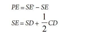
Objective: To analyze the relationship between corneal B/F ratio and postoperative refractive error in age-related cataract patients, and to explore the impact of B/F ratio on the accuracy of intraocular lens power calculation. Methods: A total of 197 age-related cataract patients (197 eyes) who were treated in the cataract center of our hospital from March 2019 to November 2019 and were going to undergo monocular cataract surgery were selected. The biological parameters of the anterior segment were measured by Pentacam anterior segment analyzer before surgery, and the patients were divided into three groups (25% below the B/F ratio, 25%~75%, and 25% below the B/F ratio) with the lower limit and the upper limit of 25%. Three months after surgery, the postoperative refractive state of patients was evaluated by automatic computerized refractometer, and the postoperative refractive error (PE) was calculated, and the percentage differences of mean refractive error (ME), mean absolute error (MAE), median absolute error (MedAE) and refractive error in the range of ±0.25, ±0.50, ±0.75, ±1.00 and < ±1.00D were evaluated. Results: The B/F ratio was moderately correlated with postoperative refractive error in age-related cataract patients (r= ?0.445, P < 0.001). With the increase of B/F ratio, the refractive state of patients shifted from hyperopia to myopia after surgery, and the MAE and MedAE were 0.55 D and 0.46 D respectively in 3 months after surgery. The percentages of refractive error in the range of ±0.25, ±0.50, ±0.75, ±1.00 and < ±1.00 D were 29.4%, 52.8%, 71.6%, 87.6% and 12.7%, respectively. After adjusting the corneal curvature according to the B/F ratio of the population based on our previous study, MAE and MedAE were 0.51 D and 0.43 D, respectively, which were lower than those before correction (P< 0.05). Conclusions: There is a correlation between B/F ratio and postoperative refractive error in age-related cataract patients. As the B/F ratio increased, the refractive state of the patient gradually drifted from farsightedness to myopia after cataract surgery, and the more the B/F ratio deviated from the normal average, the greater the absolute value of the patient's refractive error.
Alio JL, Abdelghany AA, Abdou AA, et al. Cataract surgery on the
previous corneal refractive surgery patient[ J]. Surv Ophthalmol, 2016,
61(6): 769-777.
Gale RP, Saldana M, Johnston RL, et al. Benchmark standards for
refractive outcomes after NHS cataract surgery[ J]. Eye, 2009, 23(1):
149-152.
Wu J, Gao P, Fan JW, et al. Comparison of keratometric measurements obtained by the Verion image guided system with the IOLMaster and the Pentacam before cataract extraction surgery[ J]. Zhonghua Yan Ke Za Zhi, 2020, 56(1): 47-52.
Brar S, Ganesh S, Murkey V. Interobserver agreement between central topography and Scheimpflug tomography for premium intraocular lens selection[ J]. J Cataract Refract Surg, 2023, 49(8): 790-794.
Norrby S. Sources of error in intraocular lens power calculation[ J]. J Cataract Refract Surg, 2008, 34(3): 368-376.
Eom Y, Rhim JW, Kang SY, et al. Toric intraocular lens calculations using ratio of anterior to posterior corneal cylinder power[ J]. Am J Ophthalmol, 2015, 160(4): 717-724.e2
Li HY, Li J, Song H. The distribution of the back-front corneal
radius ratio and its effect on the corneal refractive power based on
Pentacam[ J]. Recent Adv Ophthalmol, 2020, 40(11): 1065-1069.
Shi Q, Wang GY, Cheng YH, et al. Comparison of IOL-Master 700
and IOL-Master 500 biometers in ocular biological parameters of
adolescents[ J]. Int J Ophthalmol, 2021, 14(7): 1013-1017.
Bao T, Wang L, Liu C, et al. Analysis of biometric parameters of cataract
eyes measured with optical biometerLenstar LS900, IOL Master 700,
and OPD-SCAN III[ J]. PhotodiagnosisPhotodynTher, 2023, 43:
103646.
Miyata K, Otani S, Honbou N, et al. Use of Scheimpflug corneal
anterior-posterior imaging in ray-tracing intraocular lens power
calculation[ J]. Acta Ophthalmol, 2013, 91(7): e546-e549.
Wei LQ, Fu YH, Pan WH, et al. Accuracy of optimized Sirius ray-tracing
method in intraocular lens power calculation[ J]. Int J Ophthalmol,
2022, 15(2): 228-232.
Ghoreyshi M, Khalilian A, Peyman M, et al. Comparison of OKULIX
ray-tracing software with SRK-T and Hoffer-Q formula in intraocular
lens power calculation[ J]. J Curr Ophthalmol, 2018, 30(1): 63-67.
Qin M, Yuan Y, Wang Y, et al. Comparison of preoperative angle
kappa measurements in the eyes of cataract patients obtained from
PentacamScheimpflug system, optical low-coherence reflectometry,
and ray-tracing aberrometry[ J]. BMC Ophthalmol, 2022, 22(1): 153.
Hoffer KJ, Savini G. Update on Intraocular Lens Power Calculation
Study Protocols: The Better Way to Design and Report Clinical
Trials[ J]. Ophthalmology, 2021, 128(11):e115-e120.
Voytsekhivskyy OV, Hoffer KJ, Tutchenko L, et al. Accuracy of 24 IOL Power Calculation Methods. Journal of refractive surgery[ J]. J Refract Surg, 2023, 39(4): 249-256.
Abulafia A, Barrett GD, Koch DD, et al. Protocols for studies of
intraocular lens formula accuracy[ J]. Am J Ophthalmol, 2016, 164:
149-150.
Hoffer KJ, Aramberri J, Haigis W, et al. Protocols for studies of
intraocular lens formula accuracy[ J]. Am J Ophthalmol, 2015, 160(3):
403-405.e1.
Amro M, Chanbour W, Arej N, et al. Third- and fourth-generation
formulas for intraocular lens power calculation before and after phakic
intraocular lens insertion in high myopia[J]. J Cataract Refract Surg,
2018, 44(11): 1321-1325.
Wang Q, Jiang W, Lin T, et al. Accuracy of intraocular lens power
calculation formulas in long eyes: a systematic review and meta-analysis[ J]. ClinExpOphthalmol, 2018, 46(7): 738-749.
Chung J, Bu JJ, Afshari NA. Advancements in intraocular lens power
calculation formulas[ J]. Curr Opin Ophthalmol, 2022, 33(1): 35-40.
Hashemi H, Miraftab M, Panahi P, Asgari S. Biometry and intraocular
power calculation using a swept-source optical coherence tomography:
A repeatability and agreement study[ J]. Indian J Ophthalmol, 2022,
70(8): 2845-2850.
Stopyra W, Langenbucher A, Grzybowski A. Intraocular Lens Power
Calculation Formulas-A Systematic Review[ J]. OphthalmolTher, 2023,
12(6): 2881-2902.
Savini G, Hoffer KJ, Lomoriello DS, et al. Simulated keratometry versus
total corneal power by ray tracing: a comparison in prediction accuracy
of intraocular lens power[ J]. Cornea, 2017, 36(11): 1368-1372.
Ton Y, Barrett GD, Kleinmann G, et al. Toric intraocular lens power calculation in cataract patients with keratoconus[ J]. J Cataract Refract Surg, 2021, 47(11): 1389-1397.
Chen D, Lam AK. Reliability and repeatability of the Pentacam on
corneal curvatures[ J]. Clin Exp Optom, 2009, 92(2): 110-118.
Wan KH, Liao XL, Yu M, et al. Wavefrontaberrometry repeatability and
agreement-A comparison between Pentacam AXL Wave, iTrace and
OPD-Scan III[ J]. Ophthalmic Physiol Opt, 2022, 42(6):1326-1337.