





HIGHLIGHTS
1. Critical Discoveries and Outcomes
• Clinical stability alone is insufficient for surgical timing in thyroid-associated orbitopathy (TAO); thyrotropin-binding inhibitory immunoglobulin (TBII) (>15%) predicts postoperative instability. Superior oblique involvement (96%) requires preoperative imaging to prevent torsional diplopia, and excyclotropia ≥15°indicates bilateral inferior rectus restriction, guiding first-procedure bilateral surgery. Tendon elongation achieves supra-maximal correction (4.52 PD/mm) with >4-year stability, and teprotumumab reduces deviation by 28%-29%, with 46% achieving complete diplopia resolution, potentially avoiding surgery.
2. Methodological Innovations
• Preoperative evaluation integrates the Clinical Activity Score (CAS), TBII, high-resolution imaging, and cyclotorsion assessment for personalized planning. Adjustable sutures improve motor success (76% vs. 48%), the intraoperative relaxed muscle positioning (IRMP) eliminates preoperative calculations, and novel procedures(Y-split, Tenon’s recession 87% success, nasal transposition) expand options. Single-stage combined surgery is feasible in selected patients, reducing surgeries and recovery time.
3. Prospective Applications and Future Directions
• Biologics like teprotumumab offer non-surgical options during active disease; future research should optimize their timing and sequencing with surgery. Artificial Intelligence (AI) and big data hold promise for predicting surgical outcomes, yet remain unexplored for efficacy prediction. Developing AI models to forecast dose-response and reoperation risk would enable individualized decision-making, requiring rigorous validation and ethical safeguards.
Introduction
Thyroid-associated orbitopathy (TAO), the most common orbital disease in adults, presents with proptosis, eyelid retraction, extraocular muscle enlargement, and restrictive strabismus.[1] Its pathogenesis involves orbital fibroblast activation, glycosaminoglycan deposition, and muscle fibrosis, leading to mechanical restriction.[2] Epidemiologic studies show varying incidence and prevalence globally, with a general declining trend in Western countries, though some Asian regions report increased detection.[3-5] Approximately 40% of hyperthyroid patients develop TAO, most of which are mild; 10% progress to strabismus requiring surgery within 4 years, especially smokers.[4,6-7] This strabismus not only causes debilitating diplopia but also imposes a profound psychosocial burden, making effective management a critical therapeutic goal.[8-10]
Historically, the surgical management of TAO-related strabismus was guided by empirical principles and standardized techniques. However, over the past decade, this field has undergone a paradigm shift toward precision and personalized medicine. An evolution is driven by the recognition that TAO presents with highly variable phenotypes, disease activity, and individual patient needs.
This review synthesizes the current literature to critically examine this shift. Rather than cataloguing techniques, we provide a framework for understanding how contemporary advances in preoperative assessment, surgical and outcome evaluation collectively enable a more tailored approach. Key innovations include adjustable sutures, muscle elongation, and intraoperative relaxed muscle positioning, all of which improve outcomes and reduce reoperations.[11-14] We also identify critical evidence gaps that future research must address.
Unlike prior reviews, this review synthesizes advances from the past decade, not as a mere catalogue of techniques, but through the lens of the evolving paradigm toward precision and personalized medicine. We critically examine how innovations in preoperative assessment, surgical strategy, and outcome evaluation collectively enable this shift and identify key evidence gaps that future research must address.
Preoperative evaluation and timing of surgery for strabismus in TAO
Assessment of disease activity and stability
Surgery for TAO should ideally be performed during the inactive phase. The Clinical Activity Score (CAS) is the primary tool for assessing activity, with a sustained CAS ≤ 3 for ≥ 6 months indicating quiescence.[1] However, due to the subjectivity of clinical scores, imaging provides essential objective assessment. Orbital CT and MRI delineate morphological changes in the extraocular muscles (e.g., thickening, tendon involvement) and indicate active inflammation—elevated T2 signal on MRI suggests acute edema.[15]
The controversy over the stable period: is 6 months sufficient
Conventionally, stability of the strabismus angle for ≥6 months is required before surgery.[16] However, recent evidence challenges this notion. Even after this period, ~31.3% of patients still exhibit postoperative angle changes. Notably, all such cases showed elevated serum thyrotropin-binding inhibitory immunoglobulin (TBII) levels, indicating that clinical stability alone may be insufficient for optimal timing. Incorporating serological markers such as TBII may improve surgical predictability.[17] Surgery may be deferred if TBII is elevated (>15%), suggesting ongoing immunological activity. The optimal timing likely depends on achieving both sustained clinical stability and normalized serological markers.
Assessment of cyclotorsion and bilateral involvement
The study by Arnoldi and Reynolds (2015)[18] highlighted that traditional cover and forced duction tests (FDT) may underestimate contralateral inferior rectus restriction in asymmetric TAO. In contrast, excyclotropia as quantitatively measured by the double Maddox rod (DMR) test correlated significantly with the severity of bilateral restriction. Preoperative excyclotropia ≥15° strongly suggests bilateral involvement, warranting bilateral surgical intervention at the initial procedure to avoid reoperation.
Identification and significance of superior oblique muscle involvement
TAO does not exclusively affect the rectus muscles. A study by Del Porto et al. (2019)[15] used high-resolution orbital CT to demonstrate a 250% enlargement of the superior oblique muscle cross-sectional area in TAO patients compared to controls. Moreover, 96% of patients exhibited superior oblique muscle areas exceeding the control mean by >3 standard deviations. Superior oblique involvement may lead to A-pattern strabismus and incyclotorsion and increases the risk of postoperative torsional diplopia.[19] Therefore, preoperative imaging must include the oblique muscles alongside all other extraocular muscles to inform surgical planning. Intraoperatively, if a taut superior oblique is confirmed after inferior rectus disinsertion (via exaggerated traction testing), surgical adaptation is warranted. Options include superior oblique recession (to correct both pattern and torsion) or anterior tenotomy (primarily for incyclotorsion). Integrating this assessment is key to preventing complex postoperative deviations and tailoring surgery to the underlying pathophysiology.[20]
Surgical timing and multidisciplinary collaboration
A strict sequential surgical approach is widely advocated for TAO management: "orbital decompression → strabismus surgery → eyelid surgery".[21] This sequence is critical and should not be deviated from. Decompression changes globe position and extraocular muscle mechanics, potentially causing or worsening diplopia.[22-23] Performing strabismus or eyelid surgery first may compromise results due to subsequent anatomical shifts. Thus, multidisciplinary collaboration involving endocrinology, orbital surgery, strabismus, and oculoplastic specialties is essential.[24]
Is surgery advisable during the active phase
Elective strabismus surgery should be avoided during active TAO. Xu L et al. (2014)[25] reported that extraocular muscle surgery may trigger disease reactivation, even in apparently stable patients. For active cases, non-surgical options such as botulinum toxin injections serve as effective bridging therapies.[26] This approach eliminates the need for surgery in 32% of patients and reduces surgical extent in an additional 27%, especially for deviations <20 PD, effectively managing diplopia during active disease.
Surgical techniques and innovations
Traditionally, the success of strabismus surgery has been assessed primarily on the basis of ocular alignment (e.g., prism diopters). Modern studies increasingly employ composite endpoints, such as "absence of diplopia with horizontal/vertical deviation ≤10/5 PD",[27] or combine binocular single vision function with the Graves' Orbitopathy-specific Quality of Life (GO-QoL) questionnaire.[28] These criteria demand greater predictability and precision from surgical procedures, driving innovations in TAO strabismus surgery techniques. The advancements can be categorized by their primary aim: optimizing dose-response and stability in conventional recessions, managing complex scenarios (large-angle, torsion, reoperations), and utilizing novel materials to overcome anatomical limits. These refinements collectively enhance predictability and reduce reoperations.
Optimization and controversies of traditional procedures
The selection of rectus recession strategy significantly influences outcomes. Bilateral medial rectus recession offers a superior dose-response (1.4 PD/mm) compared with unilateral (1.0 PD/mm).[29] For vertical misalignment, superior rectus recession alone corrects hypertropia with 79% success,[30] whereas combining inferior rectus recession with contralateral superior rectus recession effectively addresses larger deviations (Figure 1, 2).[31] Bilateral inferior rectus recession improves elevation and reduces cyclodeviation.[32] In cases of extreme strabismus (>25 PD) unresponsive to maximum recession, rectus resection provides effective correction (mean 34.3 PD).[33] Various modified techniques continue to enhance surgical efficacy. Table 1 summarizes the core surgical options available for different clinical scenarios, with their success rates and limitations providing an evidence-based basis for formulating patient-specific surgical plans.
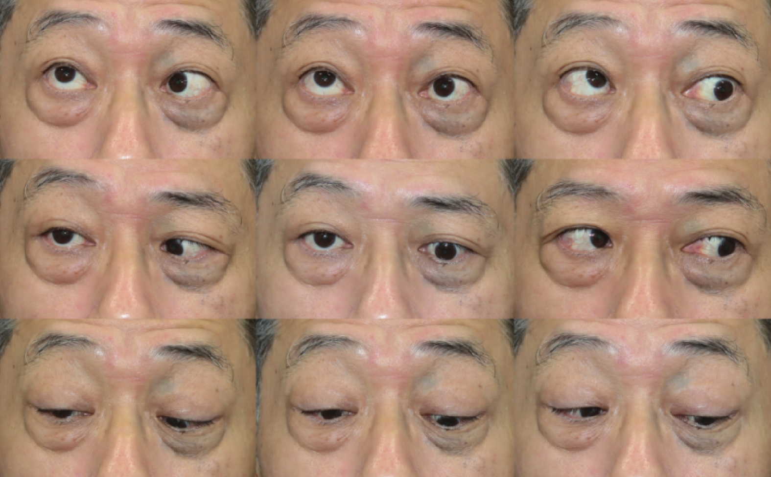
Arc perimetry showed 4° esotropia and R/L 25° in right gaze. Under general anesthesia on 2024-10-17, right superior rectus recession (8 mm) + left inferior rectus recession (6 mm) were performed.
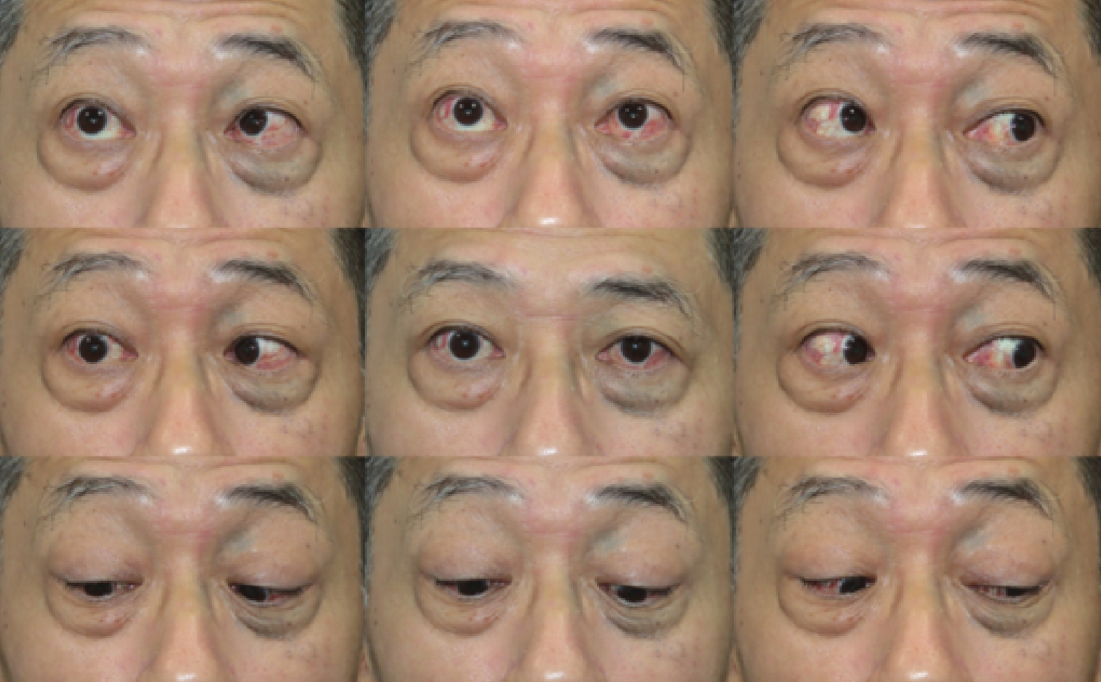
At the 3-month postoperative follow-up, the strabismus angle was reduced from preoperative measurements of 4° esotropia and R/L 20° to 0°. The patient’s diplopia has resolved, with an improvement in appearance.
|
Surgical Technique/Strategy |
Study/Data Source |
Success Rate (Definition) |
Reoperation Rate/Key Complication Data |
Main Advantages |
Main Limitations/Suitable Patient Population |
|
Vertical Rectus Recession |
Cestari et al. (2018)[35] |
89% (postoperative vertical deviation ≤5 PD) |
Reoperation rate not explicitly reported; Key difference: smaller postoperative vertical upward drift (mean 1.2 PD) |
Small postoperative vertical drift, high stability |
Primarily suitable for cases with predominant vertical deviation |
|
Vertical + Horizontal Rectus Combined Recession |
Cestari et al. (2018)[35] |
67% (postoperative vertical deviation ≤5 PD) |
Reoperation rate not explicitly reported; Key complication/difference: significantly larger postoperative vertical upward drift (mean 6.8 PD vs. 1.2 PD) |
Simultaneous correction of both vertical and horizontal deviation in a single surgery |
Higher risk of postoperative vertical drift, lower overall success rate |
|
Combined Surgery (Decompression + Strabismus ± Eyelid) |
Quaranta-Leoni et al. (2021)[38]; Choi et al. (2016)[37] |
Results "comparable" or "satisfactory" vs. staged surgery |
Eyelid subgroup: Combined surgery (1A) 0% (0/11) vs. staged surgery (2) 26.7% (4/15) required secondary surgery; Strabismus subgroup: Combined surgery (1B) 77.8% (7/9) required second-stage surgery for horizontal strabismus vs. staged surgery (3) 50% (5/10) requiring further surgery; Complications: 1 case (5%) recurrent DON (resolved with steroids); no other severe complications[38]; Another study[37] reported 1 case of CSF leak (self-resolved) |
Single-stage completion reduces total number of surgeries, treatment costs, and overall recovery time |
Requires strict patient selection: suitable for moderate-to-severe TAO patients with small asymmetry of proptosis (<2mm), simple deviation pattern, small deviation angle; Complex, asymmetric, large-angle cases remain suitable for traditional staged surgery |
|
Traditional Staged Surgery (Decompression → Strabismus → Eyelid) |
As a benchmark for comparison with combined surgery |
Success rate comparable to combined surgery (approx. 80%-90%) |
Reoperation rate varies across studies, but generally, for complex cases, staged surgery allows precise strabismus correction under stable conditions, potentially reducing secondary surgery rates |
Clear treatment steps, each stage can be performed under optimal conditions, suitable for cases of all complexities, considered the gold standard |
Long treatment duration, high total cost, patients undergo multiple surgeries and recovery periods |
|
Adjustable Suture Technique |
Volpe et al. (2012)[40]; Imburgia et al. (2016)[42] |
96% overall success rate (65% excellent - no prism; 31% good - prism <10 PD required) |
Low reoperation rate (3.7%); However, risk of overcorrection exists (incidence 20%, mean 6.2 PD), which can be partially corrected via adjustment |
Improves surgical precision, especially suitable for TAO patients (Type 2) with unpredictable muscle elasticity; Allows postoperative adjustment for tissue response |
Relies on patient cooperation during/perioperatively; Risk of overcorrection; May require prism assistance |
|
Non-absorbable Suture (for Inferior Rectus Recession) |
Kerr et al. (2011)[43] |
Overall success rate not explicitly reported |
Significantly reduces risk of early overcorrection (>5 PD): Odds Ratio (OR) = 6.0 vs. absorbable suture, p=0.041. No cases of >5 PD overcorrection in the non-absorbable suture group |
Provides more durable, secure muscle attachment, counteracts specific biomechanical stresses in the inferior rectus region in TAO (e.g., Bell's phenomenon, superior rectus contracture force), reducing early "slippage" or "stretched scar" |
Primarily used for inferior rectus recession in TAO; Suture knot may erode through conjunctiva causing irritation, constitutes a permanent intraocular foreign body |
|
IRMP Technique |
Dal Canto et al. (2006)[47]; Sarici et al. (2018)[46] |
Excellent success rate 87.5% (Dal Canto: 21/24; Sarici: 7/8); Clinically acceptable success rate 100% (Dal Canto: 24/24; Sarici: 8/8) |
Low reoperation rate (Dal Canto: 8%, 2/24 cases required second surgery); Surgical amount: Dal Canto study: average 2.4 muscles per patient; Sarici study: average 7.5mm recession for IR, 6.75mm for MR; Complications: Literature reports no severe complications, but postoperative lower lid retraction may occur |
Determines the amount of recession based on the physiological position of the relaxed muscle intraoperatively, independent of preoperative measurements or nomograms; Avoids postoperative adjustment, especially suitable for complex cases post-orbital decompression, with severe muscle fibrosis, where traditional formulas fail; Studies show significant postoperative improvement in GO-QoL scores |
Heavily dependent on surgeon's experience and intraoperative judgment, lacks objective, standardized quantitative metrics; Steep learning curve; Can be more challenging when multiple muscles are involved in deciding which muscle to recess |
Vertical rectus recession vs. combined vertical and horizontal rectus recession
Surgical strategy choice directly affects postoperative stability. A retrospective study by Cestari DM et al. (2018)[34] indicated that combined vertical and horizontal rectus recession led to more significant postoperative vertical upward drift compared with vertical recession alone (mean 6.8 PD vs. 1.2 PD). The final vertical deviation was larger and the success rate lower in the combined surgery group (67% vs. 89%). This suggests that for complex cases requiring multi-directional correction, the risks of combined surgery should be carefully evaluated, or staged surgery should be considered.
Exploration of combined surgical strategies: orbital decompression combined with strabismus surgery
The traditional staged approach (decompression → strabismus → eyelid surgery) is reliable but prolonged and costly. Combined single-stage surgery has emerged to address these limitations. In selected moderate-to-severe TAO patients, combined decompression, strabismus, and eyelid surgery achieves comparable outcomes to staged procedures while reducing the number of surgeries, cost, and recovery time.[35] Even mild to moderate cases may benefit from tailored combined surgery.[36] Success depends on careful patient selection: predictors include good symmetry (proptosis asymmetry < 2 mm), simple deviation patterns, and small angles.[37] Complex cases with large or asymmetric deviations remain better suited for traditional staged surgery. In experienced hands, combined surgery is a valuable option for optimized care (Figure 3, 4).[38]
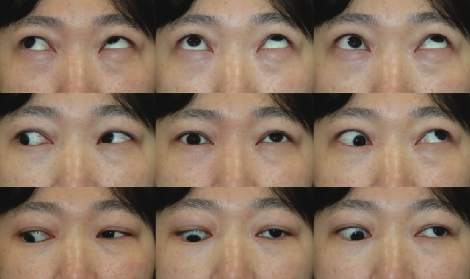
Arc perimetry showed 2° exotropia and L/R 30° in right gaze. Under general anesthesia on 2024-07-01, left orbital decompression (medial&inferior wall) + superior rectus recession (13 mm) were performed.
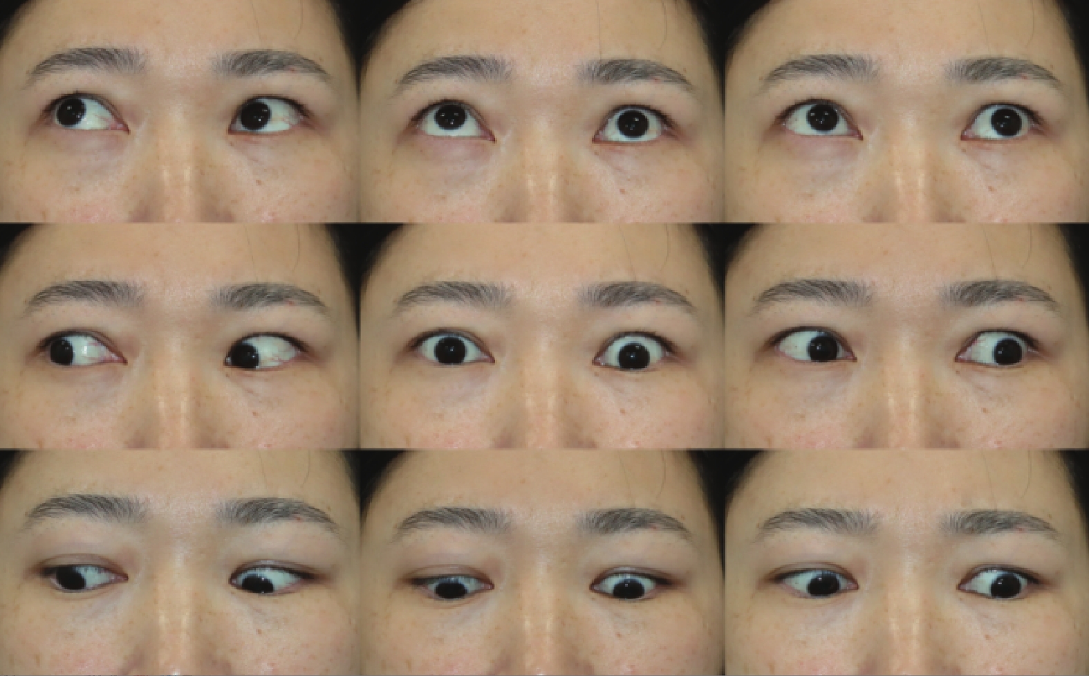
At the 3-month postoperative follow-up, the strabismus angle was reduced from preoperative measurements of 2° exotropia and L/R 30° to 0°. The patient’s diplopia and foreign body sensation have resolved, with a significant improvement in appearance.
Suture techniques
The adjustable suture technique significantly enhances precision in TAO strabismus surgery, mitigating postoperative drift from tissue reaction.[39] It improves motor success (76% vs. 48%)[40] and patient satisfaction (85%),[12] proving crucial in fibrotic Type 2 TAO by reducing residual deviation.[41] Conversely, non-absorbable sutures in inferior rectus recession reduce the risk of early overcorrection (>5 PD; OR=6.0, P=0.041),[42] highlighting the importance of suture material. Intraoperative adjustment under local anesthesia further optimizes outcomes.[43]
Intraoperative relaxed muscle positioning (IRMP) technique
The intraoperative relaxed muscle positioning (IRMP) technique, performed under general anesthesia with muscle relaxants, involves recessing the restricted extraocular muscle to a tension-free position identified by FDT, without relying on preoperative calculations or adjustments.[44] It achieves high success and low reoperation rates by physiologically releasing mechanical restriction.[45] IRMP avoids the uncertainties of postoperative suture adjustment, making it valuable for complex cases, especially after orbital decompression.[46] However, it depends heavily on surgical experience and lacks standardized metrics, limiting broader application.
Novel techniques and material applications
Beyond traditional recession, innovative techniques such as the inferior rectus Y-split reduce muscle torque, thereby better improving incomitant strabismus and reading-position diplopia.[47] For cyclotorsion, inferior rectus recession corrects mild excyclotorsion, whereas adding nasal transposition enables more substantial and predictable correction, though it may increase postoperative esodeviation.[48-49] These techniques provide a tailored strategy for TAO-related strabismus, emphasizing an individualized approach based on pathophysiology.
Tendon elongation
For extreme strabismus (e.g., esotropia ≥95 PD) uncorrectable by conventional maximum recession, tendon elongation serves as an effective alternative. This procedure uses bovine pericardium (Tutorpatch®)[50] or fascia lata[51] as an interpositional graft sutured between the muscle and its original insertion, allowing for supra-maximal elongation (10-11mm). Its advantages include: (1) the new muscle insertion can be fixed anterior to the equator, better preserving ocular motility; (2) the elastic material later fibroses, facilitating re-operation if necessary; (3) high dose-response: up to 4.52 PD/mm for medial rectus elongation;[13] (4) long-term stability (mean >4 years) has been confirmed.[52]
Tenon's recession
Tenon's capsule in TAO patients often adheres to anterior fibrotic tissue, exacerbating ocular restriction. Tenon's recession involves separating Tenon's capsule from the conjunctiva and allowing it to retract posteriorly, releasing restrictions on globe movement, improving rotational function, and expanding the field of binocular single vision, particularly for large-angle strabismus.[53] Studies have found that this technique significantly improves alignment in primary, secondary, and tertiary positions, with a success rate as high as 87%.[54]
Donor sclera as spacer
In cases of multiple surgeries or congenital strabismus where further muscle recession is not feasible, donor sclera can be used as a spacer for muscle elongation. This material offers good biocompatibility, low absorption rate, minimal inflammatory response, and ease of handling during re-operation.[55] Research indicates that it significantly reduces the deviation angle in TAO and complex strabismus patients, with high patient satisfaction (86%) and few complications.[56]
Specialized procedures and indications
Re-evaluation of extraocular muscle resection
Traditional dogma advises avoiding resection in TAO due to potential worsening of motility restriction. However, recent studies have re-evaluated its role.[57-58] Under strict patient selection criteria, it can be a safe and effective salvage strategy. Indications include inactive disease, a negative FDT indicating no mechanical restriction in the muscle planned for resection, and no significant enlargement on imaging.[59] For residual vertical strabismus, performing superior or inferior rectus resection can effectively restore binocular vision without the anticipated increase in postoperative inflammation or restriction.[60] For residual esotropia persisting after maximum rectus recession, lateral rectus resection can also significantly improve ocular alignment.[61-63] A reoperation study showed a success rate of 66.7% for resection in addressing undercorrection.[64] These findings suggest that EOM resection is an important adjunctive tool for managing large-angle residual strabismus after TAO surgery, but its success depends heavily on rigorous preoperative assessment and patient selection.
Management of torsional strabismus
TAO frequently involves torsional strabismus, primarily due to vertical rectus muscle fibrosis. Excyclotorsion occurs in 82.5% of patients. Inferior rectus recession reduces excyclotorsion (mean 0.8°/mm), and superior rectus recession addresses incyclotorsion, often eliminating the need for oblique muscle surgery.[65] However, inadvertent inferior oblique disinsertion during decompression drastically increases the risk of irreversible torsional diplopia (100% vs. 22.9%), underscoring the need for its preservation.[66] For refractory cases, inferior oblique recession serves as an effective second-line option for residual vertical strabismus with excyclotorsion that worsens in lateral gaze.[67]
Future perspectives
The management of TAO-related restrictive strabismus is advancing toward precision medicine, integrating detailed assessment, tailored surgery, and multidisciplinary care. Preoperative planning now utilizes CAS scores, serological markers, and imaging to evaluate disease activity, stability, and torsional components. Surgical timing should follow the established sequence—decompression before strabismus before eyelid surgery—with caution in active disease.
Surgical techniques continue to evolve. Traditional recessions are being refined, whereas adjustable sutures and the IRMP technique improve accuracy. Innovative procedures such as tendon elongation, rectus resection, Y-split, Tenon’s recession, and nasal transposition expand options for complex cases.
Building upon the reviewed advances in personalized assessment and tailored techniques, the future of TAO strabismus management will focus on deepening two major directions:
First, biologic therapies such as teprotumumab provide effective non-surgical intervention during active disease phases and may alter its natural history. For instance, this IGF-1R inhibitor can significantly improve strabismus in active moderate-to-severe TAO, reducing total deviation by 28% and vertical deviation by 29%, with approximately 46% of patients achieving complete diplopia resolution and potentially avoiding surgery.[68-70] Future research should therefore focus on optimizing the timing, combination, and sequencing of biological and surgical treatments to achieve integrated precision therapy.
Second, artificial intelligence and big data technologies demonstrate significant potential. Recent studies have applied machine learning models to the analysis of TAO neuroimaging for identifying abnormal functional brain connectivity and to the mining of tear fluid biomarkers for diagnostic assistance.[71-72] However, these cutting-edge explorations have not yet extended into the critical area of predicting outcomes for TAO strabismus surgery. The development of AI models capable of forecasting surgical efficacy and complication risks in TAO patients holds substantial value for enabling individualized surgical decision-making and improving long-term functional outcomes. This represents a highly promising yet critically underexplored future research direction. As this field evolves, careful attention must be paid to ethical considerations surrounding data privacy, algorithmic bias and transparency, and the rigorous clinical validation required for such predictive tools to be safely and equitably integrated into surgical practice.
Conclusion
The management of restrictive strabismus in TAO is undergoing a paradigm shift from staged, technique-centric approaches to a dynamic, precision-oriented continuum. This review has framed this shift through the interplay of predictive personalization, targeted technique selection, and integrated therapeutics. By fostering deep integration of these innovative technologies with conventional surgical techniques, we can not only improve surgical precision and safety but also develop personalized whole-course management plans—from pharmacological management to surgical intervention—for each TAO patient, ultimately achieving the therapeutic goal of dual improvement in visual function and quality of life.
Correction notice
None
Acknowledgements
None
Author Contributions
(I) Conception and design: Yiwen Zeng
(II) Administrative support: Huijing Ye, Huasheng Yang
(III) Provision of study materials or patients: Huijing Ye, Huasheng Yang
(IV) Collection and assembly of data: Yiwen Zeng, Jingjing Liu, Zhuoma Gesang
(V) Data analysis and interpretation: Yiwen Zeng, Hongru Chen, Te Zhang
(VI) Manuscript writing: All authors
(VII) Final approval of manuscript: All authors
Conflict of Interests
None of the authors has any conflicts of interest to disclose. All authors have declared in the completed the ICMJE uniform disclosure form.
Patient consent for publication
None
Ethics approval and consent to participate
None
Data availability statement
None
Open access
This is an Open Access article distributed in accordance with the Creative Commons Attribution-NonCommercial-NoDerivs 4.0 International License(CC BY-NC-ND 4.0), which permits the non-commercial replication and distribution of the article with the strict proviso that no changes or edits are made and the original work is properly cited (including links to both the formal publication through the relevant DOI and the license).