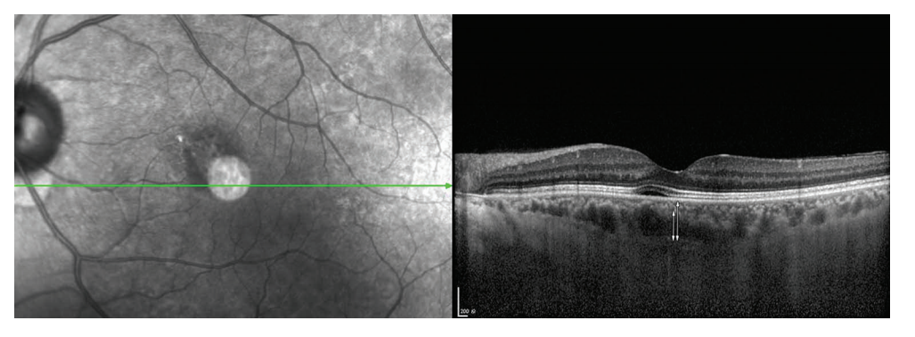
Choroidal thickness was defined as the vertical distance from outer edge of the hyperreflective RPE to the innermost hyperreflective line of the chorio-scleral interface (vertical white arrow). The thickness of the Haller layer was defined as the distance from the innermost point of the horizontal line of the large choroidal blood vessel to the innermost edge of the highly reflective line of the choroid-sclera interface (short white arrow).
Figure 2 Complete recovery of pigment epithelial detachments at the leakage point
这是1名40岁的男性患者。在疾病发作的渗漏点有1个小的PED。在随访时间内观察到完全恢复。
This is a 40-year-old male patient. There is a small pigment epithelial detachment at the leakage point on disease onset. Complete recovery was observed in the follow-up time.
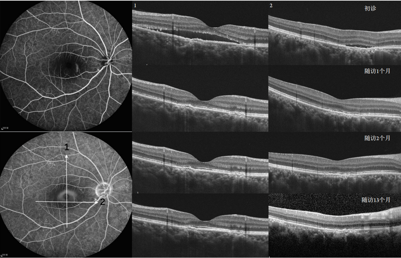
图3 渗漏点处的PED在13个月的随访时间内持续存在
Figure 3 PED at the leak point persisted during the 13-month follow-up time
This is a 33-year-old male patient. In the 13-month follow-up time, although the subretinal fluid resolved completely, separation between RPE and Bruch membrane could still be seen.
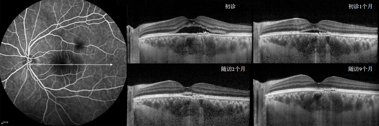
图4 渗漏点处的PED在9个月的随访时间内持续存在
Figure 4 PED at the leak point persisted during the 9-month follow-up time
This is a 39-year-old male patient. By the end of the 9-month follow-up, although the subretinal fluid resolved completely, separation between RPE and Bruch membrane could still be seen.
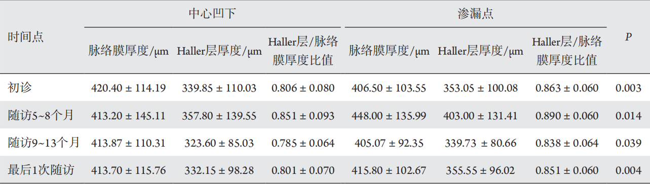
表1 中心凹及渗漏点处脉络膜厚度分析结果
Table 1 Statistical results of choroidal thickness at the fovea and leakage point
1. Zayit-Soudry S, Moroz I, Loewenstein A. Retinal pigment epithelial
detachment[ J]. Surv Ophthalmol, 2007, 52(3): 227-243.
2. Gupta V, Gupta P, Dogra MR, et al. Spontaneous closure of retinal
pigment epithelium microrip in the natural course of central serous
chorioretinopathy[ J]. Eye (Lond), 2010, 24(4): 595-599.
3. Hirami Y, Tsujikawa A, Sasahara M, et al. Alterations of retinal pigment
epithelium in central serous chorioretinopathy[ J]. Clin Experiment
Ophthalmol, 2007, 35(3): 225-230.
4. Fujimoto H, Gomi F, Wakabayashi T, et al. Morphologic changes in
acute central serous chorioretinopathy evaluated by fourier-domain
optical coherence tomography[ J]. Ophthalmology, 2008, 115(9):
1494-1500, 1500-1501.
5. Cheung C, Lee WK, Koizumi H, et al. Pachychoroid disease[ J]. Eye
(Lond), 2019, 33(1): 14-33.
7. Branchini LA, Adhi M, Regatieri CV, et al. Analysis of choroidal
morphologic features and vasculature in healthy eyes using spectral-
domain optical coherence tomography[ J]. Ophthalmology, 2013,
120(9): 1901-1908.
8. Staurenghi G, Sadda S, Chakravarthy U, et al. Proposed lexicon
for anatomic landmarks in normal posterior segment spectral-
domain optical coherence tomography: the IN?OCT consensus[ J].
Ophthalmology, 2014, 121(8): 1572-1578.
9. Chung Y, Kim JW, Choi S, et al. Subfoveal choroidal thickness
and vascular diameter in active and resolved central serous
chorioretinopathy[ J]. Retina, 2018, 38(1): 102-107.
10. Chung YR, Kim JW, Kim SW, et al. Choroidal thickness in patients
with central serous chorioretinopathy: assessment of Haller and Sattler
layers[ J]. Retina, 2016, 36(9): 1652-1657.
11. Lehmann M, Wolff B, Vasseur V, et al. Retinal and choroidal
changes observed with ‘En face’ enhanced-depth imaging OCT
in central serous chorioretinopathy[ J]. Br J Ophthalmol, 2013,
97(9): 1181-1186.
12. Ahn SJ, Kim TW, Huh JW, et al. Comparison of features on SD-OCT
between acute central serous chorioretinopathy and exudative age-
related macular degeneration[ J]. Ophthalmic Surg Lasers Imaging,
2012, 43(5): 374-382.
13. Shinojima A, Hirose T, Mori R, et al. Morphologic findings in acute
central serous chorioretinopathy using spectral domain-optical
coherence tomography with simultaneous angiography[ J]. Retina,
2010, 30(2): 193-202.
14. Daruich A, Matet A, Dirani A, et al. Central serous chorioretinopathy:
Recent findings and new physiopathology hypothesis[ J]. Prog Retin
Eye Res, 2015, 48: 82-118.
15. Daruich A, Matet A, Behar-Cohen F. Central serous chorioretinopathy[ J].
Dev Ophthalmol, 2017, 58: 27-38.
16. Kaye R, Chandra S, Sheth J, et al. Central serous chorioretinopathy:
An update on risk factors, pathophysiology and imaging modalities[ J].
Prog Retin Eye Res, 2020, 79: 100865.
'%20fill='white'%20fill-opacity='0.01'/%3e%3cmask%20id='mask0_3477_29692'%20style='mask-type:luminance'%20maskUnits='userSpaceOnUse'%20x='0'%20y='0'%20width='16'%20height='16'%3e%3crect%20id='&%23232;&%23146;&%23153;&%23231;&%23137;&%23136;_2'%20x='16'%20width='16'%20height='16'%20transform='rotate(90%2016%200)'%20fill='white'/%3e%3c/mask%3e%3cg%20mask='url(%23mask0_3477_29692)'%3e%3cpath%20id='&%23232;&%23183;&%23175;&%23229;&%23190;&%23132;'%20d='M14%205L8%2011L2%205'%20stroke='%23333333'%20stroke-width='1.5'%20stroke-linecap='round'%20stroke-linejoin='round'/%3e%3c/g%3e%3c/g%3e%3c/svg%3e)